ClickCease
+1-915-850-0900 [email protected]
Select Page
Understanding Abnormalities of the Pain System in El Paso, Tx

Understanding Abnormalities of the Pain System in El Paso, Tx

Why does localized damage or injury caused by trauma lead to chronic, intractable pain in certain patients? What’s in charge of the translation of local injury with acute pain into a chronic pain condition? Why does some pain respond to anti-inflammatory drugs and/or medications, whereas other forms of pain require opiates?


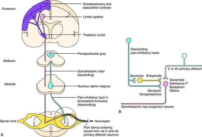
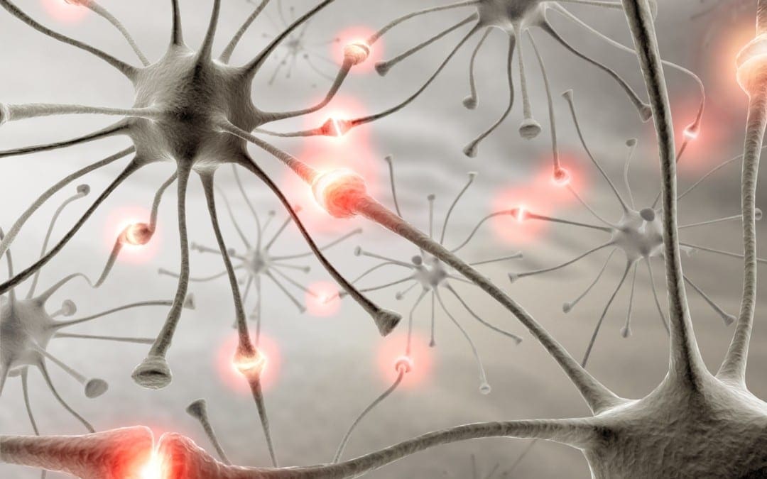
Pain is an intricate process involving both the peripheral nervous system (PNS) and the central nervous system (CNS). Tissue injury triggers the PNS, which transmits signals via the spinal cord into the brain, in which pain perception occurs. However, what causes the intense experience of pain to develop into an unremitting phenomenon? Can anything be done to prevent it? Evidence indicates that chronic pain results from a combination of mechanisms, such as neurological “memories” of preceding pain.


Nociception: The Simplest Pathway


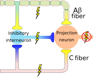
Acute or nociceptive pain is characterized as the regular experience of discomfort which occurs in response to very basic damage or injury. It is protective, warning us to move away from the origin of the insult and take care of the trauma. The mechanisms that create nociceptive pain include transduction, which extends the external traumatic stimulation into electrical activity in specialized nociceptive primary afferent nerves. The afferent nerves then conduct the sensory information from the PNS to the CNS.


In the CNS, the pain data is transmitted by the primary sensory neurons into central projection cells. After the information is transferred to all those areas of the brain which are responsible for our perception, the actual sensory experience happens. Nociceptive pain is a relatively simple reaction to a particularly simple, acute stimulus. But the mechanics in charge of nociceptive pain cannot identify phenomena, such as pain that persists despite removal or healing of the stimulation, such as in the instance of phantom limb pain.


Pain and the Inflammatory Response


In circumstances of more severe injury, such as surgical wounds, tissue damage may stimulate an inflammatory reaction. However, other conditions, especially arthritis, can also be characterized by continuing cases of inflammation associated with intense pain symptoms. The mechanisms for this type of pain related to tissue damage and an inflammatory response are different from early-warning nociceptive pain.


Observing the incision or site of other damage or injury, a cascade of hyperexcitable events occur in the nervous system. This bodily “wind-up” phenomenon begins at the skin, where it is potentiated along the peripheral nerves, and culminates at a hypersensitivity response along the spinal cord (dorsal horn) and the brain. Inflammatory cells then surround the regions of tissue damage and also produce cytokines and chemokines, substances which are intended to mediate the process of healing and tissue regeneration. But, these agents may also be considered irritants and adjust the properties of the primary sensory neurons surrounding the area of trauma.


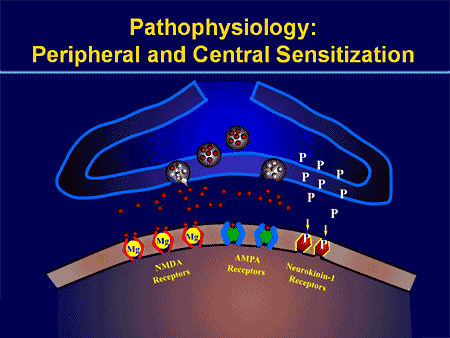
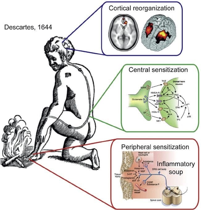
Thus, the major factors which trigger inflammatory pain include damage to the high-threshold nociceptors, known as peripheral sensitization, changes and alterations of the neurons in the nervous system, and the amplification of the excitability of neurons within the CNS. This represents central sensitization and is accountable for hypersensitivity, where areas adjacent to those of the true injury will experience pain as if these were injured. These tissues can also react to stimulation which normally doesn’t create pain, such as a touch, wearing clothing, light pressure, or even brushing your own hair, as if they were truly painful, referred to as allodynia.


Peripheral and Central Sensitization (Video)


Other Mechanisms of Pain


Neuropathic pain results from damage or injury to the nervous system, such as carpal tunnel syndrome, postherpetic neuralgia and diabetic neuropathy. Although some of the mechanisms which seem to cause neuropathic pain overlap with those responsible for inflammatory pain, many of them are different, and thus will need a different approach towards their management.


The process of peripheral and central sensitization is maintained, at least theoretically and experimentally, during the excitatory neurotransmitter, glutamate, which is believed to be released when the N-methyl-D-aspartate (NMDA) receptor is activated.


The nervous system is made up of either inhibitory or excitatory neurotransmitters. Most of what permits our nervous system to respond appropriately to damage or injury is the fine-tuning or inhibition of a variety of processes. The overexcitation of the nervous system is seen to be an issue in a number of different disorders. For instance, overactivation of an NMDA receptor can also be related to affective disorders, sympathetic abnormalities, and even opiate tolerance.


Even ordinary nociceptive pain, to some degree, activates the NMDA receptor and is believed to lead to glutamate release. Nonetheless, in neuropathic pain, oversensitivity to the NMDA receptor is key.


With other types of chronic pain, such as fibromyalgia and tension-type headaches, some of the mechanisms active in inflammatory and neuropathic pain may also create similar abnormalities in the pain system, including central sensitization, higher excitability of the somatosensory pathways, and reductions in central nervous system inhibitory mechanisms.


Peripheral Sensitization


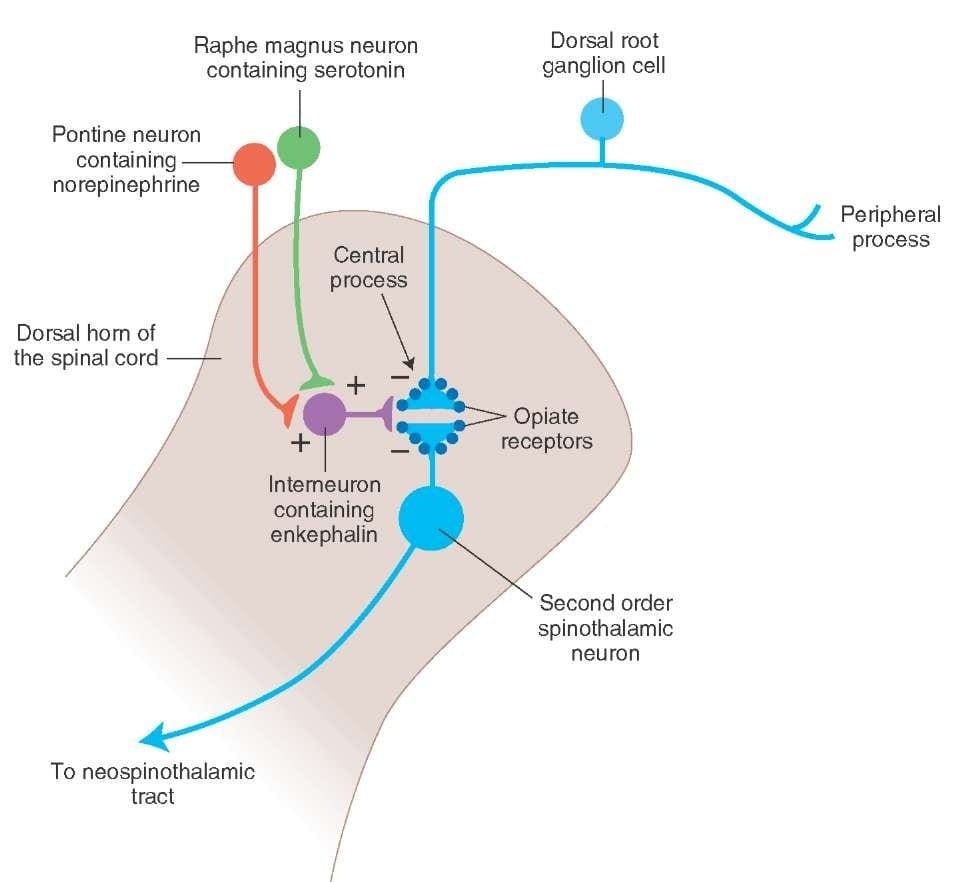
Cyclo-oxygenase (COX) also plays an essential function in both peripheral and central sensitizations. COX-2 is one of the enzymes which are induced during the inflammatory process; COX-2 converts arachidonic acid into prostaglandins, which increase the sensitivity of peripheral nociceptor terminals. Virtually, peripheral inflammation also causes COX-2 to be produced from the CNS. Signals from peripheral nociceptors are partially responsible for this upregulation, but there also seems to be a humoral component to the transduction of the pain signals across the blood-brain barrier.


For instance, in experimental models, COX-2 is generated from the CNS even if animals receive a sensory nerve block prior to peripheral inflammatory stimulation. The COX-2 that is expressed over the dorsal horn neurons of the spinal cord releases prostaglandins, which act on the central terminals, or the presynaptic terminals of nociceptive sensory fibers, to increase transmitter release. Additionally, they act postsynaptically on the dorsal horn neurons to produce direct depolarization. And finally, they inhibit the activity of glycine receptor, and this is an inhibitory transmitter. Therefore, the prostaglandins create an increase in excitability of central neurons.


Peripheral and Central Sensitization | El Paso, TX Chiropractor


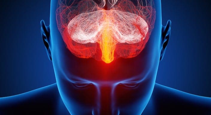
Brain Plasticity and Central Sensitization


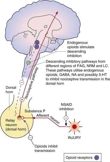
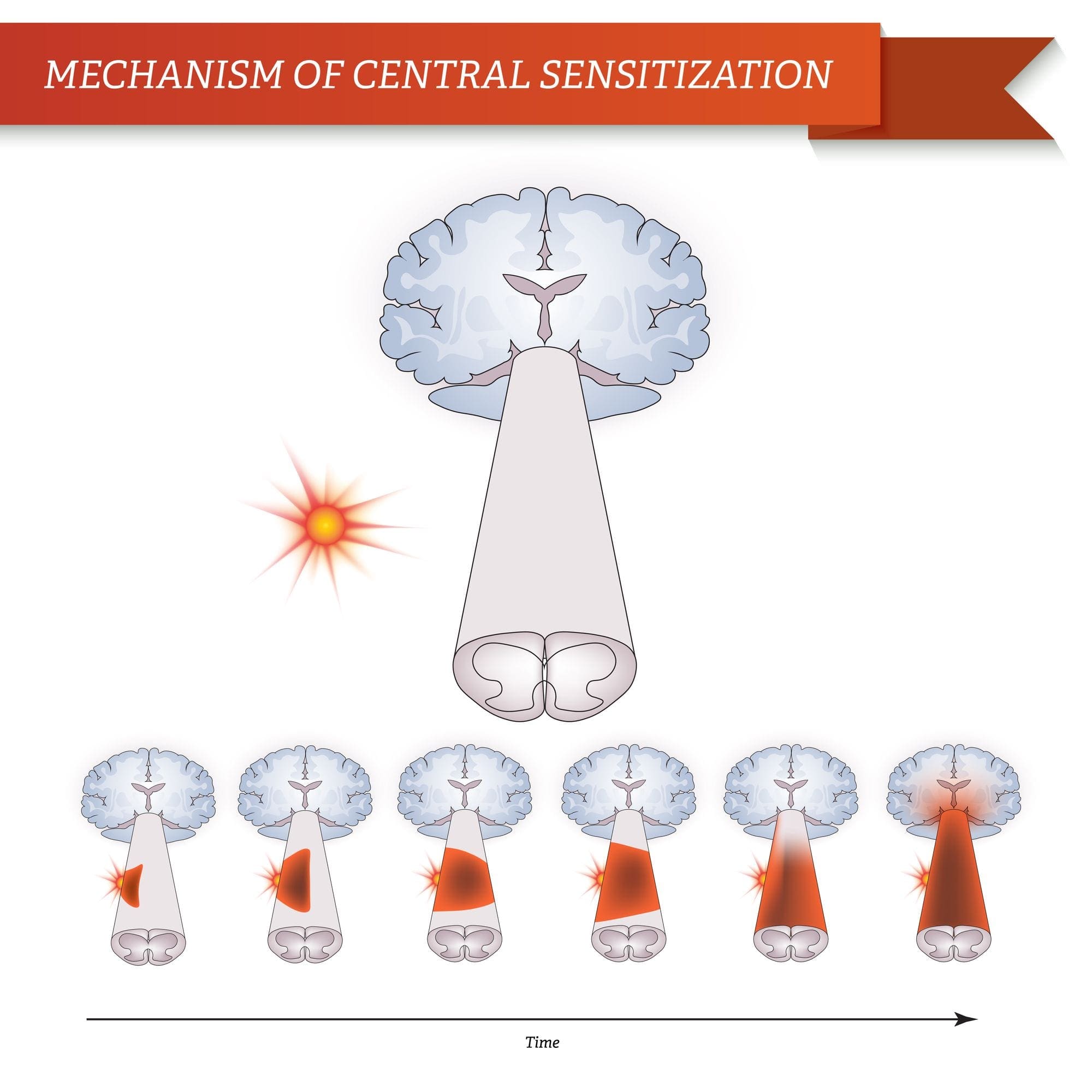
Central sensitization describes changes which happen in the brain in reaction to repeated nerve stimulation. After repeated stimuli, amounts of hormones and brain electric signals change as neurons develop a “memory’ for reacting to those signs. Constant stimulation creates a more powerful brain memory, so the brain will respond more rapidly and effectively when undergoing the identical stimulation in the future. The consequent modifications in brain wiring and reaction are referred to as neural plasticity, which describe the capability of the brain to alter itself readily, or central sensitization. Therefore, the brain is activated or sensitized by previous or repeated stimuli to become more excitable.


The fluctuations of central sensitization occur after repeated encounters with pain. Research in animals indicates that repeated exposure to a painful stimulation will change the animal’s pain threshold and lead to a stronger pain response. Researchers think that these modifications can explain the persistent pain that could occur even after successful back surgery. Although a herniated disc may be removed from a pinched nerve, pain may continue as a memory of the nerve compression. Newborns undergoing circumcision without anesthesia will react more profoundly to future painful stimulation, such as routine injections, vaccinations, and other painful processes. These children haven’t only a higher hemodynamic reaction, known as tachycardia and tachypnea, but they will also develop enhanced crying too.


This neurological memory of pain was studied extensively. In a report on his previous research studies, Woolf noted that the improved reflex excitability following peripheral tissue damage or injury doesn’t rely on continuing peripheral input signals; rather, hours after a peripheral trauma, spinal dorsal horn neuron receptive fields continued to enlarge. Researchers also have documented the significance of the spinal NMDA receptor to the induction and maintenance of central sensitization.


Mechanism of Central Sensitization | El Paso, TX Chiropractor


Cortical Reorganization | El Paso, TX Chiropractor


Significance for Pain Management


Once central sensitization is established, bigger doses of analgesics are often required to suppress it. Preemptive analgesia, or therapy before pain progresses, may lower the effects of all of these stimulation on the CNS. Woolf demonstrated that the morphine dose required to stop central hyperexcitability, given before short noxious electrical stimulation in rats, was one tenth the dose required to abolish activity after it had grown. This translates to clinical practice.


In a clinical trial of 60 patients undergoing abdominal hysterectomy, individuals who received 10 mg of morphine intravenously at the time of induction of anesthesia required significantly less morphine for postoperative pain control. Furthermore, pain sensitivity around the wound, referred to as secondary hyperalgesia, was also reduced in the morphine pretreated group. Preemptive analgesia was used with comparable success in an assortment of surgical settings, including prespinal operation and postorthopaedic operation.


A single dose of 40 or 60 mg/kg of rectal acetaminophen has a clear morphine-sparing effect in day-case surgery in children, if administered in the induction of anesthesia. Furthermore, children with sufficient analgesia with acetaminophen experienced significantly less postoperative nausea and vomiting.


NMDA receptor antagonists have imparted postoperative analgesia when administered preoperatively. Various reports exist in the literature supporting the use of ketamine and dextromethorphan in the preoperative period. In patients undergoing anterior cruciate ligament reconstruction, 24-hour patient-controlled analgesia opioid consumption was significantly less in the preoperative dextromethorphan category versus the placebo group.


In double-blind, placebo-controlled research studies, gabapentin was indicated as a premedicant analgesic for patients undergoing mastectomy and hysterectomy. Preoperative oral gabapentin reduced pain scores and postoperative analgesic consumption without gap in side effects as compared with placebo.


Preoperative administration of nonsteroidal anti-inflammatory drugs (NSAIDs) has demonstrated a significant decrease in opioid use postoperatively. COX-2s are preferable due to their relative lack of platelet effects and significant gastrointestinal safety profile when compared with conventional NSAIDs. Celecoxib, rofecoxib, valdecoxib, and parecoxib, outside the United States, administered preoperatively reduce postoperative narcotic use by more than 40 percent, with many patients using less than half of the opioids compared with placebo.


Blocking nerve conduction in the preoperative period appears to prevent the development of central sensitization. Phantom limb syndrome (PLS) has been attributed to a spinal wind-up phenomenon.�Patients with amputation
often have burning or tingling pain in the body part removed. One possible cause is that nerve fibers at the stump are stimulated and the brain interprets the signals as originating in the amputated portion. The other is the rearrangement within the cortical areas so that area say for the hand now responds to signals from other parts of the body but still interprets them as coming for the amputated hand.


However, for patients undergoing lower-extremity amputation under epidural anesthesia, not one of the 11 patients who received lumbar epidural blockade with bupivacaine and morphine for 72 hours before operation developed PLS. For people who underwent general anesthesia without prior lumbar epidural blockade, 5 of 14 patients had PLS at 6 weeks and 3 continued to experience PLS at 1 year.


Woolf and Chong have noted that perfect preoperative, intraoperative, and postoperative treatment comprises of “NSAIDs to reduce the activation/centralization of nociceptors, local anesthetics to block sensory inflow, and centrally acting drugs such as opiates.” Decreasing perioperative pain with preemptive techniques enhances satisfaction, hastens discharge, spares opioid use, along with diminished constipation, sedation, nausea, and urinary retention, and may even stop the development of chronic pain. Anesthesiologists and surgeons should consider integrating these techniques in their everyday practices.


When pain occurs as a result of damage or injury in consequence of surgery, the spinal cord can attain a hyperexcitable state wherein excessive pain reactions occur that may persist for days, weeks or even years.


Why does localized injury resulting from trauma result in chronic, intractable pain in some patients? Tissue injury leads to a constellation of changes in spinal excitability, including elevated spontaneous firing, greater response amplitude and length, decreased threshold, enhanced discharge to repeated stimulation, and expanded receptive fields. The persistence of these changes, which are collectively termed central sensitization, appears to be fundamental to the prolonged enhancement of pain sensitivity which defines chronic pain. Numerous drugs and/or medications as well as local anesthetic neural blockade may limit the magnitude of the central nervous system (CNS) windup, as evidenced by diminished pain and diminished opioid consumption in the preemptive analgesic models.


Dr-Jimenez_White-Coat_01.png

Dr. Alex Jimenez’s Insight

Chiropractic care is an alternate treatment option which utilizes spinal adjustments and manual manipulations to safely and effectively restore as well as maintain the proper alignment of the spine. Research studies have determined that spinal misalignments, or subluxations, can lead to chronic pain. Chiropractic care is commonly utilized for pain management, even if the symptoms are not associated to an injury and/or condition in the musculoskeletal and nervous system. By carefully re-aligning the spine, a chiropractor can help reduce stress and pressure from the structures surrounding the main component of out body’s foundation, ultimately providing pain relief.


Enteric Nervous System Function and Pain


When it comes to the diminished use of drugs and/or medications, including opioids, in order to prevent side-effects like gastrointestinal health issues, the proper function of the enteric nervous system may be at play.


The enteric nervous system (ENS) or intrinsic nervous system is one of the key branches of the autonomic nervous system (ANS) and consists of a mesh-like system of nerves which modulates the role of the gastrointestinal tract. It’s capable of acting independently of the sympathetic and parasympathetic nervous systems, even though it might be affected by them. The ENS can also be called the second brain.�It is derived from neural crest cells.


The enteric nervous system in humans is made up of some 500 million neurons, including the numerous types of Dogiel cells, approximately one two-hundredth of the amount of neurons in the brain. The enteric nervous system is inserted into the lining of the gastrointestinal system, beginning at the esophagus and extending down to the anus. Dogiel cells, also known as cells of Dogiel, refers to some kind of multipolar adrenal tissues within the prevertebral sympathetic ganglia.


Cells of Dogiel | El Paso, TX Chiropractor


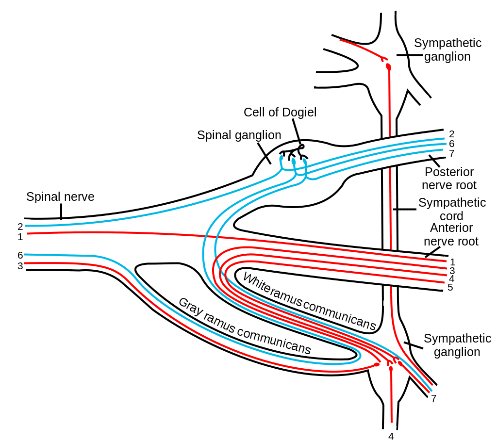
The ENS is capable of autonomous functions, such as the coordination of reflexes; even though it receives considerable innervation in the autonomic nervous system, it does and can operate independently of the brain and the spinal cord.�The enteric nervous system has been described as the “second brain” for a number of reasons. The enteric nervous system may operate autonomously. It normally communicates with the central nervous system (CNS) via the parasympathetic, or via the vagus nerve, and the sympathetic, that is through the prevertebral ganglia, nervous systems. However, vertebrate studies reveal that when the vagus nerve is severed, the enteric nervous system continues to function.


In vertebrates, the enteric nervous system includes efferent neurons, afferent neurons, and interneurons, all of which make the enteric nervous system capable of carrying reflexes and acting as an integrating center in the absence of CNS input. The sensory neurons report on mechanical and chemical conditions. The enteric nervous system has the ability to change its response based on such factors as nutrient and bulk composition. In addition, ENS contains support cells that are much like astroglia of the brain and a diffusion barrier around the capillaries surrounding ganglia that’s like the blood-brain barrier of blood vessels.


The enteric nervous system (ENS) plays a pivotal role in inflammatory and nociceptive processes. Drugs and/or medications that interact with the ENS have recently raised considerable interest because of their capacity to regulate numerous aspects of the gut physiology and pathophysiology. In particular, experiments in animals have demonstrated that�proteinase-activated receptors (PARs) may be essential to neurogenic inflammation in the intestine. Moreover, PAR2 agonists seem to induce intestinal hypersensitivity and hyperalgesic states, suggesting a role for this receptor in visceral pain perception.


Furthermore, PARs, together with the proteinases that activate them, represent exciting new targets for therapeutic intervention on the ENS. The scope of our information is limited to chiropractic as well as to spinal injuries and conditions. To discuss the subject matter, please feel free to ask Dr. Jimenez or contact us at 915-850-0900 .


Curated by Dr. Alex Jimenez


Green-Call-Now-Button-24H-150x150-2-3.png


Additional Topics: Sciatica

Sciatica is medically referred to as a collection of symptoms, rather than a single injury and/or condition. Symptoms of sciatic nerve pain, or sciatica, can vary in frequency and intensity, however, it is most commonly described as a sudden, sharp (knife-like) or electrical pain that radiates from the low back down the buttocks, hips, thighs and legs into the foot. Other symptoms of sciatica may include, tingling or burning sensations, numbness and weakness along the length of the sciatic nerve. Sciatica most frequently affects individuals between the ages of 30 and 50 years. It may often develop as a result of the degeneration of the spine due to age, however, the compression and irritation of the sciatic nerve caused by a bulging or herniated disc, among other spinal health issues, may also cause sciatic nerve pain.


blog picture of cartoon paperboy big news


EXTRA IMPORTANT TOPIC: Chiropractor Sciatica Symptoms


MORE TOPICS: EXTRA EXTRA: El Paso Back Clinic | Back Pain Care & Treatments

Pain Modulation Pathway Mechanisms in El Paso, TX

Pain Modulation Pathway Mechanisms in El Paso, TX

Most, if not all, ailments of the body trigger pain. Pain is interpreted and sensed in the brain. Pain is modulated by two key types of drugs which operate on the brain: analgesics and anesthetics. The term analgesic refers to a medication that relieves pain without loss of consciousness. The expression central anesthesia refers to a medication that depresses the CNS. It’s distinguished by the lack of all perception of sensory modalities, for instance, loss of consciousness without loss of critical functions.


Opiate Analgesia (OA)


The most successful clinically used drugs for producing temporary analgesia and relief from pain are the opioid family, which includes morphine, and heroin. There are currently no additional powerful pain therapeutic options to opiates. Several side effects caused by opiate use include tolerance and drug dependence or addiction. In general, these drugs modulate the incoming pain information in the spine and central nervous system, in addition to relieve pain temporarily, and can also be called opiate producing analgesia (OA). Opiate antagonist is a drug that antagonizes the opioid effects, such as naloxone or maltroxone, etc.. They are competitive antagonists of opiate receptors. However, the brain has a neuronal circuit and endogenous substances which modulate pain.


Endogenous Opioids


Opioidergic neurotransmission is located throughout the brain and spinal cord and is believed to influence many functions of the central nervous system, or CNS, such as nociception, cardiovascular functions, thermoregulation, respiration, neuroendocrine functions, neuroimmune functions, food consumption, sexual activity, competitive locomotor behaviour as well as memory and learning. Opioids exert marked effects on mood and motivation and produce a sense of euphoria.


Three classes of opioid receptors are identified: ?-mu, ?-delta and ?-kappa. All 3 classes are widely dispersed in the brain. The genes encoding each one of these have been cloned and found to function as members of the G protein receptors. Moreover, three major types of endogenous opioid peptides that interact with the above opiate receptors have been recognized in the central nervous system, including, ?-endorphins, enkephalins and the dynorphins. These 3 opioid peptides are derived from a large protein receptor by three different genes, such as the proopiomelanocortin, or POMC, gene, the proenkephalin gene and the prodynorphin gene.�The opioid peptides modulate nociceptive input in two ways: first, they block neurotransmitter release by inhibiting Ca2+ influx into the presynaptic terminal, or second, they open potassium channels, which hyperpolarizes neurons and inhibits spike activity. They act on various receptors within the brain and spinal cord.


Enkephalins are considered the putative ligands for the ? receptors, ? endorphins for its ?-receptors, and dynorphins for the ? receptors. The various types of opioid receptors are distributed differently within the peripheral and central nervous system, or CNS. There’s evidence for functional differences in these receptors in various structures. This explains why many undesirable side effects occur after opiate treatments. For instance, mu (?) receptors are widespread in the brain stem parabrachial nuclei, where a respiratory center and inhibition of these neurons may cause what’s known as respiratory depression.


Endogenous Opioids Diagram 4 | El Paso, TX Chiropractor


Central or peripheral terminals of nociceptive afferent fibers feature opiate receptors in which exogenous and endogenous opioids could act to modulate the capability to transmit nociceptive information. Additionally, high densities of opiate receptors are found in periaqueductal gray, or PAG, nucleus raphe magnus, or NRM, and dorsal raphe, or DR, from the rostral ventral medulla, in the spinal cord, caudate nucleus, or CN, septal nucleus, hypothalamus, habenula and hippocampus.�Systemically administered opioids at analgesic dosages activate spinal and supraspinal mechanisms via ?, ?, and ? type opioid receptors and regulate pain signals to modulate symptoms.


Neuronal Circuits and Pain Modulation


For many decades it was suggested that somewhere in the central nervous system there is a circuit which can modulate incoming pain details. The gate control theory and the ascending/descending pain transmission system are two suggestions of such a circuit. Below, we will discuss both in further detail.


Gate Control Theory


The initial pain modulatory mechanism known as the gate control theory, has been proposed by Melzack and Wall in the mid 1960’s. The notion of the gate control theory is that non-painful input closes the gates to painful input, which results in avoidance of the pain sensation from travel into the CNS, for example, non-noxious input, or stimulation, suppresses pain.


The theory implies that collaterals of the large sensory fibers carrying cutaneous sensory input activate inhibitory interneurons, which inhibit and regulate pain transmission data carried from the pain fibers. Non-noxious input inhibits pain, or sensory input, and closes the gate to noxious input. The gate control theory demonstrates that in the spinal cord level, non-noxious stimulation will create presynaptic inhibition on dorsal root nociceptor fibers that synapse on nociceptors spinal neurons (T). This presynaptic inhibition will also prevent incoming noxious information from reaching the CNS, for example, it will shut the gate to incoming toxic information.


Gate Control Theory Diagram 1 | El Paso, TX Chiropractor


The gate control theory was the rationale for the idea behind the production and utilization of the transcutaneous electrical nerve stimulation, or TENS, for pain relief. In order to be effective, the TENS unit generates two different present frequencies below the pain threshold that can be taken by the patient. This process has found a degree of achievement in chronic pain treatment.


Pain Modulation: Gate Control Theory


Stimulation Produced Analgesia (SPA)


Evidence for an inherent analgesia system was found by intracranial electrical stimulation of certain discrete brain regions. These areas would be the periaqueductal gray, or PAG, and nucleus raphe magnus, or NRM, dorsal raphe, or DR, caudate nucleus, or CN, septal nucleus, or Spt, along with other nuclei. Such stimulation or sensory signals, inhibits pain, making analgesia without behavioral suppression, while the touch, temperature and pressure sensation stays intact. According to research studies, SPA, or stimulation produced analgesia, is more pronounced and continues for a longer period of time after stimulation in humans than in experimental animals. Additionally, during SPA, the subjects, however, still respond to nonpainful stimulation like temperature and touch within the circumscribed region of analgesia. The most effective CNS, or central nervous system regions for SPA to occur, would be in the PAG and the raphe nuclei, or RN.


Electrical stimulation of PAG or NRM inhibits spinal thalamic cells, or spinal neurons that project monosynaptically to the thalamus, in laminae I, II and V to ensure the noxious information from the nociceptors which are ultimately modulated in the level of the spinal cord. Furthermore, PAG has neuronal connections to the nucleus raphe magnus, or NRM.


The activity of the PAG most likely occurs by activation of the descending pathway from NRM and likely also by activation of ascending connections acting on greater subcortical levels of the CNS. In addition, electric stimulation of PAG or NRM produces behavioral analgesia, or stimulation produced analgesia. Stimulation produced analgesia, or SPA causes the release of endorphins which can be blocked by the opiate antagonist naloxone.


During PAG and/or RN stimulation, serotonin, also medically referred to as 5-HT, can also be discharged from ascending and descending axons from subcortical nuclei, in spinal trigeminal nuclei and in the spinal cord. This release of 5-HT modulates and regulates pain transmission by inhibiting or blocking incoming neural action. Depletion of 5-HT by electrical lesion of the raphe nuclei or with a neurotoxic lesion made by local injection of a chemical agent such as parachlorophenylalanine, or PCPA, results in blocking the power of opiate, both intracranial and systemic, as well as that of electrical stimulation in order to produce analgesia.


To confirm if the electric stimulation produced analgesia via the release of opiate and dopamine, then the region is locally microinjected with morphine or 5-HT. All these microinjections ultimately create analgesia. These processes also provide a way of identifying brain areas related to pain suppression and assist to produce a map of pain centers. The most effective way of producing opiate analgesia, or OA, is by intracerebral injection of morphine into the PAG.


The PAG and RN as well as other brain structures in which analgesia is produced, are also rich in opiate receptors. Intracerebral opioid administration produced analgesia and SPA can be blocked by systemic or from local microinjections of naloxone, the morphine antagonist, into the PAG or RN. For that reason, it’s been suggested that the two, both OA and SPA, operate by a frequent mechanism.


If OA and SPA behave through the same intrinsic system, then the hypothesis that opiates activate a pain-suppression mechanism is much more likely. As a matter of fact, current evidence suggests that microinjections of an opiate into the PAG activate an efferent brainstem system which inhibits pain transmission at segmental spinal cord levels. These observations imply that analgesia elicited from the periaqueductal gray, or PAG, demands a descending pathway into the spinal cord.


Dr-Jimenez_White-Coat_01.png

Dr. Alex Jimenez’s Insight

Pain modulation occurs through the process of electrical brain stimulation which occurs due to the activation of descending inhibitory fibers, which regulate or inhibit the input and output of certain neurons. What has been described as opioid and serotonergic antagonists, is believed to reverse both local opiate analgesia and brain-stimuli generated analgesia. The sensory signals or impulses in the central nervous system are ultimately controlled by both ascending and descending inhibitory systems, utilizing endogenous opioids or other endogenous substances, such as serotonin as inhibitory mediators. Pain is a complex perception which can also be influenced by a variety of other factors, including emotional state.


Mechanisms of Pain Modulation


Ascending and Descending Pain Suppression Mechanism


The primary ascending pain fibers, such as the A ? and C fibers, reach the dorsal horn of the spinal cord from peripheral nerve areas in order to innervate the nociceptor neurons in Rexed laminae I & II. Cells from Rexed lamina II make synaptic connections in Rexed layers IV to VII. Cells, particularly within laminae I and VII of the dorsal horn, give rise to ascending spinothalamic tracts. In the spinal level, opiate receptors are located in the presynaptic endings of their nocineurons and in the interneural level layers IV to VII from the dorsal horn.


Activation of opiate receptors at the interneuronal level produces hyperpolarization of the neurons, which lead to the the inhibition of activation as well as the release of substance P, a neurotransmitter involved in pain transmission, thus preventing pain transmission. The circuit which consists of the periaqueductal gray, or PAG, matter in the upper brain stem, the locus coeruleus, or LC, the nucleus raphe magnus, or the NRM, and the nucleus reticularis gigantocellularis, or Rgc,� leads to the descending pain suppression pathway, which inhibits incoming pain data at the spinal cord level.


As stated before, opioids interact with the opiate receptors in distinct central nervous system levels. These opiate receptors are the normal target regions for hormones and endogenous opiates, such as the endorphins and enkephalins. Due to binding at the receptor in subcortical websites, secondary changes which result in some change in the electrophysiological properties of the neurons and regulation of their ascending pain information.


Ascending and Descending Pain Suppression Mechanism Diagram 2 | El Paso, TX Chiropractor


Ascending and Descending Pain Suppression Mechanism Diagram 3 | El Paso, TX Chiropractor


What activates the PAG to exert its consequences? It was discovered that noxious stimulation triggers neurons in the nucleus reticularis gigantocellularis, or RGC. The nucleus Rgc innervates both PAG and NRM. The PAG sends axons into the NRM, and nerves in the NRM send their axons to the spinal cord. Additionally, bilateral dorsolateral funiculus, or DLF, lesions, referred to as DLFX, block the analgesia produced by both electrical stimulation and by microinjection of opiates directly into the PAG and NRM, but they just attenuate the systemic analgesic effects of opiates. These observations support the hypothesis that discrete descending pathways from the DLF are necessary for both OA and SPA.


The DLF is comprised of fibers originating from several brainstem nuclei, which can be serotonergic, or 5-HT, from nerves located inside the nucleus raphe magnus, or NRM; dopaminergic neurons originating from ventral tegmental area, or VTA, and adrenergic neurons originating from the locus coeruleus, or LC. These descending fibers suppress noxious input in the nociceptive spinal cord neurons in laminae I, II, and V.


Opiate receptors have also been discovered in the dorsal horn of the spinal cord, chiefly in Rexed laminae I, II, and V, and such spinal opiate receptors mediate inhibitory effects on dorsal horn neurons transmitting nociceptive information. The action of morphine seems to be exerted equally in the spinal cord and brainstem nuclei, including the PAG and NRM. Systemic morphine acts on both brain stem and spinal cord opiate receptors to produce analgesia. Morphine binds the brainstem opiate receptors, which triggers the brainstem descending serotonergic pathway into the spinal cord as well as the DLF, and these have an opioid-mediated synapse at the level of the spinal cord.


This observation demonstrates that noxious stimuli, instead of non-noxious stimulus, determine the gate control theory, which are critical for the activation of the descending pain modulation circuit where pain inhibits pain via the descending DLF pathway. In addition, there are ascending connections in the PAG and the raphe nuclei into the PF-CM complex. These thalamic regions are a part of the ascending pain modulation at the diencephalon degree.


Stress Induced Analgesia (SIA)


Analgesia may be produced in certain stressful circumstances. Exposure to many different stressful or painful events generates an analgesic response. This phenomenon is known as stress induced analgesia, or SIA. Stress induced analgesia has been believed to give insight into the physiological and psychological factors that trigger endogenous pain control and opiate systems. By way of instance, soldiers injured in battle or athletes hurt in sports sometimes report that they don’t feel pain or discomfort during the battle or game, nevertheless, they will go through the pain afterwards once the specific situation has stopped. It’s been demonstrated in animals that electrical shocks cause stress-induced analgesia. Based on these experiments, it is assumed that the pressure the soldiers and the athletes experienced suppressed the pain which they would later experience.


It’s believed that endogenous opiates are produced in response to stress and inhibit pain by triggering the midbrain descending system. Furthermore, some SIA exhibited cross tolerance with opiate analgesia, which indicates that this SIA is mediated via opiate receptors. Experiments using different parameters of electrical shock stimulation demonstrate such stress induced analgesia and some of those anxieties that produce analgesia could be blocked by the opioid antagonist naloxone, whereas others were not blocked by naloxone. In conclusion, these observations lead to the decision that both opiate and non-opiate forms of SIA exist.


Somatovisceral Reflex


The somatovisceral reflex is a reflex in which visceral functions are activated or inhibited by somatic sensory stimulation. In experimental animals, both noxious and innocuous stimulation of somatic afferents are proven to evoke reflex changes in sympathetic efferent activity and, consequently, effector organ function. These phenomena have been shown in such regions as the gastrointestinal tract, urinary tract, adrenal medulla, lymphatic cells, heart and vessels of the brain and peripheral nerves.


Most frequently, incisions are elicited experimentally by stimulation of cutaneous afferents, even though some work has also been conducted on muscle and articular afferents, including those of spinal cells. The ultimate responses will represent the integration of multiple tonic and reflex influences and might exhibit laterality and segmental trends as well as variable excitability in line with the afferents involved. Given the complexity and multiplicity of mechanisms involved in the last expression of the reflex response, attempts to extrapolate to clinical situations should most likely be conducted in favor of further systematic physiological studies.


The scope of our information is limited to chiropractic as well as to spinal injuries and conditions. To discuss the subject matter, please feel free to ask Dr. Jimenez or contact us at 915-850-0900 .


Curated by Dr. Alex Jimenez


Green-Call-Now-Button-24H-150x150-2-3.png


Additional Topics: Sciatica

Sciatica is medically referred to as a collection of symptoms, rather than a single injury and/or condition. Symptoms of sciatic nerve pain, or sciatica, can vary in frequency and intensity, however, it is most commonly described as a sudden, sharp (knife-like) or electrical pain that radiates from the low back down the buttocks, hips, thighs and legs into the foot. Other symptoms of sciatica may include, tingling or burning sensations, numbness and weakness along the length of the sciatic nerve. Sciatica most frequently affects individuals between the ages of 30 and 50 years. It may often develop as a result of the degeneration of the spine due to age, however, the compression and irritation of the sciatic nerve caused by a bulging or herniated disc, among other spinal health issues, may also cause sciatic nerve pain.


blog picture of cartoon paperboy big news


EXTRA IMPORTANT TOPIC: Chiropractor Sciatica Symptoms


MORE TOPICS: EXTRA EXTRA: El Paso Back Clinic | Back Pain Care & Treatments

What is Central Sensitization? | El Paso, TX Chiropractor

What is Central Sensitization? | El Paso, TX Chiropractor

Central sensitization is a state of the nervous system that’s related to the development and maintenance of chronic pain. When central sensitization occurs, the nervous system goes through a procedure known as wind-up and gets regulated in a constant condition of increased reactivity. This persistent, or regulated, state of reactivity decreases the threshold for what causes pain and subsequently learns to keep pain after the initial injury has healed. Central sensitization has two major characteristics. Both have an increased sensitivity to pain and to the feeling of touch. These are referred to as allodynia and hyperalgesia.


Allodynia occurs when an individual experiences pain with circumstances that are normally not supposed to be painful. For instance, chronic pain patients often experience pain even with things as simple as touch or a massage. In these situations, nerves in the region which has been touched sends signals through the nervous system into the brain. Because the nervous system is in a constant condition of heightened reactivity, the brain doesn’t generate a mild feeling of touch as it should, given that the stimulus that initiated it was an easy touch or massage. Instead, the brain produces a feeling of pain and discomfort.


Hyperalgesia occurs when a stimulus that’s usually considered to be somewhat painful is perceived as a much more debilitating pain than it ought to be. For instance, chronic pain patients that experience a simple bump, which generally would be mildly painful, will often feel intense pain. Again, once the nervous system is in a constant condition of high reactivity, it amplifies pain.


Peripheral and Central Sensitization


Chronic pain patients sometimes believe they might be suffering from a mental health issue because they understand from common sense that touch or simple bumps produce tremendous amounts of pain or discomfort. Other times, it’s not the patients themselves who feel this way, but their friends and family members. Individuals who don’t suffer with chronic pain may witness others who have central sensitization experience pain at the slightest touch or cry out at the simplest bump. However, because they don’t have the condition, it may be difficult for them to understand what someone who does is going through.


In addition to allodynia and hyperalgesia, central sensitization has other well-known features, though they may occur less commonly. Central sensitization may lead to heightened sensitivities throughout all senses, not only the feeling of touch. Chronic pain patients can sometimes report sensitivities to light, smell and sound. As such, regular levels of light may seem overly bright or even the perfume aisle in the department shop can produce a headache. Central sensitization can also be associated with cognitive deficits, such as poor concentration and poor short-term memory. Central sensitization also interferes with increased levels of psychological distress, particularly fear and axiety. After all, the nervous system is responsible for not merely senses, like pain, but also emotions. If the nervous system is trapped in a constant condition of reactivity, patients are going to be nervous or anxious. Lastly, central sensitization is also correlated with sick role behaviors, such as resting and malaise, and pain behavior.


Central sensitization has long been known as a potential consequence of stroke and spinal cord injury. However, it is increasingly believed that it plays a part in several different chronic pain disorders. It may happen with chronic low back pain, chronic neck pain, whiplash injuries, chronic tension headaches, migraine headaches, rheumatoid arthritis, osteoarthritis of the knee, endometriosis, injuries sustained in an automobile accident, and even following surgeries. Fibromyalgia, irritable bowel syndrome, and chronic fatigue syndrome, all appear to occur due to central sensitization as well.


Central Sensitization and C Fibers


What Causes Central Sensitization?


Central sensitization involves specific changes to the nervous system. Changes in the dorsal horn of the spinal cord and in the brain occur, particularly at the cellular level, such as at the receptor sites. As mentioned previously, it has long been proven that fractures and spinal cord injuries can cause central sensitization. It stands to reason. Strokes and spinal cord injuries cause harm to the central nervous system, including the brain, in the event of strokes, and the spinal cord, in the case of spinal cord injuries. These injuries change the sections of the nervous system which are involved in central sensitization.


However, what about the other, more prevalent, types of chronic pain disorders, recorded above, such as headaches, chronic back pain, or pain in the extremities? The accidents or conditions which lead to these kinds of chronic pain are not direct injuries to the brain or spinal cord. Rather, they include injuries or condition which affect the peripheral nervous system, particularly in that are of the nervous system which lies outside the spinal cord and brain. How can health issues associated with the peripheral nervous system contribute to modifications in the central nervous system and cause chronic pain in the isolated area of the initial injury? In summary, how can isolated migraine headaches eventually become chronic daily headaches? How can an acute low back lifting injury become chronic low back pain? How does an injury to the hand or foot turn into a complex regional pain syndrome?


There are probably multiple factors that cause the development of central sensitization in these ‘peripheral’ chronic pain disorders. These variables may be divided into two classes:


  • Factors that are associated with the state of the central nervous system before onset of the initial pain or injury condition
  • Factors that are associated with the central nervous system following onset of the initial pain or injury condition


The first group involves those factors that might predispose individuals to developing central sensitization once an accident occurs and the next group involves antecedent factors that boost central sensitization once pain begins.


Dr-Jimenez_White-Coat_01.png

Dr. Alex Jimenez’s Insight

Chronic pain can often modify the way the central nervous system itself functions, so much so that a patient may become more sensitive to pain with less provocation. This is what’s referred to as central sensitization and it generally involves changes in the central nervous system, or CNS, more specifically, in the brain and the spinal cord. Central sensitization has been associated with several common diseases and it’s even been reported to develop with something as simple as a muscle ache. Central sensitization has also been documented to persist and worsen even in the absence of obvious provocation. Several factors have also been attributed with the development of central sensitization, although the true cause is still unknown.


Predisposing Factors for Central Sensitization


There are probably biological, emotional, and environmental predisposing factors for central sensitization. Low and higher sensitivity to pain, or pain thresholds, are perhaps in part due to numerous genetic factors. While there’s absolutely no research as of yet to support a causal link between pre-existing pain thresholds and following development of central sensitization after an incident, it’s largely assumed that it will be eventually found.


Psychophysiological factors, like the stress-response, are also apt to play a part in the development of central sensitization. Direct experimental evidence on animals and humans, as well as prospective studies on humans, have demonstrated a connection between stress and the decrease of pain thresholds. Similarly, different kinds of pre-existing anxiety about pain is consistently related to higher pain sensitivities. All these psychophysiological aspects suggest that the preexisting state of the nervous system is also an important determinant of creating central sensitization after the onset of pain. If the stress response has made the nervous system responsive prior to injury, then the nervous system might be more prone to become sensitized once onset of pain happens.


There is considerable indirect evidence for this theory as well. A prior history of anxiety, physical and psychological trauma, and depression are predictive of onset of chronic pain later in life. The most common denominator between chronic pain, anxiety, nervousness, injury, and depression, is the nervous system. They’re all states of the nervous system, especially a persistently changed, or dysregulated, nervous system.


It’s not that such pre-existing health issues make individuals more vulnerable to injury or the onset of illness, as injury or illness is apt to happen on a somewhat random basis across the populace. Instead, these pre-existing health issues are more inclined to make people prone to the development of chronic pain once an injury or disease occurs. The dysregulated nervous system, at the time of injury, for instance, may interfere with the normal trajectory of healing and thereby stop pain from subsiding once tissue damage is healed.


Factors Resulting in Central Sensitization After Onset of Pain


Predisposing factors may also be part of the development of central sensitization. The onset of pain is frequently associated with subsequent development of conditions, such as depression, fear-avoidance, nervousness or anxiety and other phobias. The stress of those responses can, in turn, further exacerbate the reactivity of the nervous system, leading to central sensitization.�Inadequate sleep is also a frequent effect of living with chronic pain. It’s associated with increased sensitivity to pain as well. In what’s technically known as operant learning, interpersonal and environmental reinforcements have long been proven to lead to pain behaviors, however, it is also evident that such reinforcements may lead to the development of central sensitization.


Mayo Clinic Discusses Central Sensitization


Treatments of Central Sensitization


Treatments for chronic pain syndromes that involve fundamental sensitization typically target the central nervous system or the inflammation which corresponds with central sensitization. All these often generally include antidepressants and anticonvulsant medications, and cognitive behavioral treatment. While usually not considered to target the central nervous system, regular mild aerobic exercise changes structures in the central nervous system and contributes to reductions in the pain of many ailments which are mediated by central sensitization. As such, moderate aerobic exercise is used to treat chronic pain syndromes marked by central sensitization. Non-steroidal anti-inflammatories are utilized for the inflammation associated with central sensitization.


Finally, chronic pain rehabilitation programs are a standard, interdisciplinary treatment that employs each of the above-noted therapy strategies in a coordinated manner. They also make the most of the research on the role of operant learning from central sensitization and also have developed behavioral interventions to reduce the pain and discomfort associated with the health issue. Such applications are typically considered the most effective treatment option for chronic pain syndromes.�The scope of our information is limited to chiropractic as well as to spinal injuries and conditions. To discuss the subject matter, please feel free to ask Dr. Jimenez or contact us at 915-850-0900 .


Curated by Dr. Alex Jimenez

Green-Call-Now-Button-24H-150x150-2-3.png

Additional Topics: Sciatica

Sciatica is medically referred to as a collection of symptoms, rather than a single injury and/or condition. Symptoms of sciatic nerve pain, or sciatica, can vary in frequency and intensity, however, it is most commonly described as a sudden, sharp (knife-like) or electrical pain that radiates from the low back down the buttocks, hips, thighs and legs into the foot. Other symptoms of sciatica may include, tingling or burning sensations, numbness and weakness along the length of the sciatic nerve. Sciatica most frequently affects individuals between the ages of 30 and 50 years. It may often develop as a result of the degeneration of the spine due to age, however, the compression and irritation of the sciatic nerve caused by a bulging or herniated disc, among other spinal health issues, may also cause sciatic nerve pain.


blog picture of cartoon paperboy big news


EXTRA IMPORTANT TOPIC: Chiropractor Sciatica Symptoms


MORE TOPICS: EXTRA EXTRA: El Paso Back Clinic | Back Pain Care & Treatments

Blank
References

1.�Phillips, K. & Clauw, D. J. (2011). Central pain mechanisms in chronic pain states � maybe it is all in their head.�Best Practice Research in Clinical Rheumatology, 25, 141-154.

2.�Yunus, M. B. (2007). The role of central sensitization in symptoms beyond muscle pain, and the evaluation of a patient with widespread pain.�Best Practice Research in Clinical Rheumatology, 21, 481-497.

3.�Curatolo, M., Arendt-Nielsen, L., & Petersen-Felix, S. (2006). Central hypersensitivity in chronic pain: Mechanisms and clinical implications.�Physical Medicine and Rehabilitation Clinics of North America, 17, 287-302.

4.�Wieseler-Frank, J., Maier, S. F., & Watkins, L. R. (2005). Immune-to-brain communication dynamically modulates pain: Physiological and pathological consequences.�Brain, Behavior, & Immunity, 19, 104-111.

5.�Meeus M., & Nijs, J. (2007). Central sensitization: A biopsychosocial explanation for chronic widespread pain in patients with fibromyalgia and chronic fatigue syndrome.�Clinical Journal of Rheumatology, 26, 465-473.

6. Melzack, R., Coderre, T. J., Kat, J., & Vaccarino, A. L. (2001). Central neuroplasticity and pathological pain.�Annals of the New York Academy of Sciences, 933, 157-174.

7.�Flor, H., Braun, C., Elbert, T., & Birbaumer, N. (1997). Extensive reorganization of primary somatosensory cortex in chronic back pain patients.�Neuroscience Letters, 224, 5-8.

8. O�Neill, S., Manniche, C., Graven-Nielsen, T., Arendt-Nielsen, L. (2007). Generalized deep-tissue hyperalgesia in patients with chronic low-back pain.�European Journal of Pain, 11, 415-420.

9.�Chua, N. H., Van Suijlekom, H. A., Vissers, K. C., Arendt-Nielsen, L., & Wilder-Smith, O. H. (2011). Differences in sensory processing between chronic cervical zygapophysial joint pain patients with and without cervicogenic headache.�Cephalalgia, 31, 953-963.

10.�Banic, B, Petersen-Felix, S., Andersen O. K., Radanov, B. P., Villiger, P. M., Arendt-Nielsen, L., & Curatolo, M. (2004). Evidence for spinal cord hypersensitivity in chronic pain after whiplash injury and fibromyalgia.�Pain, 107, 7-15.

11.�Bendtsen, L. (2000). Central sensitization in tension-type headaches � possible pathophysiological mechanisms.�Cephalalgia, 20, 486-508.

12. Coppola, G., DiLorenzo, C., Schoenen, J. & Peirelli, F. (2013). Habituation and sensitization in primary headaches. Journal of Headache and Pain, 14, 65.

13.�Stankewitz, A., & May, A. (2009). The phenomenon of changes in cortical excitability in migraine is not migraine-specific � A unifying thesis.�Pain, 145, 14-17.

14.�Meeus M., Vervisch, S., De Clerck, L. S., Moorkens, G., Hans, G., & Nijs, J. (2012). Central sensitization in patients with rheumatoid arthritis: A systematic literature review.�Seminars in Arthritis & Rheumatism, 41, 556-567.

15.�Arendt-Nielsen, L., Nie, H., Laursen M. B., Laursen, B. S., Madeleine P., Simonson O. H., & Graven-Nielsen, T. (2010). Sensitization in patients with painful knee osteoarthritis.�Pain, 149, 573-581.

16.�Bajaj, P., Bajaj, P., Madsen, H., & Arendt-Nielsen, L. (2003). Endometriosis is associated with central sensitization: A psychophysical controlled study.�The Journal of Pain, 4, 372-380.

17.�McLean, S., Clauw, D. J., Abelson, J. L., & Liberzon, I. (2005). The development of persistent pain and psychological morbidity after motor vehicle collision: Integrating the potential role of stress response systems into a biopsychosocial model.�Psychosomatic Medicine, 67, 783-790.

18.�Fernandez-Lao, Cantarero-Villanueva, I., Fernandez-de-Las-Penas, C, Del-Moral-Avila, R., Arendt-Nielsen, L., Arroyo-Morales, M. (2010). Myofascial trigger points in neck and shoulder muscles and widespread pressure pain hypersensitivity in patients with post-mastectomy pain: Evidence of peripheral and central sensitization.�Clinical Journal of Pain, 26, 798-806.

19.�Staud, R. (2006). Biology and therapy of fibromyalgia: Pain in fibromyalgia syndrome.�Arthritis Research and Therapy, 8, 208.

20. Verne, V. N., & Price, D. D. (2002). Irritable bowel syndrome as a common precipitant of central sensitization.�Current Rheumatology Reports, 4, 322-328.

21.�Meeus M., & Nijs, J. (2007). Central sensitization: A biopsychosocial explanation for chronic widespread pain in patients with fibromyalgia and chronic fatigue syndrome.�Clinical Journal of Rheumatology, 26, 465-473.

22.�Schwartzman, R. J., Grothusen, R. J., Kiefer, T. R., & Rohr, P. (2001). Neuropathic central pain: Epidemiology, etiology, and treatment options.�Archives of Neurology, 58, 1547-1550.

23.�Alexander, J., DeVries, A., Kigerl, K., Dahlman, J., & Popovich, P. (2009). Stress exacerbates neuropathic pain via glucocorticoid and NMDA receptor activation.�Brain, Behavior and Immunity, 23, 851-860.

24. Imbe, H., Iwai-Liao, Y., & Senba, E. (2006). Stress-induced hyperalgesia: Animal models and putative mechanisms.�Frontiers in Bioscience, 11, 2179-2192.

25. Kuehl, L. �K., Michaux, G. �P., Richter, S., Schachinger, H., & Anton F. (2010). Increased basal mechanical sensitivity but decreased perceptual wind-up in a human model of relative hypocortisolism.�Pain, 194, 539-546.

26. Rivat, C., Becker, C., Blugeot, A., Zeau, B., Mauborgne, A., Pohl, M., & Benoliel, J. (2010). Chronic stress induces transient spinal neuroinflammation, triggering sensory hypersensitivity and long-lasting anxiety-induced hyperalgesia.�Pain, 150, 358-368.

27.�Slade, G. D., Diatchenko, L., Bhalang, K., Sigurdsson, A., Fillingim, R. B., Belfer, I., Max, M. B., Goldman, D., & Maixner, W. (2007). Influence of psychological factors on risk of temporomandibular disorders.�Journal of Dental Research, 86, 1120-1125.

28.�Hirsh, A. T., George, S. Z., Bialosky, J. E., & Robinson, M. E. (2008). Fear of pain, pain catastrophizing, and acute pain perception: Relative prediction and timing of assessment.�Journal of Pain, 9, 806-812.

29. Sullivan, M. J. Thorn, B., Rodgers, W., & Ward, L. C. (2004). Path model of psychological antecedents to pain experience: Experimental and clinical findings.�Clinical Journal of Pain, 20, 164-173.

30.�Nahit, E. S., Hunt, I. M., Lunt, M., Dunn, G., Silman, A. J., & Macfarlane, G. J. (2003). Effects of psychosocial and individual psychological factors on the onset of musculoskeletal pain: Common and site-specific effects.�Annals of Rheumatic Disease, 62, 755-760.

31. Talbot, N. L., Chapman, B., Conwell, Y., McCollumn, K., Franus, N., Cotescu, S., & Duberstein, P. R. (2009). Childhood sexual abuse is associated with physical illness burden and functioning in psychiatric patients 50 years of age or older.�Psychosomatic Medicine, 71, 417-422.

32. McLean, S. A., Clauw, D. J., Abelson, J. L., & Liberzon, I. (2005). The development of persistent pain and psychological morbidity after motor vehicle collision: Integrating the potential role of stress response systems into a biopsychosocial model.�Psychosomatic Medicine, 67, 783-790.

33. Hauser, W., Galek, A., Erbsloh-Moller, B., Kollner, V., Kuhn-Becker, H., Langhorst, J… & Glaesmer, H. (2013). Posttraumatic stress disorder in fibromyalgia syndrome: Prevalence, temporal relationship between posttraumatic stress and fibromyalgia symptoms and impact on clinical outcome.�Pain, 154, 1216-1223.

34.�Diatchenko, L., Nackley, A. G., Slade, G. D., Fillingim, R. B., & Maixner, W. (2006). Idiopathic pain disorders � Pathways of vulnerability.�Pain, 123, 226-230.

35.�Azevedo, E., Manzano, G. M., Silva, A., Martins, R., Andersen, M. L., & Tufik, S. (2011). The effects of total and REM sleep deprivation on laser-invoked potential threshold and pain perception.�Pain, 152, 2052-2058.

36. Chiu, Y. H., Silman, A. J., Macfarlane, G. J., Ray, D., Gupta, A., Dickens, C., Morris, R., & McBeth, J. (2005). Poor sleep and depression are independently associated with a reduced pain threshold: Results of a population based study.�Pain, 115, 316-321.

37.�Holzl, R., Kleinbohl, D. & Huse, E. (2005). Implicit operant learning of pain sensitization.�Pain, 115, 12-20.

38. Baumbauer, K. M., Young, E. E., & Joynes, R. L. (2009). Pain and learning in spinal system: Contradictory outcomes from common origins.�Brain Research Reviews, 61, 124-143.

39. Becker, S., Kleinbohl, D., Baus, D., & Holzl, R. (2011). Operant learning of perceptual sensitization and habituation is impaired in fibromyalgia patients with and without irritable bowel syndrome.�Pain, 152, 1408-1417.

40.�Hauser, W., Wolfe, F., Tolle, T., Uceyler, N. & Sommer, C. (2012). The role of antidepressants in the management of fibromyalgia: A systematic review and meta-analysis.�CNS Drugs, 26, 297-307.

41.�Hauser, W., Bernardy, K., Uceyler, N., & Sommer, C. (2009). Treatment of fibromyalgia syndrome with gabapentin and pregabalin � A meta-analysis of randomized controlled trials.�Pain, 145, 169-181.

42. Straube, S., Derry, S., Moore, R. A., & McQuay, H. J. (2010). Pregabalin in fibromyalgia: Meta-analysis of efficacy and safety from company clinical trial reports.�Rheumatology, 49, 706-715.

43. Tzellos, T. G., Toulis, K. A., Goulis, D. G., Papazisis, G., Zampellis, Z. A., Vakfari, A., & Kouvelas, D. (2010). Gabapentin and pregabalin in the treatment of fibromyalgia: A systematic review and meta-analysis.�Journal of Clinical Pharmacy and Therapeutics, 35, 639-656.

44.�Thieme, K. Flor, H., & Turk, D. C. (2006). Psychological pain treatment in fibromyalgia syndrome: Efficacy of operant behavioral and cognitive behavioral treatments.�Arthritis Research & Therapy, 8, R121.

45. Lackner, J. M., Mesmer, C., Morley, S., Dowzer, C., & Hamilton, S. (2004). Psychological treatments for irritable bowel syndrome: A systematic review and meta-analysis.�Journal of Clinical and Consulting Psychology, 72, 1100-1113.

46. Salomons, T. V., Moayedi, M. Erpelding, N., & Davis, K. D. (2014). A brief cognitive-behavioral intervention for pain reduces secondary hyperalgesia. Pain, 155, 1446-1452. doi: 10.1016/j.pain.2014,02.012

47.�Erickson, K. I., Voss., M. W., Prakesh, R. S., et al. (2011). Exercise training increases size of hippocampus and improves memory.�Proceedings of the National Academy of Sciences, 108, 3017-3022.

48. Hilman, C. H., Erickson, K. I., & Kramer, A. F. (2008). Be smart, exercise your heart: Exercise effects on brain and cognition.�Nature Reviews Neuroscience, 9, 58-65.

49.�Busch, A. J., Barber, K. A., Overend, T. J., Peloso, P. M., & Schachter, C. L. (Updated August 17, 2007). Exercise for treating fibromyalgia. In Cochrane Database Reviews, 2007, (4). Retrieved May 16, 2011, from The Cochrane Library, Wiley Interscience.

50.�Fordyce, W. E., Fowler, R. S., Lehmann, J. F., Delateur, B. J., Sand, P. L., & Trieschmann, R. B. (1973). Operant conditioning in the treatment of chronic pain.�Archives of Physical Medicine and Rehabilitation, 54, 399-408.

51. Gatzounis, R., Schrooten, M. G., Crombez, G., & Vlaeyen, J. W. (2012). Operant learning theory in pain and chronic pain rehabilitation.�Current Pain and Headache Reports, 16, 117-126.

52.�Hauser, W., Bernardy, K., Arnold, B., Offenbacher, M., & Schiltenwolf, M. (2009). Efficacy of multicomponent treatment in fibromyalgia syndrome: A meta-analysis of randomized controlled clinical trials.�Arthritis & Rheumatism, 61, 216-224.

53. Flor, H., Fydrich, T. & Turk, D. C. (1992). Efficacy of multidisciplinary pain treatment centers: A meta-analytic review.�Pain, 49, 221-230.

54. Gatchel, R., J., & Okifuji, A. (2006). Evidence-based scientific data documenting the treatment and cost-effectiveness of comprehensive pain programs for chronic non-malignant pain.�Journal of Pain, 7, 779-793.

55. Turk, D. C. (2002). Clinical effectiveness and cost-effectiveness of treatments for patients with chronic pain.�The Clinical Journal of Pain, 18, 355-365.

Close Accordion
Mastodon