If you are experiencing any of these situations, then you might be experiencing some gut and liver issues in your body.
The role of the gut-liver axis in NAFLD (non-alcoholic fatty liver disease) has been examining probiotics and have found some new information on the gut microbiome and how probiotics work in NAFLD. The new information that future research found was quite interesting. It stated that there were about 26 major randomized controlled trials that used probiotics for NALFD that ranged between 20 to 200 individuals in four weeks to 1 year. The laboratory assessments included liver enzymes and anthropometric parameters in the body. Some of the studies added cardiovascular risk factors like C-reactive proteins and lipid profiles as markers for insulin resistance. Furthermore, most of the studies have used a probiotic formulation that includes multiple species, although a few were conducted by using a single strain.
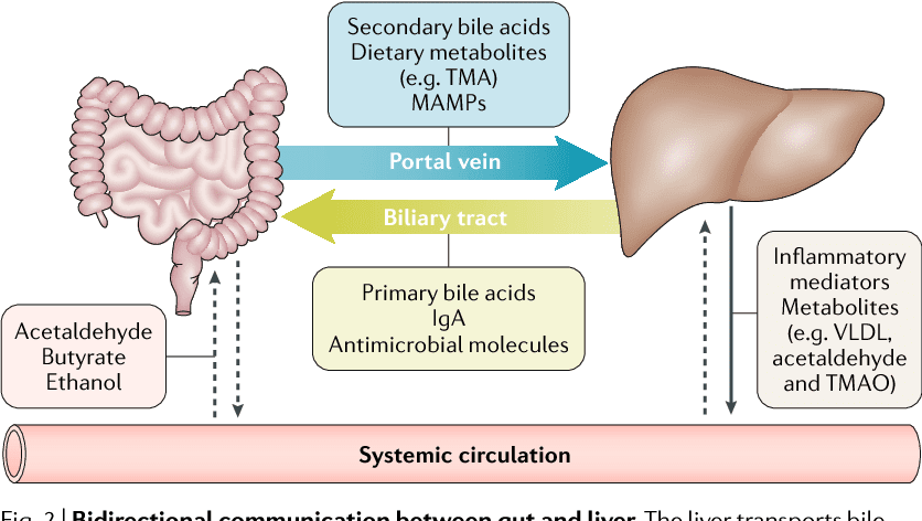
What NAFLD does to the body is that it becomes a hepatic consequence of metabolic syndrome. This includes obesity, diabetes, and dyslipidemia. What is interesting about the connection between gut microbiota and NAFLD has been attracting a significant amount of attention in recent years. The data has shown that the gut microbiota can affect the hepatic lipid metabolism while also influencing the balance between pro/anti-inflammatory effectors in the liver.
Chronic Liver Diseases
Even though chronic liver disease is the primary cause of morbidity and mortality worldwide. Studies showed that gut dysbiosis was identified as an essential factor in the pathogenesis of the liver disease. The relationship between the gut microbiota and the liver is still not understood, but the dysfunction of leaky gut and an increased bacterial translocation into the liver. Another study showed that immense importance is a massive advancement in understanding the roles of the gut and liver microbiome that is driven by a high DNA sequencing and improving them.
There are many stages of liver disease that can happen, but when it comes to excessive alcohol in the liver. Studies show that excessive alcohol consumption is the leading cause of chronic liver disease worldwide. The stages of alcoholic liver disease are hepatic steatosis, steatohepatitis, and, ultimately, liver cirrhosis. One of the main characteristics of alcohol liver disease is that there is an increased gut permeability due to a direct toxic effect of alcohol on the epithelial cell in the gastrointestinal tract and a decreases expression of the tight-junction protein.
Probiotic Supplements
For probiotic supplementation, they have demonstrated a significant decrease in liver enzymes, which were compared to the placebo group. Studies found that probiotics were shown to have synergistic effects with metformin on liver enzymes for patients that have NASH. Any products that contained both prebiotics and probiotics can demonstrate a similar effect in the probiotic groups. In another study, it showed a reduction of intrahepatic fat that is measured by MRI, but the improvement in liver enzymes in the body did not reach any clinical significance. It is essential to know that liver enzymes can have a highly variable and do not always have a direct correlation with disease progression that they may encounter.
Research shows that there are five meta-analyses included and that they all demonstrated that probiotics and synbiotics have improved on AST and ALT levels in the body significantly. Surprisingly, several other studies have assisted probiotics by countering hepatic steatosis, fibrosis, and liver stiffness in the body. Ultrasound imaging can help assist these parameters and show some positive clinical outcomes with these two supplements.
When probiotics help restore the gastrointestinal barrier function in the body, they can eliminate the harmful bacteria that has interacted with the gastrointestinal system. Not only that, but probiotics can also be beneficial by modulating the immune system, reduce liver fats, and improve the liver enzymes as well. By using probiotics, they are most likely to be more productive by helping the body and preventing bacterial translocation in the gut, thus reducing the effects of the intestinal microbiota on the liver to prevent chronic illnesses from forming and causing havoc.
Conclusion
For individuals that have NAFLD, they will already establish the disease and required a higher nutrient intake demand than what can be obtained from any diets alone. So using dietary supplements should be considered to help reduce the NAFLD disease’s progression, thus improving the liver and its functions. The gut-liver axis is connected to the body since if anything happens to the liver like chronic diseases, it can affect the gut as well. Using probiotics to help the liver is essential to make sure that the liver is functioning correctly and that the body is being as healthy as possible. Some products are here to offer gastrointestinal and metabolic support while also supporting multiple aspects of the biliary system.
The scope of our information is limited to chiropractic, musculoskeletal, and nervous health issues or functional medicine articles, topics, and discussions. We use functional health protocols to treat injuries or disorders of the musculoskeletal system. Our office has made a reasonable attempt to provide supportive citations and has identified the relevant research study or studies supporting our posts. We also make copies of supporting research studies available to the board and or the public upon request. To further discuss the subject matter above, please feel free to ask Dr. Alex Jimenez or contact us at 915-850-0900.
References:
Jurgelewicz, Michael. �New Review Demonstrates the Role of the Gut Microbiome and Probiotics in Nonalcoholic Fatty Liver Disease.� Designs for Health, 25 Nov. 2019, blog.designsforhealth.com/node/1160.
Konturek, Peter Christopher, et al. �Gut?Liver Axis: How Do Gut Bacteria Influence the Liver?� Medical Sciences (Basel, Switzerland), MDPI, 17 Sept. 2018, www.ncbi.nlm.nih.gov/pmc/articles/PMC6165386/.
Tripathi, Anupriya, et al. �The Gut-Liver Axis and the Intersection with the Microbiome.� Nature Reviews. Gastroenterology & Hepatology, U.S. National Library of Medicine, July 2018, www.ncbi.nlm.nih.gov/pmc/articles/PMC6319369/.
Xie, Chencheng, and Dina Halegoua DeMarzio. “Role of Probiotics in Nonalcoholic Fatty Liver Disease: Does Gut Microbiota Matter?” MDPI, Multidisciplinary Digital Publishing Institute, 19 Nov. 2019, www.mdpi.com/2072-6643/11/11/2837.
There are all kinds of tools for all kinds of jobs. However, using the correct tool can mean the difference between a job well done and a job that got done but also generated injuries and pain.
Examples:
Long tools are best for when you need leverage, sparing the need for massive physical force.
Vice grips and clamps can grip/stabilize objects rather than trying to hold objects with your hands.
Tilt objects to avoid overbending the wrists.
Use a cart/dolly/arm straps to carry heavy loads.
Take some time to think about how to make the job easier on yourself and look up youtube tutorials to find innovative ways to do these jobs making it less stressful both mentally and physically.
The National Institute for Occupational Safety & Health guidelines hand tool use
Keep the wrists straight &� Avoid bending/rotating the wrists.
Instead bend the tool, not the wrist and there are a variety of bent-handle tools just for this reason. Using the handshake wrist position is a good way to approach a job, as it is a neutral position for the wrist.
Don’t stand still in one place for an extended time when using a heavy tool.
Instead, reduce the size and weight of the tool which will help avoid strain, keeping the elbows low and slightly bent.
Tools that place pressure on the base of the palm stress the soft tissues of the hands and fingers interrupting circulation and nerve function.
Instead, opt for one with finger grooves that fit the hand. Short-handles help by reducing stress on the soft tissues.
Don’t use tools that need a lot of grip force to use or hold.
Instead, use one with a grip that compresses like memory foam and shapes to the hand. This is far better than hard plastic.
Don’t use tools that need the fingers to grip.
Instead, use tools that utilize a full-hand power grip.
Do not use tools that have sharp-edged handles or areas where the hands could get pinched.
Instead, use tools that keep the hands/fingers safe.
Trigger-finger operational tools should be avoided as this can easily cause repetitive finger/hand/wrist injury from the constant on-off motion.
Instead, use tools with large switches that can be operated using all four fingers.
Excessive temperatures affect manual dexterity, therefore keep hands free from extreme heat and cold.
If possible, do a different job that’s away from the extreme weather and if not wear properly insulated work gloves.
Keep excessive vibration to a minimum. Excessive vibration can affect circulation.
Use tools with control�features that limit vibration to the extremities and whole body.
Wear gloves that fit. If they are too tight they will place extreme pressure on the hands. Loose-fitting gloves reduce grip strength and the ability to grip properly.
Instead have a selection that is designed for different jobs.
Safely Operating Tools that Cause Whole-Body Vibration��
There are power tools that vibrate no matter what and transmit vibration into the operator’s arms and hands, legs, and feet. Using a tool like this can cause a condition called white finger or Raynaud’s Phenomenon to present.
The symptoms include:
Aching in the wrists and muscles of the forearm
Tingling sensations
Numbness
Whiteness in the fingers from restricted circulation
This type of vibration from riveting tools, grinders, pneumatic hammers, drills, and chain saws will affect the whole body’s well being.
Suggestions to help reduce the risk of musculoskeletal disorders
Choose power tools with anti-vibration controls and handles coated with suppressant/cushioned material to help with vibration.
Maintenance power tools by making sure they are balanced, clean, and lubricated.
Use gloves designed to absorb or reduce vibration.
Ask for help if the job requires equipment or tools that vibrate.
Whether using a hand or power tool to get a job done, the whole body is involved. Executing proper posture and body mechanics, along with proper tool choice and how it is used is vital to injury prevention.
El Paso, TX Chiropractor Recommended
Chiropractic care can help keep bodies flexible and help with range of motion. It is a very effective, non-invasive treatment for pain and can help with joint and muscular problems as well. Regular chiropractic treatments can help you better manage your body�s response to your work environment. It can also undo many of the ill effects that that type of work can cause.
NCBI Resources
Standing for extended or frequent periods of time without any breaks (such as walking or stretching) can cause the joints in the feet, knees, hips, and spine to become locked or immobilized temporarily. If the behavior continues, it can cause degenerative damage, leading to rheumatic diseases because the ligaments and tendons become damaged.
Blood vessels in the brain protect us from “harmful” components in the bloodstream. This is known as the blood-brain barrier. A Science Translational Medicine research study demonstrated how inflammation and cognitive impairment in a group of aging mice may be associated with the breakdown of the blood-brain barrier. In the following article, we will discuss how restoring the breakdown of the blood-brain barrier may improve overall brain health. �
What is a Leaky Blood-Brain Barrier?
Scientists determined that the breakdown of the blood-brain barrier can ultimately trigger a signaling protein in brain cells, known as astrocytes. They then created and evaluated a drug/medication that blocked the activation of the astrocytes, or the signaling protein known as the transforming growth factor-beta (TGF-beta). Following the treatment, the group of aging mice demonstrated reduced brain inflammation and improved cognitive function. �
“Our team of scientists associates the aging brain in the same way we associate neurodegeneration. The aging brain is characterized by loss of cognitive function and dead brain cells,” stated Daniela Kaufer, co-senior author and professor of integrative biology at the University of California, Berkeley. “However, our research study determined a different story about why the aging brain doesn’t function properly: It is because of brain fog,” she concluded. �
According to the research study, reducing inflammation and brain fog by restoring the breakdown of the blood-brain barrier can help improve the overall health and wellness of the aging brain. The results may also help scientists understand how cognitive impairment associated with inflammation and brain fog can accompany the aging brain and neurodegeneration as well as how the blood-brain barrier may be associated with improved brain function. �
Inflammation, Brain Fog, and Cognitive Impairment
A variety of research studies, including imaging research studies performed by Alon Friedman, co-senior author of Ben-Gurion University of the Negev in Israel and Dalhousie University in Canada, discussed the breakdown of the blood-brain barrier and how it can ultimately become less effective with age. A “leaky” blood-brain barrier can make it easier for “harmful” compounds to penetrate the brain and damage cells and tissue from the bloodstream. �
Kaufer and Friedman are also co-senior authors of another Science Translational Medicine research study that evaluated inflammation and brain fog in leaky blood-brain barriers. By way of instance, patients with Alzheimer’s disease may experience epileptic episodes, however, they may not be aware of them. Aging is a risk factor for Alzheimer’s disease and epilepsy where research studies have associated a connection between the two brain health issues. �
For the second research study, the team of scientists evaluated EEG readings from patients with Alzheimer’s disease and determined an EEG signature for what is ultimately known as paroxysmal slow wave events (PSWEs). From the EEGs, the scientists demonstrated how the rate of PSWEs appeared to match the level of cognitive impairment of the patients. In EEGs of patients with epilepsy, they demonstrated that PSWEs that happened between seizures matched cases of leaky blood-brain barriers. They determined the same match in aging mice, mice prone to Alzheimer’s disease, and rats with induced epilepsy. �
Further research studies in young rats also demonstrated that the blood-brain barrier can start to breakdown by introducing the protein albumin to the brain. According to the results, this ultimately caused an increased rate of PSWEs. Friedman and Kaufer also demonstrated that the protein albumin can penetrate the blood-brain barrier following trauma. The protein albumin can attach itself to the TGF-beta receptor of astrocytes and cause brain health issues. The team of scientists also concluded that a leaky blood-brain barrier may ultimately be a cause of inflammation, brain fog, and cognitive impairment. �
Leaky Blood-Brain Barrier Biomarkers
The scientists suggest that the results of the various research studies on leaky blood-brain barriers and brain health ultimately help offer a variety of biomarkers that could possibly help healthcare professionals recognize these type of problems by using MRI, which can detect leaky blood-brain barriers, and using EEG, which can detect abnormal brain rhythms. The outcome measures may also help develop the treatment that they may use as a way to restore a leaky blood-brain barrier to reduce and even reverse several of the brain health issues it may ultimately cause, including neurodegeneration. �
“Our team of scientists now use several biomarkers that demonstrate leaky blood-brain barriers, so healthcare professionals can choose patients for treatment,” stated professor Daniela Kaufer. “These research studies ultimately support the effects of leaky blood-brain barriers on a variety of brain health issues, including dementia and Alzheimer’s disease, associated with inflammation, brain fog, and cognitive impairment as well as offer possible results for future research studies,” stated Diego Gomez-Nicola, an associate professor of neuroscience at the University of Southampton in the United Kingdom, �
After the discoveries of the German scientist Paul Ehrlich during the late 1800s, a collection of experiments on a group of mice demonstrated how the brain regulates what to permit passage to and what to block from entering its blood vessels through the blood-brain barrier. The brain is ultimately protected by the blood-brain barrier, however, this security system can frequently prevent drugs and/or medications from being able to effectively treat brain health issues. Scientists have started working towards developing successful ways to allow treatments to penetrate the blood-brain barrier. Other research studies have demonstrated that by the aging brain, as well as neurodegeneration, can cause the breakdown of the blood-brain barrier. A leaky blood-brain barrier can ultimately cause inflammation, brain fog, and cognitive impairment. However, research studies have demonstrated ways to restore and even reverse leaky blood-brain barriers and several brain health issues. – Dr. Alex Jimenez D.C., C.C.S.T. Insight
Neurotransmitter Assessment Form
The following Neurotransmitter Assessment Form can be filled out and presented to Dr. Alex Jimenez. The following symptoms listed on this form are not intended to be utilized as a diagnosis of any type of disease, condition, or any other type of health issue. �
Blood vessels in the brain protect us from “harmful” components in the bloodstream. This is known as the blood-brain barrier. A Science Translational Medicine research study demonstrated how inflammation and cognitive impairment in a group of aging mice may be associated with the breakdown of the blood-brain barrier. In the following article, we will discuss how restoring the breakdown of the blood-brain barrier may improve overall brain health. �
The scope of our information is limited to chiropractic, musculoskeletal, and nervous health issues or functional medicine articles, topics, and discussions. We use functional health protocols to treat injuries or disorders of the musculoskeletal system. Our office has made a reasonable attempt to provide supportive citations and has identified the relevant research study or studies supporting our posts. We also make copies of supporting research studies available to the board and or the public upon request. To further discuss the subject matter above, please feel free to ask Dr. Alex Jimenez or contact us at 915-850-0900.�
Curated by Dr. Alex Jimenez �
References:
Catharine Paddock, Ph.D. �Repairing Leaky Blood-Brain Barrier May Rejuvenate Brain Function.� Medical News Today, MediLexicon International, 6 Dec. 2019, www.medicalnewstoday.com/articles/327248.php#1.
Additional Topic Discussion: Chronic Pain
Sudden pain is a natural response of the nervous system which helps to demonstrate possible injury. By way of instance, pain signals travel from an injured region through the nerves and spinal cord to the brain. Pain is generally less severe as the injury heals, however, chronic pain is different than the average type of pain. With chronic pain, the human body will continue sending pain signals to the brain, regardless if the injury has healed. Chronic pain can last for several weeks to even several years. Chronic pain can tremendously affect a patient’s mobility and it can reduce flexibility, strength, and endurance. �
Neural Zoomer Plus for Neurological Disease
�
Dr. Alex Jimenez utilizes a series of tests to help evaluate neurological diseases. The Neural ZoomerTM Plus is an array of neurological autoantibodies which offers specific antibody-to-antigen recognition. The Vibrant Neural ZoomerTM Plus is designed to assess an individual�s reactivity to 48 neurological antigens with connections to a variety of neurologically related diseases. The Vibrant Neural ZoomerTM Plus aims to reduce neurological conditions by empowering patients and physicians with a vital resource for early risk detection and an enhanced focus on personalized primary prevention. �
Food Sensitivity for the IgG & IgA Immune Response
�
Dr. Alex Jimenez utilizes a series of tests to help evaluate health issues associated with food sensitivities. The Food Sensitivity ZoomerTM is an array of 180 commonly consumed food antigens that offers very specific antibody-to-antigen recognition. This panel measures an individual�s IgG and IgA sensitivity to food antigens. Being able to test IgA antibodies provides additional information to foods that may be causing mucosal damage. Additionally, this test is ideal for patients who might be suffering from delayed reactions to certain foods. Utilizing an antibody-based food sensitivity test can help prioritize the necessary foods to eliminate and create a customized diet plan around the patient�s specific needs. �
Gut Zoomer for Small Intestinal Bacterial Overgrowth (SIBO)
�
Dr. Alex Jimenez utilizes a series of tests to help evaluate gut health associated with small intestinal bacterial overgrowth (SIBO). The Vibrant Gut ZoomerTM offers a report that includes dietary recommendations and other natural supplementation like prebiotics, probiotics, and polyphenols. The gut microbiome is mainly found in the large intestine and it has more than 1000 species of bacteria that play a fundamental role in the human body, from shaping the immune system and affecting the metabolism of nutrients to strengthening the intestinal mucosal barrier (gut-barrier). It is essential to understand how the number of bacteria that symbiotically live in the human gastrointestinal (GI) tract influences gut health because imbalances in the gut microbiome may ultimately lead to gastrointestinal (GI) tract symptoms, skin conditions, autoimmune disorders, immune system imbalances, and multiple inflammatory disorders. �
Formulas for Methylation Support
XYMOGEN�s Exclusive Professional Formulas are available through select licensed health care professionals. The internet sale and discounting of XYMOGEN formulas are strictly prohibited.
Proudly,�Dr. Alexander Jimenez makes XYMOGEN formulas available only to patients under our care.
Please call our office in order for us to assign a doctor consultation for immediate access.
If you are a patient of Injury Medical & Chiropractic�Clinic, you may inquire about XYMOGEN by calling 915-850-0900.
�
�
For your convenience and review of the XYMOGEN products please review the following link. *XYMOGEN-Catalog-Download �
* All of the above XYMOGEN policies remain strictly in force. �
Creating a healthy, safe ergonomic work environment is important to protect your back, neck and whole body. Just by taking simple breaks and enhancing your workspace with a sit-stand desk will protect your spine and general health.
Break Taking
Working in the same position and using the same muscles, joints, and ligaments for hours is not good for any part of your body. Ergonomists are lifestylescientists that design spaces/equipment/tools to reduce discomfort, fatigue, and injury, agree that taking frequent and brief rest breaks is essential for total and optimal body health. And, it�s not just your legs that need a break every now and then.
At work, start practicing:
Eye breaks:Looking at the computer screen for a long time changes how the eyes work. What happens is you blink less and expose the eyes to the air. Therefore, every 15 minutes look away from the screen for a minute or two to a distant area that is at least 20 feet away or further. This allows the muscles in the eye to relax. Also, blink your eyes real quick for a few seconds. This refreshes the tear ducts and clears dust from the surface of the eyes.
Micro-breaks: These are breaks that are less than two minutes and perfect to utilize between office jobs. Most people work in bursts rather than continuously. So in between these bursts�take a rest in a:
Relaxed
Flat
Straight posture
These breaks are short but perfect for stretching, standing up, and moving around, or switching to a different task like making a phone call or making some copies, etc. These types of breaks are a break from using the same set of muscles over and over.
Rest breaks: These you want to do every 30 to 60 minutes. This is the break to, get up, move around, and do something else non-office related. Go get a beverage, quick conversation with a coworker, or take a walk around the office or building. As long it’s within reason. This allows your body and mind to empty and workout different muscles. Practice this and the feeling of tiredness will be a thing of the past.
Exercise breaks: This is purely a stretching and gentle exercise break to do to relieve muscle fatigue. These should be done every one to two hours.
Ergonomic software: It is easy to lose track of how long you’ve been working. There is software that monitors how long you’ve been on the computer and will alert you to take a break at different intervals and offer easy ergonomic exercises to keep your muscles loose and in top office shape.
Ergonomic Products
There are plenty of products out there to improve your workplace environment and promote top spine health. Consider an ergonomic chair, computer accessories, or sit-stand desk to help maintain proper posture.
These products can be adjusted and customized to your needs. They encourage healthy long-term habits that can reduce and prevent various types of injuries. Sit-to-stand desks allow transition from sitting to a standing position. Varying your posture throughout the day is highly beneficial to general health and even helps to burn extra calories.
These ergonomic products are for creating a healthy/safe work environment. Therefore take some time to research the product you are interested in before buying.� Here are a few questions to think about:
Do the manufacturer’s claims make sense or are too good to be true?
Is there evidence that can support their claims?
Is it a cheap knock-off? Knock off products should be avoided as they can worsen and create more injuries. However, when it comes to the brand name products, don’t go for the fully loaded models that could cost quite a bit, instead find something in the middle but that still meets ergonomic standards.
Are you comfortable using the product?
What do experts/reviews say about the product? If it’s not recommended then don’t use it.
Some products can feel strange or uncomfortable because they make you change the way you work. Don’t panic, as this is the point of the product that you have to get used to. But it will be beneficial to your overall health. However, if a product continues to feel uncomfortable or causes pain after short use, then discontinue using and try something else.
Improving spinal health and hygiene at work is as simple as taking breaks for light stretching, walking around, and utilizing ergonomic office products to stay fit and injury-free. Whatever you choose, understand how your back and neck are moving/functioning during the workday/night by using ergonomic practices that will keep your body/mind healthy and prevent office injuries.
Control *FOOT MOTION & POSTURE* with Functional Foot Orthotics | El Paso, Tx (2019)
NCBI Resources
Ergonomics is a scientific discipline that�s been in existence for many years. Keeping their work environments safe and efficient and traditionally concerned with factory workers, ergonomic professionals have expanded their work to include all types of workers from laborers to seniors to office workers & students.
It looks for means to improve our environment to lower the risks of illness and harm, enhance productivity, and improve the caliber of our work life.
If you are experiencing any of these situations, then you might have pain and inflammation in your body. Here are some ways to help naturally ease the pain in your body.
There is some evidence the conventional go-to pain relievers and anti-inflammatory medicine can help by delivering their benefits along with a host of potentially dangerous effects to anyone. Both medical professionals and patients are looking for alternative medicines that have the same effects as pharmaceutical medicine but that are safe and more effective. Fortunately, numerous natural compounds are backed up by clinical research and studies that are available to help anyone that have a variety of issues that are caused by pain and inflammation.
Factors Involving Inflammation
Research from the Cleveland Clinic had warned patients that NSAIDs (nonsteroidal anti-inflammatory drugs) are intended for short-term use. That person should never use it continuously for fever for more than three days and at least ten days for pain without the consultant from a healthcare provider. Studies show that for any fish oils or other sources of long-chain omega-3 fatty acids that have the same effect since it is the precursor to anti-inflammatory prostaglandins and resolvins for the body.
Natural Products for Inflammation
If there is a long-term consumption food that is high in EPA and DHA that are dangerous, there will be a substantial fish-eating population in East Asia and Scandinavia; then, there would be a considerable problem long ago. This, however, is the opposite of what epidemiological studies found since fish consumption is beneficial for the person’s health and improving the biomarker for cardiovascular risk. For anyone that is allergic to fish, eggs, flaxseeds, and it is oil or algal oil is a rich source of omega-3s for the body.
There are other various foods and herbal extracts that been shown to be beneficial in managing pain and inflammation. One of them is ginger. Ginger has been used for a long time as a remedy for upset stomach and indigestion, while also being effective for reducing menstrual cycle pain like NSAIDs. Studies show the herb Boswellia can reduce the pain and stiffness that are associated with osteo- and rheumatoid arthritis and one of the mechanics that are responsible for the body that inhibits the inflammatory 5-LOX (5-lipoxygenase).
What is interesting about Boswellia is that studies show that the herb is different from NSAIDs because NSAIDs may induce potential life-threating GI tract bleeding. The evidence shows that the Boswellia herb can improve inflammatory conditions in the gastrointestinal tract, like chronic colitis. There are more natural compounds that are the potential to ease pain and inflammation like cannabis. With the expanding legalization throughout the US, medicinal and recreational cannabis can help many people deal with their pain with this impressive plant.
Incorporating Changes To Stop Inflammation
Besides incorporating these natural herbs and anti-inflammatory foods into a person�s diet to help alleviate pain and inflammation, no one should neglect the non-diet strategies. Changing food and supplements are not the only changes when a person is seeking alternatives. A past article started to explore how laughter, positive thinking, and maintaining physical activity has the potential to manage chronic pain that the body may encounter. Even though the old saying states that “laughter is the best medicine,” the old saying is contradicted now and days. Even though laughter is not the best medicine, but laughter can undoubtedly be a form of medicine.
Another way a person can reduce or resolve inflammation is by their diet. This can affect what a person does not eat than what they do eat. Studies show that ketogenic diets, which are high in fats and very low in carbohydrates, have been known to increase pain tolerance and reduce acute inflammation. The results were astounding as patients with type 2 diabetes following a ketogenic diet for one year, experienced a 39% reduction in hsCRP, which is a massive indicator for inflammation. Another research study observed patients who followed a ketogenic diet for two years, had a 37% reduction from their baseline.
More research studies show that patients that have metabolic syndrome and follow a carbohydrate-restricted Paleolithic diet for four weeks have experienced a 39% reduction in their hsCRP as well as 35% and 29% reduction in their TNF-a and IL-6. When low-carb Paleo diets are combined with intensity exercises can help improve the body by dampening the inflammatory markers significantly.
Conclusion
For anyone that is living with inflammation in their body, using pharmaceutical medicines is not always the answer. Combining a carb-restricted diet and other natural interventions can help with inflammation. By making small changes for the body, the chronic pain and inflammation can be reduced, thus the body can start healing naturally. Some products are designed to help relax the tight muscles in the body to prevent inflammation while also helping the body to relax and provide a better night’s sleep.
The scope of our information is limited to chiropractic, musculoskeletal, and nervous health issues or functional medicine articles, topics, and discussions. We use functional health protocols to treat injuries or disorders of the musculoskeletal system. Our office has made a reasonable attempt to provide supportive citations and has identified the relevant research study or studies supporting our posts. We also make copies of supporting research studies available to the board and or the public upon request. To further discuss the subject matter above, please feel free to ask Dr. Alex Jimenez or contact us at 915-850-0900.
References:
Alhassan, Abeer, et al. �Consumption of Fish and Vascular Risk Factors: A Systematic Review and Meta-Analysis of Intervention Studies.� Atherosclerosis, US National Library of Medicine, Nov. 2017, www.ncbi.nlm.nih.gov/pubmed/28992469.
Ammon, H P T. �Boswellic Acids in Chronic Inflammatory Diseases.� Planta Medica, US National Library of Medicine, Oct. 2006, www.ncbi.nlm.nih.gov/pubmed/17024588.
Athinarayanan, Shaminie J, et al. �Long-Term Effects of a Novel Continuous Remote Care Intervention Including Nutritional Ketosis for the Management of Type 2 Diabetes: A 2-Year Non-Randomized Clinical Trial.� Frontiers in Endocrinology, Frontiers Media SA, 5 June 2019, www.ncbi.nlm.nih.gov/pmc/articles/PMC6561315/.
Bhanpuri, Nasir H, et al. �Cardiovascular Disease Risk Factor Responses to a Type 2 Diabetes Care Model Including Nutritional Ketosis Induced by Sustained Carbohydrate Restriction at 1�Year: an Open Label, Non-Randomized, Controlled Study.� Cardiovascular Diabetology, BioMed Central, 1 May 2018, www.ncbi.nlm.nih.gov/pmc/articles/PMC5928595/.
Calder, Philip C. �Omega-3 Fatty Acids and Inflammatory Processes.� Nutrients, Molecular Diversity Preservation International, Mar. 2010, www.ncbi.nlm.nih.gov/pmc/articles/PMC3257651/.
Commissioner, Office of the. �The Benefits and Risks of Pain Relievers: Q & A on NSAIDs.� US Food and Drug Administration, FDA, 24 Sept. 2015, www.fda.gov/consumers/consumer-updates/benefits-and-risks-pain-relievers-q-nsaids-sharon-hertz-md.
Drug Evaluation and Research, Center for. �FDA Drug Safety Communication.� US Food and Drug Administration, FDA, 9 July 2015, www.fda.gov/drugs/drug-safety-and-availability/fda-drug-safety-communication-fda-strengthens-warning-non-aspirin-nonsteroidal-anti-inflammatory.
Gupta, I, et al. �Effects of Gum Resin of Boswellia Serrata in Patients with Chronic Colitis.� Planta Medica, US National Library of Medicine, July 2001, www.ncbi.nlm.nih.gov/pubmed/11488449.
Gyorkos, Amy, et al. �Carbohydrate-Restricted Diet and High-Intensity Interval Training Exercise Improve Cardio-Metabolic and Inflammatory Profiles in Metabolic Syndrome: A Randomized Crossover Trial.� Cureus, Cureus, 8 Sept. 2019, www.ncbi.nlm.nih.gov/pmc/articles/PMC6822889/.
Hosomi, Ryota, et al. �Seafood Consumption and Components for Health.� Global Journal of Health Science, Canadian Center of Science and Education, 28 Apr. 2012, www.ncbi.nlm.nih.gov/pmc/articles/PMC4776937/.
Masino, Susan A, and David N Ruskin. �Ketogenic Diets and Pain.� Journal of Child Neurology, US National Library of Medicine, Aug. 2013, www.ncbi.nlm.nih.gov/pmc/articles/PMC4124736/.
Nandivada, Prathima, et al. �Eucaloric Ketogenic Diet Reduces Hypoglycemia and Inflammation in Mice with Endotoxemia.� Lipids, US National Library of Medicine, June 2016, www.ncbi.nlm.nih.gov/pubmed/27117864.
Team, Cleveland Clinic. �NSAIDs: What You Need to Know.� Cleveland Clinic, 2016, my.clevelandclinic.org/health/drugs/11086-non-steroidal-anti-inflammatory-medicines-nsaids.
Team, DFH. �3 Non-Pharmacological Daily Practices for Managing Pain.� Designs for Health, 1 Feb. 2019, blog.designsforhealth.com/node/942.
Team, DFH. �Ginger � as Effective as NSAIDs for Menstrual Pain.� Designs for Health, 5 Jan. 2018, blog.designsforhealth.com/ginger-as-effective-as-nsaids-for-menstrual-pain.
If you are experiencing any of these situations, then why not try these botanical herbs in your food dish.
The holiday season brings out the joy out of people with holiday traditions and its unique, universal foods, drinks, seasonal herbs, and spices to the table. The colder seasons are where everyone in the U.S. indulges in hearty soups, pumpkin spice products, root vegetables, and turkey leftovers. Many holiday menu items will incorporate sweet and savory herbs and spices in their food dishes. What people do not realize is that even though these herbs and spices enhance the flavors of the food, they also provide a plethora of health benefits for the body. So using these herbs and spices in the kitchen can help increase the person’s chances to receive their beneficial properties, hence “the more, the merrier.”
Throughout the centuries, medicinal plants have been traditionally used by a variety of cultures. Some of nature’s most potent herbs are most used in culinary dishes, while also providing the beneficial properties to the human body; they are rosemary, sage, and clove.
Rosemary
Being commonly used as a condiment and a food preservative, rosemary is a native herb in the Mediterranean region. A review did an animal study of in vivo and in vitro that showed that rosemary had demonstrated similar beneficial effects to any medications that are for a variety of physiological disorders. Some of the physiological disorders include:
Lead hepato-nephrotoxicity
Stress
Anxiety
Bodyweight and dyslipidemia
Pain
Cerebral ischemia
Another review was looking at how rosemary has antimicrobial and antioxidant properties and that its abundance in isoprenoid quinones. The review stated that rosemary could �act as chain terminators for free radicals and as chelators for ROS (reactive oxygen species.)” The antioxidative properties in rosemary have about the majority of phenolic diterpenes; like carnosic acid and carnosol, that are responsible for about 90% of this herb. Since it inhibits lipid peroxidation, scavenge radicals, and helps reduce cytochrome c by activating the redox-dependent signaling pathways in the body.
With carnosic acid, it has been shown to provide superior antimicrobial actions to other significant constituents that are found in rosemary. Furthermore, there have been studies shown that rosemary has antibacterial effectiveness against resistant bacteria in the body.
Sage
Sage is another herb that is native to the Mediterranean and Middle East regions and has been used in traditional folk medicine to treat a variety of disorders in the body. There is recent research that has suggested that sage can possess a wide range of beneficial properties for the body due to the presence of carnosic acid and carnosol in this herb. Another study showed that sage contains high contents of glycosidic flavones that provide functional inhibitory capacity against xanthine oxidase activity in the body. Some of the beneficial properties include:
Anti-inflammatory
Antinociceptive
Antibacterial
Hypoglycemic
Antioxidative
There is further research that demonstrates how sage has powerful cognitive enhancing and neuroprotective properties. Studies show that patients with Alzheimer�s disease took a 4-month supplementation of sage, and the results are remarkable. Alzheimer patients experienced significant improvements in their cognitive function and mood enhancements. In another study, sage extract can help reduce the severity of physical and psychological systems that are experienced in premenstrual syndrome in the body. A recent article showed how freshly harvested sage leaves can become a potent modulator for neuroreceptor pathways that involves serotonin transporters that may help normalize thermoregulation and mental impairment for menopausal women.
Surprisingly though, both rosemary and sage have been proven and shown that they can help improve gabaergic pathways in the brain and can help decrease the neuronal activities that are associated with anxiety disorders. Studies show that rosemary and sage extracts have hepatoprotective and antioxidative roles that can increase catalase and glutathione levels and decrease lipid peroxidation in the body.
Cloves
Clove buds were initially found in east Indonesia, and it plays a role as the herb has a potent antimicrobial and antioxidative botanical. Research shows that clove oil possesses a bactericidal effect against pathogenic species that can harm the body. Earlier this year, a research study found out that clove oil extract can enhance anti-inflammatory activity by reducing myeloperoxidase activity in human neutrophils significantly. This herb can help reduce ROS and a variety of other inflammatory mediators that can promote significant damage to the body at the site of inflammation.
Conclusion
So for the holidays, adding these three powerful herbs to the next holiday feast are not just there to help enhance the flavors of the dishes, but they are beneficial to the body. Since they provide anti-inflammatory properties to the body by decreasing the inflammatory responses. So for the colder seasons, add that extra dash of herbs into the dish recipe will make anyone’s day merry. Some products are specialized in countering the metabolic effects of temporary stress and can support the body.
The scope of our information is limited to chiropractic, musculoskeletal, and nervous health issues or functional medicine articles, topics, and discussions. We use functional health protocols to treat injuries or disorders of the musculoskeletal system. Our office has made a reasonable attempt to provide supportive citations and has identified the relevant research study or studies supporting our posts. We also make copies of supporting research studies available to the board and or the public upon request. To further discuss the subject matter above, please feel free to ask Dr. Alex Jimenez or contact us at 915-850-0900.
References:
Chniguir, Amina, et al. �Syzygium Aromaticum Aqueous Extract Inhibits Human Neutrophils Myeloperoxidase and Protects Mice from LPS-Induced Lung Inflammation.� Pharmaceutical Biology, Taylor & Francis, Dec. 2019, www.ncbi.nlm.nih.gov/pmc/articles/PMC6366422/#!po=2.63158.
Choukairi, Zineb, et al. �Effect of Salvia Officinalis L. and Rosmarinus Officinalis L. Leaves Extracts on Anxiety and Neural Activity.� Bioinformation, Biomedical Informatics, 15 Mar. 2019, www.ncbi.nlm.nih.gov/pmc/articles/PMC6637401/.
de Oliveira, Jonatas Rafael, et al. �Rosmarinus Officinalis L. (Rosemary) as Therapeutic and Prophylactic Agent.� Journal of Biomedical Science, BioMed Central, 9 Jan. 2019, www.ncbi.nlm.nih.gov/pmc/articles/PMC6325740/.
Lopresti, Adrian L. �Salvia (Sage): A Review of Its Potential Cognitive-Enhancing and Protective Effects.� Drugs in R&D, Springer International Publishing, Mar. 2017, www.ncbi.nlm.nih.gov/pmc/articles/PMC5318325/.
Nieto, Gema, et al. �Antioxidant and Antimicrobial Properties of Rosemary (Rosmarinus Officinalis, L.): A Review.� Medicines (Basel, Switzerland), MDPI, 4 Sept. 2018, www.ncbi.nlm.nih.gov/pmc/articles/PMC6165352/.
Pavi?, Valentina, et al. �Extraction of Carnosic Acid and Carnosol from Sage (Salvia Officinalis L.) Leaves by Supercritical Fluid Extraction and Their Antioxidant and Antibacterial Activity.� Plants (Basel, Switzerland), MDPI, 9 Jan. 2019, www.ncbi.nlm.nih.gov/pmc/articles/PMC6359053/.
Pereira, Ol�via R, et al. �Salvia Elegans, Salvia Greggii and Salvia Officinalis Decoctions: Antioxidant Activities and Inhibition of Carbohydrate and Lipid Metabolic Enzymes.� Molecules (Basel, Switzerland), MDPI, 1 Dec. 2018, www.ncbi.nlm.nih.gov/pmc/articles/PMC6321363/.
Team, DFH. �Spice Up the Holidays with Medicinal Botanicals.� Designs for Health, 25 Nov. 2019, blog.designsforhealth.com/node/1156.
Tober, Carsten, and Roland Schoop. �Modulation of Neurological Pathways by Salvia Officinalis and Its Dependence on Manufacturing Process and Plant Parts Used.� BMC Complementary and Alternative Medicine, BioMed Central, 13 June 2019, www.ncbi.nlm.nih.gov/pmc/articles/PMC6567565/.
All individuals that participate in some form of sports or athletic training, professionals to weekend sports enthusiasts are at risk for back and neck injuries. Common injuries include strains, and sprains, pulls, and tears especially around the low back area. If left untreated these injuries can lead to chronic back pain or more severe conditions.
Although we can’t prevent all sports injuries, here are some sports tips to keep your spine healthy.
1.� Warm-Up and Stretch
Properly warming up with stretching exercises increases blood circulation and improves the flexibility of muscles and ligaments. This is not only for helping enhance athletic performance but prevents injuries by keeping the muscles/ligaments loose so if any type of collision, tear, or pull occurs the stretched muscles stay relaxed and do not tense up or contract, which helps reduce the severity of an injury. To stretch properly:
Stretch time on each part of the body is also 10-30 seconds
Stretch after the game to relieve sore or tight muscles
2. Use Proper Sport Equipment
All sports have a risk of injury. In general, the more contact there is the higher the risk of injury.
To reduce the risk of injury athletes should wear protective equipment that goes with their sport like neck rolls, shoulder, elbow and knee pads.
Well made and supportive shoes combined with custom orthotics are a must.
Other types of equipment include:
Helmet
Pads elbow, wrist, chest, knee, shins
Mouthpiece
Faceguard
Protective cup
Eye protection
3. Stay Hydrated
Injuries caused by heat occur as a result of�high temperatures, humidity and excessive/overdoing it. To avoid these serious injuries:
Drink plenty of water before, during, and after playing.
Try to avoid play or practice during extreme heat and humidity.
Wear lightweight clothing/uniform with maximum ability to allow sweat to evaporate.
Take plenty of breaks or periods of rest to allow the body to recover and recuperate.
4. Don’t Overwork/Overdo it
Repetitive Motion Disorders like tennis elbow, bursitis, and tendonitis, happen when movements e.g. swinging motions that go with the sport like tennis, bowling, golf, etc are repeated over and over and cause injury/damage to those parts of the body. To avoid overuse injury try:
Take plenty of breaks during practice and games. Do not power through it!
Use proper/correct form and techniques. If unsure then take lessons to make sure you are doing it correctly.
See a doctor if any pain or muscle fatigue, inflammation, swelling, or compression of nerve tissue present.
Cross-training can strengthen muscle groups and those areas that take the most force.
5. Stay Ready for Play with a Healthy Lifestyle
Besides sports, try to find ways to improve general health through a healthy lifestyle:
Get plenty of sleep, the body/mind needs to recover from all the activity.
Consult a doctor before beginning any new exercise program.
Staying fit, healthy and ready for play means preventing injuries from happening.� By being aware of how to prevent injuries with these basic tips, which feel free to take it further and raise the probability of avoiding back and all sports injuries.
As El Paso�s Chiropractic Rehabilitation Clinic & Integrated Medicine Center,�we passionately are focused on treating patients after frustrating injuries and chronic pain syndromes. We focus on improving your ability through flexibility, mobility and agility programs tailored for all age groups and disabilities.
*BEST* Heel Spurs Treatment | El Paso, Tx (2020)
NCBI Resources
Extension sports like gymnastics, tennis, swimming, diving, football, volleyball, basketball, track and field, cricket have the most pronounced extension/rotation on the spine. With a normal extension of the lumbar spine (or backward bending), the facet joints begin to approximate each other and compress. This is a normal biomechanical movement. However, if the extension ranges are excessive, the procedures will impinge quite aggressively and damage to the cartilage surfaces within the facet joint can result.
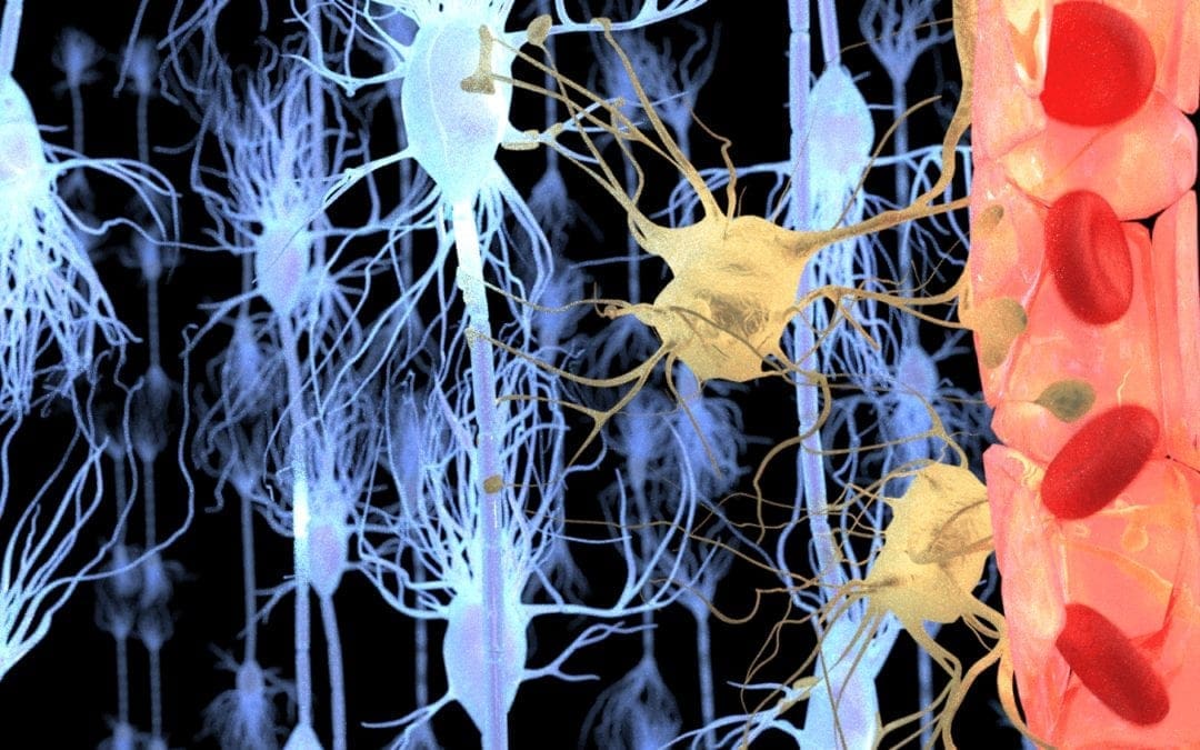
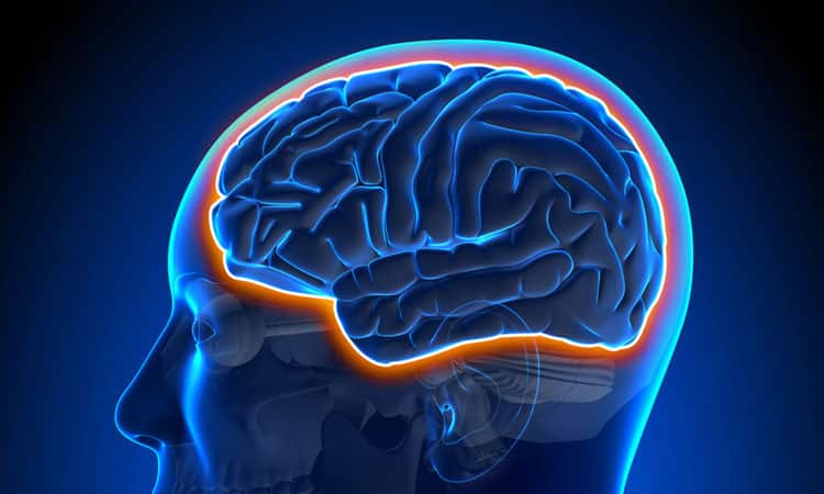
Our brain is a complex organ protected by our 7mm thick skull, a protective membrane, known as the meninges, and cerebrospinal fluid. These essential brain structures help protect the brain against physical damage or injury. Another brain structure that protects the brain from harm is the blood-brain barrier. The blood-brain barrier is the shield between the brain’s blood vessels and the cells in the brain’s tissue. While the skull, meninges, and cerebrospinal fluid protect the brain from physical damage or injury, the blood-brain barrier protects the brain from toxins and pathogens in the bloodstream. �
Moreover, the presence of the blood-brain barrier in the human brain was first discovered in the late 1800s by the German scientist Paul Ehrlich when he injected blue dye in the bloodstream of a group of mice during an experiment. The blue dye strained all of the animals’ tissues and organs with the exception of the brain and spinal cord. Although the outcome measures of the research study demonstrated the existence of the blood-brain barrier, it wasn’t until the 1960s that researchers were able to use much more powerful technologies to ultimately prove the presence of the blood-brain barrier in the human brain. �
Anatomy of the Blood-Brain Barrier
The main structure of the blood-brain barrier that helps protect the brain from toxins and pathogens in the bloodstream is known as the endothelial tight junction. The endothelial cells cover the inside of the blood vessels in the brain. The blood-brain barrier is such an effective security system because these endothelial cells in the blood vessels in the brain are connected extremely close to each other in “tight junctions”. The blood-brain barrier only permits small, fat-soluble molecules and several types of gases to pass freely between the blood vessels and the brain. Furthermore, bigger molecules, such as glucose and insulin, are only permitted passage through transporter proteins which act like “bouncers” that only open the doors for certain necessary molecules. �
The purpose of the blood-brain barrier is to protect the brain against toxins and pathogens circulating in the bloodstream that could cause brain health issues while also permitting passage to fundamental nutrients that are necessary for the brain. Other functions of the blood-brain barrier include regulating and managing consistent levels of nutrients, hormones, and water in the human brain. Changes in these may affect the homeostasis of the brain. �
The homeostasis of the brain can commonly be affected by bacterial infections, such as meningococcal disease. Meningococcal bacteria can attach to the endothelial cells of the blood vessels in the brain and cause the tight junctions to slightly open. This causes the blood-brain barrier to become more porous which can then permit passage to toxins and pathogens that can infect the brain tissue, leading to inflammation and sometimes even death. It�s also believed that the blood-brain barrier can decrease in a variety of other brain health issues. In multiple sclerosis, by way of instance, decreased blood-brain barrier function may permit white blood cells to pass into the brain and attack the structures that transmit messages from one neuron to another. �
Blood-Brain Barrier Treatment
The blood-brain barrier is so effective at protecting the brain from toxins and pathogens that it can even block necessary treatments from reaching the brain. The vast majority of potential drugs and/or medications that could help treat a variety of brain health issues aren’t able to readily penetrate the blood-brain barrier which may become a tremendous problem in treating neurological diseases. However, one possible way to penetrate the blood-brain barrier is to �trick� the security system into permitting passage to certain medicines. The blood-brain barrier can also be temporarily opened using ultrasound. �
One research study demonstrated that using ultrasound to temporarily open the blood-brain barrier in a mouse with Alzheimer�s disease can improve cognition and decrease toxic plaques in the brain. But most importantly, using ultrasound to temporarily open the blood-brain barrier didn�t damage or injure the brain. In another research study, researchers demonstrated that by temporarily opening the blood-brain barrier, ultrasound can permit the passage of drugs and/or medications into the brain, improving cognition and Alzheimer’s disease more than when using ultrasound or medicines alone. �
After the discoveries of the German scientist Paul Ehrlich during the late 1800s, a collection of experiments on a group of mice demonstrated how the brain regulates what to permit passage to and what to block from entering its blood vessels through the blood-brain barrier. The brain is ultimately protected by the blood-brain barrier, however, this security system can frequently prevent drugs and/or medications from being able to effectively treat brain health issues. Scientists have started working towards developing successful ways to allow treatments to penetrate the blood-brain barrier. Other research studies have demonstrated that by using ultrasound, the blood-brain barrier can be temporarily opened to permit passage for medicines to help treat a variety of brain health issues and neurological diseases, among other problems.�- Dr. Alex Jimenez D.C., C.C.S.T. Insight
Neurotransmitter Assessment Form
The following Neurotransmitter Assessment Form can be filled out and presented to Dr. Alex Jimenez. The following symptoms listed on this form are not intended to be utilized as a diagnosis of any type of disease, condition, or any other type of health issue. �
Our brain is a complex organ that is protected by our 7mm thick skull, a protective membrane, known as the meninges, and cerebrospinal fluid. These essential brain structures ultimately help protect the brain against physical damage or injury. Another brain structure that protects the brain from harm is the blood-brain barrier. The blood-brain barrier is the shield between the brain’s blood vessels and the cells in the brain’s tissue. While the skull, meninges, and cerebrospinal fluid protect the brain from physical damage or injury, the blood-brain barrier protects the brain from toxins and pathogens in the bloodstream. �
Moreover, the presence of the blood-brain barrier in the human brain was first discovered in the late 1800s by the German scientist Paul Ehrlich when he injected blue dye in the bloodstream of a group of mice during an experiment. The blue dye strained all of the animals’ tissues and organs with the exception of the brain and spinal cord. Although the outcome measures of the research study demonstrated the existence of the blood-brain barrier, it wasn’t until the 1960s that researchers were able to use much more powerful technologies to ultimately prove the presence of the blood-brain barrier in the human brain. �
The scope of our information is limited to chiropractic, musculoskeletal, and nervous health issues or functional medicine articles, topics, and discussions. We use functional health protocols to treat injuries or disorders of the musculoskeletal system. Our office has made a reasonable attempt to provide supportive citations and has identified the relevant research study or studies supporting our posts. We also make copies of supporting research studies available to the board and or the public upon request. To further discuss the subject matter above, please feel free to ask Dr. Alex Jimenez or contact us at 915-850-0900.�
Curated by Dr. Alex Jimenez �
References:
Woodruff, Alan. �What Is the Blood-Brain Barrier?� Queensland Brain Institute, 11 Jan. 2018, qbi.uq.edu.au/brain/brain-anatomy/what-blood-brain-barrier.
Additional Topic Discussion: Chronic Pain
Sudden pain is a natural response of the nervous system which helps to demonstrate possible injury. By way of instance, pain signals travel from an injured region through the nerves and spinal cord to the brain. Pain is generally less severe as the injury heals, however, chronic pain is different than the average type of pain. With chronic pain, the human body will continue sending pain signals to the brain, regardless if the injury has healed. Chronic pain can last for several weeks to even several years. Chronic pain can tremendously affect a patient’s mobility and it can reduce flexibility, strength, and endurance. �
Neural Zoomer Plus for Neurological Disease
Dr. Alex Jimenez utilizes a series of tests to help evaluate neurological diseases. The Neural ZoomerTM Plus is an array of neurological autoantibodies which offers specific antibody-to-antigen recognition. The Vibrant Neural ZoomerTM Plus is designed to assess an individual�s reactivity to 48 neurological antigens with connections to a variety of neurologically related diseases. The Vibrant Neural ZoomerTM Plus aims to reduce neurological conditions by empowering patients and physicians with a vital resource for early risk detection and an enhanced focus on personalized primary prevention. �
Food Sensitivity for the IgG & IgA Immune Response
Dr. Alex Jimenez utilizes a series of tests to help evaluate health issues associated with food sensitivities. The Food Sensitivity ZoomerTM is an array of 180 commonly consumed food antigens that offers very specific antibody-to-antigen recognition. This panel measures an individual�s IgG and IgA sensitivity to food antigens. Being able to test IgA antibodies provides additional information to foods that may be causing mucosal damage. Additionally, this test is ideal for patients who might be suffering from delayed reactions to certain foods. Utilizing an antibody-based food sensitivity test can help prioritize the necessary foods to eliminate and create a customized diet plan around the patient�s specific needs. �
Gut Zoomer for Small Intestinal Bacterial Overgrowth (SIBO)
Dr. Alex Jimenez utilizes a series of tests to help evaluate gut health associated with small intestinal bacterial overgrowth (SIBO). The Vibrant Gut ZoomerTM offers a report that includes dietary recommendations and other natural supplementation like prebiotics, probiotics, and polyphenols. The gut microbiome is mainly found in the large intestine and it has more than 1000 species of bacteria that play a fundamental role in the human body, from shaping the immune system and affecting the metabolism of nutrients to strengthening the intestinal mucosal barrier (gut-barrier). It is essential to understand how the number of bacteria that symbiotically live in the human gastrointestinal (GI) tract influences gut health because imbalances in the gut microbiome may ultimately lead to gastrointestinal (GI) tract symptoms, skin conditions, autoimmune disorders, immune system imbalances, and multiple inflammatory disorders. �
Formulas for Methylation Support
� XYMOGEN�s Exclusive Professional Formulas are available through select licensed health care professionals. The internet sale and discounting of XYMOGEN formulas are strictly prohibited.
Proudly,�Dr. Alexander Jimenez makes XYMOGEN formulas available only to patients under our care.
Please call our office in order for us to assign a doctor consultation for immediate access.
If you are a patient of Injury Medical & Chiropractic�Clinic, you may inquire about XYMOGEN by calling 915-850-0900.
�
For your convenience and review of the XYMOGEN products please review the following link. *XYMOGEN-Catalog-Download �
* All of the above XYMOGEN policies remain strictly in force. �
Our brain is protected by the blood-brain barrier, a connection of blood vessels that permits the passage of important nutrients while blocking other compounds. However, this security system is so efficient at guarding the brain that it can frequently prevent many drugs and/or medications from being able to effectively treat a variety of brain health issues. Scientists are working towards developing ways to allow treatments to penetrate the blood-brain barrier. �
The brain depends on a variety of precise communication signals from the nerve cells to perform many fundamental functions. And it’s surrounding environment must remain stable to successfully send and receive transmissions. During the late 1800s, the outcome measures of a research study on the brain started a collection of experiments that demonstrated how the brain ultimately protects itself from the natural changes that happen in the body. �
Understanding the Blood-Brain Barrier
Paul Ehrlich, a German scientist that later earned a Nobel Prize for developing a cure for syphilis, was first interested in the way that several different types of cells and tissues absorb various chemical dyes. The purpose of Ehrlich’s experiments was to discover new components that could protect the brain from other “harmful” compounds. However, Paul Ehrlich made an unexpectedly strange discovery throughout the course of his research studies. �
During a research study in 1885, Ehrlich injected blue dye into the bloodstream of a group of mice. The dye stained all of the animals’ organs blue with the exception of one essential organ: their brains. During a follow-up research study in 1913, the same blue dye previously used for the last experiment was directly injected into the brains of another group of mice. But this time, the animals’ brains turned blue while the other organs did not change color. �
While these experiments discovered the blood-brain barrier, there was no such equipment at the time that could have demonstrated the existence of this security system. As a matter of fact, it wasn’t until the 1960s that scientists detected the actual blood-brain barrier for the first time.� Scientists were able to see the detailed connection of blood vessels in the brain using a microscope that was about 5,000 times more powerful than the one Paul Ehrlich used. �
Scientists also discovered that, much like all of the other blood vessels in the body, the blood vessels in the brain are filled with endothelial cells which function as a sort of connection between the blood and vessel wall. Unlike all of the other blood vessels in the body, however, the endothelial cells in the brain are closely attached together which forms an almost impermeable barrier between the brain and the bloodstream: the blood-brain barrier. �
The blood-brain barrier permits the passage of nutrients and hormones, such as glucose and amino acids, while blocking “harmful” components, such as bacteria and toxins, from entering the brain. In order to demonstrate how the brain’s security system determines which molecules to permit passage to and which molecules to block, scientists injected certain compounds into the bloodstream of animals and then measured the amounts that arrived in the brain. �
Blood-Brain Barrier and Brain Health Issues
Over the course of several research studies, scientists demonstrated that certain compounds that are very small and/or fat-soluble, including anti-anxiety medicines, antidepressants, cocaine, alcohol, and many different types of hormones can slip through the endothelial cells of the blood-brain barrier. Bigger, more desirable molecules, including glucose and/or insulin, must be selectively carried across the blood-brain barrier by transporter proteins. �
Nerve cells found on either side of the blood-brain barrier are constantly communicating with one another regarding which molecules they should permit passage to and which components to block. By way of instance, nerve cells working hard within the brain may ultimately transmit signals to the blood vessels on either side of the blood-brain barrier for them dilate and allow nutrients to rapidly travel from the bloodstream to the nerve cells in need. �
However, several brain health issues, including injuries, underlying conditions, infections, and certain types of cancers, can cause the blood-brain barrier to become damaged and break down, resulting in tiny ruptures to the blood vessels in the brain. A variety of components that would normally be blocked by the blood-brain barrier can then be permitted passage, causing various problems for the brain, including a variety of neurological diseases. �
Moreover, outcome measures from several research studies suggest that the weakening of the blood-brain barrier may even be associated with a number of neurodegenerative disorders. For example, scientists suggest that a leaky blood-brain barrier permits the passage of too many white blood cells into the brains of patients with multiple sclerosis (MS). These cells attack the myelin sheath between the nerve cells, causing multiple sclerosis (MS) symptoms. �
Through new treatment technologies, scientists are starting to create new ways to open up the blood-brain barrier to allow drugs and/or medications to reach the brain without interfering with its functions. Although numerous challenges probably still lie ahead, many scientists are becoming hopeful that new knowledge on the structure of the blood-brain barrier will one day lead to better treatments for several of the most challenging brain health issues. �
After the discoveries Paul Ehrlich during the late 1800s, a collection of experiments started to help demonstrated how the brain regulates what to permit passage to and what to block from entering its blood vessels. The brain is protected by the blood-brain barrier, however, this security system can frequently prevent drugs and/or medications from being able to effectively treat brain health issues. Scientists have started working towards developing successful ways to allow treatments to penetrate the blood-brain barrier.�- Dr. Alex Jimenez D.C., C.C.S.T. Insight
Neurotransmitter Assessment Form
The following Neurotransmitter Assessment Form can be filled out and presented to Dr. Alex Jimenez. The following symptoms listed on this form are not intended to be utilized as a diagnosis of any type of disease, condition, or any other type of health issue. �
Our brain is protected by the blood-brain barrier, a connection of blood vessels that permits the passage of important nutrients while blocking other compounds. However, this security system is so efficient at guarding the brain that it can frequently prevent many drugs and/or medications from being able to effectively treat a variety of brain health issues. Scientists are working towards developing ways to allow treatments to penetrate the blood-brain barrier. �
The brain depends on a variety of precise communication signals from the nerve cells to perform many fundamental functions. And it’s surrounding environment must remain stable to successfully send and receive transmissions. During the late 1800s, the outcome measures of a research study on the brain started a collection of experiments that demonstrated how the brain ultimately protects itself from the natural changes that happen in the body. �
The scope of our information is limited to chiropractic, musculoskeletal, and nervous health issues or functional medicine articles, topics, and discussions. We use functional health protocols to treat injuries or disorders of the musculoskeletal system. Our office has made a reasonable attempt to provide supportive citations and has identified the relevant research study or studies supporting our posts. We also make copies of supporting research studies available to the board and or the public upon request. To further discuss the subject matter above, please feel free to ask Dr. Alex Jimenez or contact us at 915-850-0900.�
Curated by Dr. Alex Jimenez �
References:
Bates, Mary. �The Blood-Brain Barrier.� BrainFacts.org, 2 July 2014, www.brainfacts.org/brain-anatomy-and-function/anatomy/2014/blood-brain-barrier.
Additional Topic Discussion: Chronic Pain
Sudden pain is a natural response of the nervous system which helps to demonstrate possible injury. By way of instance, pain signals travel from an injured region through the nerves and spinal cord to the brain. Pain is generally less severe as the injury heals, however, chronic pain is different than the average type of pain. With chronic pain, the human body will continue sending pain signals to the brain, regardless if the injury has healed. Chronic pain can last for several weeks to even several years. Chronic pain can tremendously affect a patient’s mobility and it can reduce flexibility, strength, and endurance. �
Neural Zoomer Plus for Neurological Disease
�
Dr. Alex Jimenez utilizes a series of tests to help evaluate neurological diseases. The Neural ZoomerTM Plus is an array of neurological autoantibodies which offers specific antibody-to-antigen recognition. The Vibrant Neural ZoomerTM Plus is designed to assess an individual�s reactivity to 48 neurological antigens with connections to a variety of neurologically related diseases. The Vibrant Neural ZoomerTM Plus aims to reduce neurological conditions by empowering patients and physicians with a vital resource for early risk detection and an enhanced focus on personalized primary prevention. �
Food Sensitivity for the IgG & IgA Immune Response
�
Dr. Alex Jimenez utilizes a series of tests to help evaluate health issues associated with food sensitivities. The Food Sensitivity ZoomerTM is an array of 180 commonly consumed food antigens that offers very specific antibody-to-antigen recognition. This panel measures an individual�s IgG and IgA sensitivity to food antigens. Being able to test IgA antibodies provides additional information to foods that may be causing mucosal damage. Additionally, this test is ideal for patients who might be suffering from delayed reactions to certain foods. Utilizing an antibody-based food sensitivity test can help prioritize the necessary foods to eliminate and create a customized diet plan around the patient�s specific needs. �
Gut Zoomer for Small Intestinal Bacterial Overgrowth (SIBO)
Dr. Alex Jimenez utilizes a series of tests to help evaluate gut health associated with small intestinal bacterial overgrowth (SIBO). The Vibrant Gut ZoomerTM offers a report that includes dietary recommendations and other natural supplementation like prebiotics, probiotics, and polyphenols. The gut microbiome is mainly found in the large intestine and it has more than 1000 species of bacteria that play a fundamental role in the human body, from shaping the immune system and affecting the metabolism of nutrients to strengthening the intestinal mucosal barrier (gut-barrier). It is essential to understand how the number of bacteria that symbiotically live in the human gastrointestinal (GI) tract influences gut health because imbalances in the gut microbiome may ultimately lead to gastrointestinal (GI) tract symptoms, skin conditions, autoimmune disorders, immune system imbalances, and multiple inflammatory disorders. �
Formulas for Methylation Support
XYMOGEN�s Exclusive Professional Formulas are available through select licensed health care professionals. The internet sale and discounting of XYMOGEN formulas are strictly prohibited.
Proudly,�Dr. Alexander Jimenez makes XYMOGEN formulas available only to patients under our care.
Please call our office in order for us to assign a doctor consultation for immediate access.
If you are a patient of Injury Medical & Chiropractic�Clinic, you may inquire about XYMOGEN by calling 915-850-0900.
�
For your convenience and review of the XYMOGEN products please review the following link. *XYMOGEN-Catalog-Download �
* All of the above XYMOGEN policies remain strictly in force. �
As doctor Mark Hyman says, “Food is medicine. You can not find anything in a bottle that is more powerful than what you put on your fork”. The best diet for pain relief depends on the individual but ultimately comes down to reducing inflammation and restoring the healthy bacteria in the gut.
Methylation Diet
Methylation is a natural process in the body that controls the replication of DNA. In turn, methylation is responsible for the aging of each cell and has been thought to play a role in the onset of, or a lack of, chronic disease.� Methylation is also responsible to control the unhealthy compound that can damage blood vessels called homocysteine, recycles molecules for optimal detoxification and keeps inflammation under control. The methylation diet focuses around adding foods into the diet that enhance the role in DNA methylation. The process behind methylation is to remove the obstacles the body faces that drain the natural methylation process while providing the body with a wide variety of nutrients for optimal balanced activity. This results in a gut that is not as irritated, leading to less gut inflammation and ultimately will reduce inflammation in the overall body, leading to less pain.
An example of breakfast following the methylation diet provided by Lara Zakaria found in the book “Everyday MDL” is Cranberry-Apple- Cinnamon Oatmeal. This meal consists of steel-cut oats, coconut oil, fresh cranberries, a crisp apple, and honey for topping!
Avoiding Nightshades
What is a nightshade?� A nightshade is a vegetable that contains Alkaloids, which are substances containing nitrogen. These nightshades are thought to increase inflammation in the gut and lead to other autoimmune diseases. Many nightshades are a rich source of nutrients and do not cause people issues. However, to reduce overall pain in the body,� avoiding these foods reduces inflammation and offers health benefits.
Examples of common nightshades to avoid include Eggplant, Peppers, Potatoes, and Tomatoes.
Gluten-Free
Joint pain and inflammation are two common symptoms when it comes to celiac disease. Celiac disease is when an individual has an autoimmune response to gluten. Gluten is the protein commonly found in wheat. For those with celiac disease, gluten does not get properly digested, causing damage to the small intestine and the inability to absorb the nutrients.
This leads to painful stomach aches, diarrhea, and inflammation of the intestines and tissues. This causes problems inside the gut, and outside the gut. By reducing gluten in the diet, patients see a reduction of depression, joint pain, headaches, and skin rashes. With the reduction of these symptoms, comes the reduction of pain. The less inflammation being caused from the inside of the body, the less pain felt by the patient.
Phytonutrient Diet
Phytonutrients come from plants. The word “Phyto” refers to the plant, in Greek. Phytonutrients are not essential to live, but they have been shown to help prevent disease and reduce inflammation by providing the body with natural compounds that are produced by plants.
Phytonutrients offer the body antioxidants, carotenoids to support immune health, flavonoids to aid in healthy cell communication that lead to detoxification, and glucosinolates to help eliminate toxins.
On top of all these diets, one of the best things to aid in pain relief is to add all-natural simple supplements into your daily lifestyle. Fish oils will help the body’s cardiovascular system as well as support healthy mental function as well as glucose and insulin metabolism.
Pre/Probiotics will feed the healthy bacteria along with providing a natural immune response, bowel regularity, and lactose digestion.
Vitamin D is a vitamin that almost everyone is deficient in. By adding this supplement to your diet it will support bone health and cardiovascular health.
Curcumin is derived from Tumeric. This is an all-natural supplement that provides antioxidant and cell activity, supports the joints and helps to relieve minor pain, provides the health of organs and their systems while promoting healthy cell growth.
Eating healthy and providing your body with a diet to allow it to work optimally, does not have to be repetitive, plain or boring. These foods and diets allow individuals to enjoy their life and their foods while ultimately reducing inflammation and experiencing pain relief. -Kenna Vaughn, Senior Health Coach
*The scope of our information is limited to chiropractic, musculoskeletal, and nervous health issues or functional medicine articles, topics, and discussions. We use functional health protocols to treat injuries or disorders of the musculoskeletal system. Our office has made a reasonable attempt to provide supportive citations and has identified the relevant research study or studies supporting our posts. We also make copies of supporting research studies available to the board and or the public upon request.�
References:
Hodges, Romilly. �Breakfast.� Everyday MDL Recipes for the Methylation Diet & Lifestyle Program for Optimal Genetic Expression, edited by Kara Fitzgerald , p. 35.
IFM's Find A Practitioner tool is the largest referral network in Functional Medicine, created to help patients locate Functional Medicine practitioners anywhere in the world. IFM Certified Practitioners are listed first in the search results, given their extensive education in Functional Medicine