Only about 5 to 10 percent of cancer is hereditary, although most cancer scientists have thought that cancer was a disease, states Dr D’Agostino.
A metabolic disorder is one that interrupts normal metabolism, the process of converting food to energy on a cellular level. The mitochondria create the energy that our cells will need to perform their job, and these are usually known as the powerhouses of the cells.
When carbohydrates (composed of glucose) are consumed, they cause the blood glucose levels to rise. The hormone insulin, responsible for regulating energy use, is secreted by the pancreas because it damages the structure of all proteins, as a high blood sugar concentration is toxic for human tissues.
Based on Dr Fettke, we could simply metabolise about one teaspoon (4 grams) of glucose at once and the remainder is stored in the liver and muscles as glycogen, or if this cannot happen, it’s stored as fat.
The longer carbs are ingested, the more glucose is produced, the more our body becomes resistant.
Insulin resistance occurs when the body does not respond to insulin properly. This results in increased blood glucose levels, which can not be saved in muscles or the liver must store it as fat, as discussed by Prof Noakes.
Relation of Insulin and Health
Insulin is consequently the fat storing hormone, which leads to an expanding waist. In case a high carb diet is followed, and if unchecked, it can cause obesity, metabolic syndrome (a combination of hypertension, obesity and hypertension) and to type 2 Diabetes.
The long-term impairment which occurs in Diabetes is because of the effect of always high blood glucose levels on a lot of different organs. If blood glucose levels are high, so too will insulin amounts be, and will consequently add to the damage.
“The more I read the more I’m convinced of the connection between diet and cancer. A lot hinges on stimulating factors involved in metabolism and cellular division, says Dr Gary Fettkesaid
In his study, Dr Elio Riboli notes the higher risk of late onset breast cancer, colon, rectum, endometrial, oesophageal and kidney disorders together with obesity. He explains the link between endometrial cancer and obesity: “Essentially, endometrial cancer is quite closely connected with oestrogen levels. So the tissue there is, the more oestrogens. So there are two outcomes. One is that in the obese, oestrogens are produced by the tissue, converts androgens to oestrogens. The second one is that down-regulating sex hormone binding globulin, insulin, makes oestrogen more bioavailable.
According to Dr Gary Fettke, in his lecture at the LCHF Convention before this season, cancer could be tied up with sugar metabolism. Cancer cells cannot use any additional fuel for growth, except for sugar. Without sugar they starve to death. This theory is based upon the Warburg effect, by Dr Otto Warburg, who won the 1931 Nobel Prize for discovering aerobic glycolysis – a flaw in subcutaneous sugar metabolism which diverts glucose away from energy production to cell development and causes fermentation of sugar. In other words, he discovered that cancer cells thrive on glucose and have mitochondria. Dr Gary Fettke also thinks that the problem with modern cancer treatment is that it ignores the glucose metabolism.
“We also haven’t fully recognised the institution of diet in the causation of cancer. The problem is sugar, especially fructose, refined fats and polyunsaturated seed oils. The modern diet is inflammatory and it generates masses of oxygen free radicals.”
Ketogenic Diet Health Benefits
A low carb, high fat Ketogenic diet (that is in nature the Banting diet, but with carb consumption below 25g per day) has successfully treated many different ailments like obesity, epilepsy, Diabetes, Alzheimer’s and cardiovascular disease. Dr Seyfried requires it a single metabolic procedure for a profusion of ailments that are distinct.
By maintaining carbs below 25g a day, your system moves from a carb burning state to a fat burning state. Ketones are formed when the liver for energy breaks down fatty acids. Ketosis is reached when ketones are formed through withdrawal of carbs within the body. These compounds are generated throughout metabolism — and are a sign that your body is presently using fat for energy. This process forces the body. Prof Noakes explains this in more detail in the Beginner Banting Online Program, in which you may find the tools to stick to a way of life.
“Virtually all the wholesome cells in our body have the metabolic versatility to utilize glucose, fat and ketones to survive, but cancer cells lack this metabolic versatility and require large quantities of sugar and can’t survive on ketones. Therefore by limiting carbohydrates, we could reduce insulin and glucose, and thus limit the key fuel for cancer cell growth.” Says Dr Seyfried. Dr Gary Fettke has a vested interest in this study as he had brain cancer 15 decades ago. He switched to a diet plan and shattered the cancer.
Prof Noakes says, “When fighting cancer, just the finest will do. Grass-fed beef, pasture-reared chickens, organic vegetables, etc.. Since hormones and tainted foods have been fed to animals, pesticides sprayed on veg and genetically modified soya and corn is routinely fed to cows and livestock, one must be dedicated to quality in order to avoid the dangers of the substances, highly carcinogenic independently.”
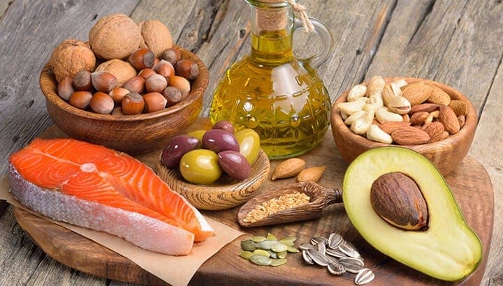
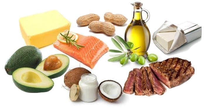
What to eat and drink on a Ketogenic diet
Animal protein
Saturated fat
Olive oil
Avocado
Above the ground vegetables
Water
What to avoid on a Ketogenic diet
Processed food
Fizzy drinks
Toxic oils
Processed meat
Fast food
Cancer Fighting Foods
Tomatoes: cooking enhances cancer-fighting and anti inflammatory properties. Lycopene was found to prevent cancer cell growth in a study in Cancer and Nutrition.
Chilli: capsaicin that gives chillies their powerful, spicy personality is anti-bacterial, anti-carcinogenic and anti-diabetic.
Cruciferous vegetables: such as cabbage, cauliflower, broccoli, spinach, Brussels sprouts and kale have powerful anti-carcinogens. Cabbage in particular contain anti-oxidants known to help protect against prostate, colon and breast cancers. Broccoli is the only one having a sizable quantity of sulforaphane, an especially potent chemical that boosts the body enzymes and flushes compounds out .
Mushrooms: include the amino acid ergothioneine, which is an anti-oxidant and an anti-inflammatory, it protects against free radicals and boosts the immune system.
Aubergine: that the epidermis is rich in anti-oxidants known as anthocyanins, which are believed to fight cancer, inflammation, aging and neurological diseases.
Turmeric: includes curcumin that’s a powerful anti-oxidant and anti inflammatory. According to Cancer Research UK, it seems to have the ability to kill cancer cells and stop more from growing. It’s the very best consequences on breast cancer, bowel cancer, stomach cancer and skin cancer cells.
Berries: the idea of berries as anticarcinogens began in the late 1980s, when it was discovered that berries, and specifically black peppers, comprised ellagic acid, which is believed to inhibited the genesis of tumours.
Garlic: belongs to the Allium class of bulb-shaped plants, which also includes onions, chives, leeks, and scallions. It’s an strong and excellent neutraliser of free radicals. It contains good levels of selenium and, in several studies, selenium has been shown to decrease cancers. Phytochemicals in garlic have been found to stop the formation of nitrosamines, carcinogens formed in the stomach.
In summary, from the evidence that we have collected from all of the various sources, it’s obvious to see that the link between diet and health is a serious one and that what we consume really has an impact in the long term. Dr D’Agostino goes as far as to state, “let food be thy medicine.”
The scope of our information is limited to chiropractic and spinal injuries and conditions. To discuss options on the subject matter, please feel free to ask Dr. Jimenez or contact us at 915-850-0900 .�
By Dr. Alex Jimenez
Additional Topics: Wellness
Overall health and wellness are essential towards maintaining the proper mental and physical balance in the body. From eating a balanced nutrition as well as exercising and participating in physical activities, to sleeping a healthy amount of time on a regular basis, following the best health and wellness tips can ultimately help maintain overall well-being. Eating plenty of fruits and vegetables can go a long way towards helping people become healthy.
The benefits that come from a ketogenic diet are much like those of any strict low-carb diet. The effect may be greater since protein is significantly more restricted. This raises ketones more, and reduces insulin (the fat-storing hormone).
Weight Loss
Turning your body to some fat-burning machine has clear benefits for weight loss. Fat burning is significantly increased while insulin, the hormone that focuses on fat-storing, drops considerably. This produces the perfect circumstances.
About 20 scientific research of the maximum category (RCTs) reveal that, compared to other diets, low-fat and ketogenic diets result in more effective weight reduction.
Reverse Type 2 Diabetes
A ketogenic diet is excellent for reversing type 2 diabetes, because it lowers blood-sugar levels as well as also helping to reverse the negative effect of elevated insulin levels from this condition.
Improved Mental Focus
Ketosis ends in a steady stream of gas (ketones) to the brain. And on a ketogenic diet you stay away from swings in blood glucose. This contributes to the experience of concentration and attention.
A lot of people use keto diets specifically for improved mental performance. Interestingly, there is a frequent misperception that eating a great deal of carbs6 is necessary for proper brain functioning. When ketones aren’t available but this is only true.
Following a couple of times (up to a week) of keto adaptation, through that people can experience some difficulty concentrating, have headaches and be easily irritated, both the human body and mind can run smoothly on ketones.
Inside this state, lots of men and women experience more energy and enhanced mental focus.
Increased physical endurance
Ketogenic diets may vastly increase your physical endurance, by giving you constant access to all of the energy of your own fat stores.
The body’s source of stored carbohydrates (glycogen) only lasts for a few hours of intense exercise, or less. But your fat stores hold sufficient energy to easily last for weeks or perhaps months.
When you’re accommodated to burning primarily carbs — like most individuals are now — that your fat stores aren’t readily accessible, and they can not fuel your brain. This results in needing to fill up by eating before, during and after exercise sessions that are longer. Or even simply to fuel your everyday activities and prevent “hanger” (hungry and irritable). On a ketogenic diet this dilemma is solved. As the body and brain can be fueled 24/7 from the stores that are powerful, you can keep going.
Whether you are competing in a bodily endurance event, or just trying to remain focused on reaching some other target, your body gets the fuel it needs to keep you going and going.
Two Problems
So how is it possible that the majority of people feel that carbohydrates are essential to do exercise? There are just two reasons. Not, and to unlock the power of ketogenic diets for bodily endurance rather suffer reduced performance, you’ll need:
Enough fluid and salt
Fourteen days of adaptation into burning fat — it does not happen immediately
Metabolic Syndrome
There are many studies demonstrating that low-carb diets improve markers of metabolic syndrome, such as blood lipids, insulin levels, HDL-cholesterol, LDL particle size and fasting blood sugar levels. Improvements have been demonstrated to be greater when carbs and protein are limited to some the point of becoming.
Epilepsy
The ketogenic diet is a proven medical therapy for epilepsy that’s been utilized since the 1920s. Traditionally it has been used in children with uncontrolled epilepsy despite drugs.
More recently it has also been tested successfully by adults with epilepsy, with similar good results. There are randomized controlled trials that demonstrate the potency of the ketogenic diet in seizures in patients with epilepsy.
Employing a ketogenic diet in epilepsy is that usually enables people to take less anti-epileptic drugs, while staying seizure-free. It is not uncommon to even be in a position to completely stop taking these drugs.
As a number of medications have side effects, such as nausea, reduced concentration, personality changes or even reduced IQ — being able to shoot less or no medications can be enormously beneficial.
More Prevalent Advantages
The advantages will be the most frequent ones. However there are many others that are potentially even more unexpected and, at least for some people, lifechanging.
The scope of our information is limited to chiropractic and spinal injuries and conditions. To discuss options on the subject matter, please feel free to ask Dr. Jimenez or contact us at 915-850-0900 .�
By Dr. Alex Jimenez
Additional Topics: Wellness
Overall health and wellness are essential towards maintaining the proper mental and physical balance in the body. From eating a balanced nutrition as well as exercising and participating in physical activities, to sleeping a healthy amount of time on a regular basis, following the best health and wellness tips can ultimately help maintain overall well-being. Eating plenty of fruits and vegetables can go a long way towards helping people become healthy.
A ketogenic diet, or keto diet, is a diet, which turns your system into a fat-burning machine. It has some initial side effects towards health and functionality, as well as many advantages for weight loss.
A ketogenic diet is comparable to other rigorous low-carb diets, like the Atkins diet plan or LCHF (low carb, higher fat). These diets wind up being ketogenic more or less by accident. The main difference between LCHF and keto is that protein is restricted in the latter.
A keto diet plan is made specifically to lead to ketosis. It’s possible to measure and adapt to achieve optimal ketone amounts for wellness or for bodily and psychological performance. Below, you can learn how to use keto to achieve your personal goals.
What is Ketosis?
The keto in a ketogenic diet stems in the fact that it leaves the body to create small fuel molecules known as ketones. This is an alternate fuel for your body, used when blood sugar (glucose) is in short supply.
Ketones are produced if you eat hardly any carbs (that are quickly broken down into blood sugar) and only moderate levels of protein (excess protein can also be converted to blood sugar). Ketones are produced in the liver, from fat. They are then used throughout the entire body as fuel. The brain is an organ which requires a lot of energy to function and fat can’t be used for energy by it. The brain can only run on glucose or ketones.
On a ketogenic diet your entire body switches its fuel source to operate almost entirely on fat. Insulin levels become very low and fat burning increases dramatically. It becomes easy to get into your fat stores to burn them off. If you are trying to drop weight, this is obviously excellent, but in addition, there are other benefits, such as less appetite and a continuous supply of energy.
Once the body produces ketones, it’s supposedly in ketosis. The quickest way to get there is by fasting, not eating anything, but obviously, it is not feasible to fast. A ketogenic diet, on the other hand, can be eaten forever and also results in ketosis. Without even having to fast, it has many of the benefits of fasting. including weight loss.
What to Eat on a Ketogenic Diet
Here are typical foods to enjoy on a ketogenic diet. The amounts are net carbs per 100 g. To remain in ketosis, lower is generally better:
The most essential thing to achieve ketosis is to stay away from eating most carbohydrates. You will need to keep intake ideally under 20 grams but under 50 grams per day of carbs is accepted. The fewer carbs the more successful.
Try to avoid
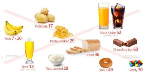
Here is what you shouldn’t eat on a keto diet, meals full of sugar and starch, including starchy foods such as bread, rice, pasta and potatoes. These foods are much higher in carbohydrates, as you can see.
The amounts are g of digestible carbs per 100 g (3.5 oz), unless otherwise noticed.
This usually means you will want to completely prevent sweet sugary foods, also starchy foods such as bread, pasta, rice and potatoes. Basically follow the guidelines to get a diet that is low-carb that is rigorous, and remember it is assumed to be full of fat, not high in protein.
A rough guideline is under 10 percent energy from carbs (the fewer carbs, the more successful), 15 to 25 percent protein (the lower end is more successful), and 70 percent or more from fat.
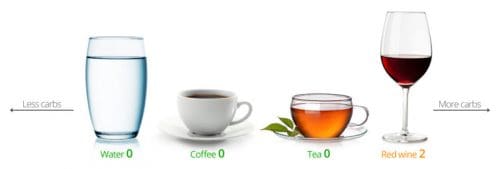
What to Drink on a Ketogenic Diet
So what do you drink on a keto diet? Water is ideal, and so is tea or coffee. Use no additives. A small amount of milk or cream is OK (but beware of caffe latte!) . The glass of wine is fine.
How Low is Keto?
The fewer carbohydrates you consume, the larger the effects on fat and blood sugar will be. A keto diet is a strict low-carb diet, and consequently highly effective.
We recommend following the dietary advice as strictly as you can. When you are contented with your weight and health, you might carefully try eating more liberally (if you would like to).
The scope of our information is limited to chiropractic and spinal injuries and conditions. To discuss options on the subject matter, please feel free to ask Dr. Jimenez or contact us at 915-850-0900 .�
By Dr. Alex Jimenez
Additional Topics: Wellness
Overall health and wellness are essential towards maintaining the proper mental and physical balance in the body. From eating a balanced nutrition as well as exercising and participating in physical activities, to sleeping a healthy amount of time on a regular basis, following the best health and wellness tips can ultimately help maintain overall well-being. Eating plenty of fruits and vegetables can go a long way towards helping people become healthy.
There’s been a lot of discussion about the advantages of intermittent fasting (IF) in the community lately. Paul Jaminet mentions its role in the function of the metabolism as well as it’s role in boosting the immune system. In his novel, Health Diet, he discusses how IF may be helpful for those attempting to shed weight, among other benefits.
Intermittent fasting is an eating pattern where you cycle between times of fasting and eating. It does not state anything about which foods to eat, but instead when they should be eaten by you. There are numerous different fasting methods, all of that which split the days and weeks to eating intervals and fasting intervals.
Most people fast; daily, while they sleep. Fasting is often as easy as stretching that. You can drink water, coffee, tea and other non-caloric drinks, although no food is permitted during the fasting period. Some forms of intermittent fasting allow small amounts of low-calorie foods . Taking supplements is generally allowed while fasting, as long as there are no calories in them.
From an evolutionary perspective, intermittent fasting has been likely the ordinary state of affairs. There were no restaurants, no grocery stores or convenience stores, and food was not nearly as readily accessible or easy to come by as it is today. Nor were there watches, lunch breaks, programs or the sort of construction and routine we have in today’s world. This means it is very likely that our paleo ancestors had days when they ate lightly or did not eat, and possibly did move 12-16 hours between meals on a regular basis.
Therefore, while intermittent fasting is a part of our legacy, and that it can be useful in certain situations, Its not believed to be a suitable strategy for everyone. Why? Because cortisol levels can be elevated due to fasting. One of cortisol’s effects is that it raises blood glucose. In someone with blood glucose regulation difficulties, fasting can make them worse.
This has been seen again and again with many patients. Many patients have blood sugar imbalances. And it is usually not as straightforward as “high blood sugar” or “low blood sugar”. They frequently have a combination of both (reactive hypoglycemia), or strange blood sugar patterns which, on the outside, do not make much sense. These folks are not currently eating a Standard American Diet. Most of them are on a paleo-type or low-carb dietplan. Yet they have blood sugar issues.
In these cases, cortisol dysregulation is almost always the culprit. Whenever these patients try fasting, their blood sugar management gets worse. Its been frequently observed that blood sugar readings in the 90s and even low 100s are recorded from fasting, in spite of the fact that they are eating a low-carb, paleo-type diet.
That’s why intermittent fasting is often not recommended for those who have blood sugar regulation issues. Instead, its suggested that they eat every 2-3 hours. This helps to maintain stable blood sugar during the day and prevents cortisol and other stress hormones like epinephrine and norepinephrine from becoming involved. When patients that have been fasting and experiencing high blood sugar readings switch to eating this way, their blood sugar numbers almost always hastens.
There’s a controversial standpoint about how eating every 2-3 hours is “normal” in the evolutionary view. But neither is driving in traffic or staying up till 2:00am on Facebook. It’s not a set of rules, although the paleo template is there to direct us. This should also be a reminder that there’s no “one size fits all” approach when it comes to healthcare. Successful therapy depends on addressing them and identifying the underlying mechanisms.
The scope of our information is limited to chiropractic and spinal injuries and conditions. To discuss options on the subject matter, please feel free to ask Dr. Jimenez or contact us at 915-850-0900 .�
By Dr. Alex Jimenez
Additional Topics: Wellness
Overall health and wellness are essential towards maintaining the proper mental and physical balance in the body. From eating a balanced nutrition as well as exercising and participating in physical activities, to sleeping a healthy amount of time on a regular basis, following the best health and wellness tips can ultimately help maintain overall well-being. Eating plenty of fruits and vegetables can go a long way towards helping people become healthy.
Intermittent fasting is not a diet, but a diet program that is supposed to accelerate fat loss and muscle development compared to traditional eating schedules. It is promoted primarily from the scientific community, however, there are no scientific research (as of February 2014) who have affirmed intermittent fasting to gaining muscle while losing weight.
With metabolic restriction, intermittent fasting may lead to weight reduction. In a recent review (Varady, 2011) and a recent randomized clinical trial (Harvie et al., 2011), many writers concluded that intermittent fasting and daily caloric restriction are equally effective at promoting weight loss in overweight and obese individuals. No research to date has been performed with athletes that require upkeep of strength, muscular size, and function.
Intermittent Fasting Approach & Scientific Support
There have been many suggested methods for intermittent fasting, from skipping a single meal daily to eating only every other day. The majority of these diets are encouraged through webpages, blogs, and books published by exercise and diet enthusiasts.
Up to now, regardless of the focused marketing of intermittent fasting into the athletic community, there are just a few well-controlled, scientific research exploring the effects of intermittent fasting on the body composition and performance in athletes. Currently, the majority of the scientific evidence for the health benefits of intermittent fasting has arrived from animal studies (Longo and Mattson, 2014) and the unwanted effects of intermittent fasting have originated from Muslim athletes throughout Ramadan (review: (Chaouachi et al., 2009), both with restricted ability to be translated into the overall athletic community. Even more and more human research have been conducted to confirm claims discovered in animals, many studies have been with patients with a certain illness or condition (ex. Rheumatoid arthritis, hypertension, and obesity) rather than in healthy, energetic individuals.
Similarities Among Strategies
The different intermittent fasting approaches tend to emphasize their differences (and therefore purported superiority) however, there are also many similarities. Among the advantages of the form of caloric control is that it allows people. Instead of linking “appetite” with “panic” or even “want” (Ganley 1989), “hunger” can theoretically be newly associated with “achievement” or “pride”, or simply dismissed.
Really, with any method, there’s a critical transition period of approximately 3-6 weeks through which the human body and mind adapt to the new eating schedule (Longo and Mattson, 2014). This period can be extremely uncomfortable, as restricted eating was anecdotally associated with intense hunger, irritability, loss of stamina, loss of libido, along with other unwanted side effects (Dirks and Leeuwenburgh 2006; Johnstone 2007; Heilbronn, Smith, et al. 2005). When the body is accustomed, however, the hunger levels may decrease and disposition might become more favorable in contrast to prior to the program began. Elevated mood and diminished hunger on caloric restrictive diets are noted in some (Wing et al. 1991) although not all (ex. (Heilbronn, Smith, et al., 2005) research.
Intermittent fasting isn’t a weight loss program per se; only if calories are restricted will somebody lose weight. Although intermittent fasting is one way to limit intake of total calories to achieve weight loss (Varady et al., 2009; Varady, 2011; Harvie et al., 2011), there have not been any studies to date on athletes who prioritize maintenance of muscle size and strength. In actuality, there are conflicting views on whether intermittent caloric limitation. daily calorie restriction greatest maintain lean muscle mass (Varady, 2011; Johnstone, 2007).
All of these approaches emphasize the value of the nutritional quality of the meals that are consumed. Nutrients like fiber, fat, protein, vitamins, and minerals are crucial for good health and, because nutrients aren’t consumed while fasting, they are especially important when breaking the fast. Additionally, drinking a lot of water has been encouraged both to stay hydrated and to alleviate hunger. John Berardi of Precision Nutrition (see “More info”) allows green powders, green tea, and branched chain amino acids during his quick, but it’s unknown how these supplements influence appetite, energy levels, muscle synthesis/breakdown, or the general advantages of intermittent fasting.
Exercising and Intermittent Fasting
All intermittent fasting approaches can be damaging to athletic gains for several reasons. To begin with, meals in close proximity to your workout are essential for optimal performance, healing, and muscle gain (Aragon and Schoenfeld, 2013). Secondly, greater appetite sensations can hinder compliance in addition to increase the potential to over-consume meals when it becomes available (Hawks and Gas,t 1998). Despite the frequent belief that you will burn off more fat if you exercise while firming, performing aerobic exercise in the fasted state is not recommended (review: (Schoenfeld 2011)). Actually:
Performing aerobic exercise following consuming carbohydrates doesn’t hinder fat oxidation (Febbraio et al., 2000; p Bock et al., 2008),
Performing aerobic exercise fasted will also promote reduction of lean muscle mass, since muscle will be burned for fuel (Lemon and Mullin, 1980),
Exercising at a fasted state often does not result in an optimal exercise. In contrast, having readily available energy will allow optimal performance that will burn off more calories overall and lead to the Greatest gains (Loy et al., 1986; Schabort et al., 1999),
Exercising at the fasted condition, fed state reduces static and dynamic balance and can increase the risk of harm (Johnson and Leck, 2010).
There are fewer studies investing the effects of doing resistance training in the fasted vs. fed state, but it is anticipated that the same points hold true.
Intermittent fasting recommends consuming at least 5 grams BCAAs before a workout when exercising during your fasting period. There’s no proof substantiating that claim, although this bolus of BCAAs in your blood flow theoretically could help maintain muscle protein throughout the exercise. In one study, a BCAA infusion prior to a workout in the fasted state failed to enhance performance in one set of individuals onto a rated incremental exercise test (Varnier et al., 1994). So that you can enjoy a complete meal that is post-workout advocates also advise to program your schedule, but they tend to dismiss the importance for pre-workout nourishment.
As you can see, all diet programs share a frequent theme of compartmentalizing “fasting” and “eating” periods. Because there is no one method that’s best so many versions of those diets exist. Further, some individuals who attempt fasting use a hybrid of present approaches to discover a technique that is successful.
Most importantly, intermittent fasting isn’t suggested for pregnant women, women that are breastfeeding, people with diabetes, or other people who wish to closely regulate their blood sugar. In addition, there has not been a study on participants that are underweight, very old, or very young.
The scope of our information is limited to chiropractic and spinal injuries and conditions. To discuss options on the subject matter, please feel free to ask Dr. Jimenez or contact us at 915-850-0900 .�
By Dr. Alex Jimenez
Additional Topics: Wellness
Overall health and wellness are essential towards maintaining the proper mental and physical balance in the body. From eating a balanced nutrition as well as exercising and participating in physical activities, to sleeping a healthy amount of time on a regular basis, following the best health and wellness tips can ultimately help maintain overall well-being. Eating plenty of fruits and vegetables can go a long way towards helping people become healthy.
Though the truth has a tendency to get distorted by time, diet and exercise tendencies have origins in science. Benefits are exaggerated. Hazards are downplayed. Meanwhile science takes a back seat to marketing.
An individual need not look any farther than the emerging tendency of intermittent fasting for a prime illustration. Advocates for taking breaks from eating, for up to 24 hours a week, tout it as a safe and effective method for improving health and wellness. That message has been reaching more and more ears as of late.
“Right now, we are in a vital juncture for fasting,” states Brad Pilon, an expert on occasional fasting and author of the novel Eat Stop Eat. “It’s becoming extremely popular.”
Intermittent Fasting: Know the Facts
Such may become so popular, in fact, that it’s moving into trend territory, indicates Pilon. And if something becomes a fad, only for a short period although intensely popular, several problems normally ensue. For one, he states, nutrition experts and many doctors have a tendency toward dismissing fads from hand. So clients and their patients, while shielded from the claims of dieting evangelists that are overzealous, can lose out on the advantages of fasting right.
Yet another concern is that promoters of intermittent fasting will, perhaps unintentionally, encourage extreme behaviour, such as bingeing. This is reflected in the photos accompanying many recent new articles on “the fast diet” or even the “5:2 diet.” Often, they portray people eating loads of high-calorie, high-fat foods, like hamburgers, french fries and cake. The implication being that if you fast two days a week, you are able to consume a lot during the remaining five days.
Not so, say more moderate proponents of fasting. Their take on intermittent fasting: eat sensibly the majority of the time, eat nothing for a protracted period every now and then, gratify only on occasion (maybe once a week, state, on a predetermined “cheat day”). There is study, they assert, to back up the health benefits of wisely.
There is indeed a large body of research to support the health benefits of fasting, although most of it has been conducted on animals, not humans. Still, the results have been promising. Fasting has been shown to improve biomarkers of illness, decrease stress and preserve memory and learning function, based on Mark Mattson, senior investigator at the National Institute on Aging, part of the US National Institutes of Health. Mattson has investigated the health advantages of intermittent fasting on the cardiovascular system and brain in rodents, also has known for “well-controlled human studies” in people “across a assortment of body mass indexes” (J Nutr Biochem 2005;16:129–37).
There are several theories about why fasting offers physiological advantages, says Mattson. “The one which we have researched a great deal, and designed experiments to examine, is the hypothesis that during the fasting period, cells are under a mild strain,” he states. “And they respond to the stress adaptively by enhancing their ability to deal with additional stressors and, possibly, to resist illness.”
Although the term “stress” is frequently used in a negative sense, taxing the body and mind has benefits. Consider exercise, which stresses, in particular, muscles and the cardiovascular system. So long as you give the body time to recover, it will grow stronger. “There is considerable similarity between how cells react to the pressure of exercise and how cells react to intermittent fasting,” says Mattson.
Additional Research: Intermittent Fasting
Mattson has contributed to several additional research on intermittent fasting and caloric restriction. In one, obese adults with moderate asthma consumed only 20% of their normal calorie intake on alternative days (Free Radical Bio Med 2007;42:665–74). Participants that stuck to the diet lost 8 percent of their initial body weight over eight months. They also found a decrease in markers of oxidative stress and inflammation, and improvement of several indicators and asthma-related symptoms.
In another study, Mattson and colleagues researched the effects of intermittent and continuous energy limitation on weight loss and various biomarkers (such as conditions such as breast cancer, diabetes and cardiovascular disease) among young obese woman (Int rheumatoid arthritis 2011;35:714–27). They discovered for improving weight loss, insulin sensitivity and other wellness biomarkers that restriction was as effective as continuous restriction.
Mattson has also investigated the protective benefits of fasting to neurons. Should youn’t eat for 10 – 16 hours, then your body will go for energy, and fatty acids called ketones will probably be released into the bloodstream. It has been demonstrated to protect memory and learning says disease processes in the mind, as well as Mattson.
But maybe it isn’t so much that the fasting that creates health benefits, per se, because the consequent overall reduction in calorie intake (if, in other words, you don’t overeat on nonfasting days, which could cause a caloric surplus rather than a deficit). That seems, at least, to be true in slowing diseases like cancer in mice, according to Dr. Stephen Freedland, associate professor of urology and pathology at the Duke University Medical Center in Durham, North Carolina.
“Caloric restriction, undernutrition without malnutrition, is the only experimental approach consistently shown to prolong survival in animal models,” Freedland and colleagues stated in a study on the effects of intermittent fasting on prostate cancer growth in mice (Prostate Cancer Prostatic Dis 2010; 13:350–5). From the study, mice fasted twice weekly for 24 hours, but were otherwise permitted to eat at liberty. During days, the mice overate. They did not lose weight. Intermittent fasting using compensatory overeating “did not enhance mouse success nor did it delay prostrate tumor growth,” the study concluded.
To improve health, the aim should be to shed weight by reducing the entire amount of calories consumed, indicates Freedland, rather than focusing on if those calories are consumed. “If you [do not] eat two days every week, and restrict what you eat another five days, you will lose weight. It is one approach to losing weight,” he says. “I am not sure it works any better than trimming down marginally seven days each week.”
People should also be skeptical of books written for wide audiences that describe the science behind fasting or another health trend, ” he says. One purpose of writing a novel for the consumer market, after all, would be to sell as many copies as you can. Authors tend to present evidence, while ignoring the facts and their impact. “It has a whole lot of spin when you compose a book.”
The scope of our information is limited to chiropractic and spinal injuries and conditions. To discuss options on the subject matter, please feel free to ask Dr. Jimenez or contact us at 915-850-0900 .�
By Dr. Alex Jimenez
Additional Topics: Wellness
Overall health and wellness are essential towards maintaining the proper mental and physical balance in the body. From eating a balanced nutrition as well as exercising and participating in physical activities, to sleeping a healthy amount of time on a regular basis, following the best health and wellness tips can ultimately help maintain overall well-being. Eating plenty of fruits and vegetables can go a long way towards helping people become healthy.
Studies have demonstrated that reducing typical calorie consumption, usually by 30 to 40 percent, extends life span by a third or more in many animals, including fruit flies, nematodes and rodents. But the jury remains out, when it comes to calorie restriction in primates and individuals.
Although some studies have suggested that primates that eat less live longer, research concluded that restriction does not extend average life span in some species of monkeys. A section of the data confirms the idea that limiting food intake reduces the dangers of diseases common in older age and lengthens the period of life spent in good health, even if the research concludes that restriction does not help people live longer.
If only one could claim those advantages without being hungry all the time. In recent years researchers have concentrated on a strategy known as intermittent fasting as a promising option to continuous calorie restriction.
Intermittent fasting, including everything from regular multi-day fasts to skipping a meal or 2 on particular days of the week, may encourage a number of the identical health benefits that uninterrupted calorie restriction promises. The idea of intermittent fasting is palatable to people since somebody does not need to renounce the joys of eating. Studies suggest that rodents live as long as rats eating foods every moment consuming fewer calories overall than they would normally.
In a 2003 mouse analysis controlled by Mark Mattson, head of the National Institute on Aging’s neuroscience lab, mice that fasted regularly were healthier by some measures than mice subjected to constant calorie limitation; they had reduced levels of glucose and insulin in their blood, by way of instance, which signified increased sensitivity to insulin and a reduced risk of diabetes.
The First Fasts
Religions have long claimed that fasting is good for the soul, but its bodily benefits weren’t widely known until the early 1900s, when doctors began recommending it to treat different disorders, such as diabetes, obesity and epilepsy.
Associated research on calorie restriction took off in the 1930’s, following Cornell University nutritionist Clive McCay found that rats exposed to stringent daily dieting in an early age lived longer and were less likely to develop cancer and other ailments as they elderly, compared with animals that ate at will. Research on calorie restriction and periodic fasting intersected in 1945, when University of Chicago scientists reported that alternate-day feeding extended the life span of rats as much as exercising in McCay experiments. Additionally, intermittent fasting “appears to delay the development of the disorders that cause death,” that the Chicago researchers wrote.
Within the upcoming decades study into anti-aging diets took a backseat to more powerful clinical advances, like the continued development of antibiotics and coronary artery bypass operation. However, researchers also have resisted the idea that intermittent fasting lowers the risks of degenerative brain diseases in later life. Mattson and his colleagues have shown that fasting protects neurons from several types of harmful stress, at least in rodents. Among the earliest studies demonstrated that alternate-day feeding made the rats’ brains as they age, that induce damage akin to the kind cells endure. In follow-up rodent research, his team discovered that fasting slows cognitive decline in mice genetically engineered to mimic the signs of Alzheimer’s, suppresses motor deficits in a mouse model of Parkinson’s disease and protects against stroke damage. The 55-year-old researcher, who has a Ph.D. in biology although not a medical degree, has written or co-authored over 700 posts.
Mattson believes that intermittent fasting functions in part as a kind of moderate stress that continually revs up mobile defenses against molecular damage. For instance, occasional fasting increases the degrees of “chaperone proteins,” which forbid the incorrect assembly of other molecules at the cell. Additionally, fasting mice have greater degrees of brain-derived neurotrophic factor (BDNF), a protein that prevents nervous nerves from dying. Low levels of BDNF are linked to Alzheimer’s, although it is still unclear whether these findings reflect cause and effect. Fasting also ramps up a sort of system in cells which eliminates damaged molecules, autophagy, including ones that have been tied to Parkinson’s, Alzheimer’s and other neurological diseases.
One of intermittent fasting’s major effects seems to be raising the body’s responsiveness to insulin, the hormone which regulates blood glucose. Sensitivity to insulin accompanies and obesity has been associated with diabetes and heart failure; people and long-lived animals tend to have unusually low insulinbecause their cells are more sensitive to the endocrine and therefore require less of it. A recent study in the Salk Institute for Biological Studies in La Jolla, Calif., revealed that mice that feasted on fatty foods for 2 hours each day and subsequently fasted for the remainder of each day did not become obese or show dangerously high insulin levels.
The idea that periodic fasting may offer some of the same health benefits as continuous calorie restriction–and allows for a few feasting while shedding down–has persuaded an increasing number of people to attempt it, says Steve Mount, a University of Maryland genetics professor who has moderated a Yahoo discussion group on intermittent fasting for more than seven decades. Intermittent fasting “isn’t a panacea–it’s always hard to lose weight,” adds Mount, who has fasted three days per week since 2004. “But the concept [that it activates the identical signaling pathways in cells as calorie restriction] makes sense.”
Further Research Still Needed
Despite the increasing excitement for intermittent fasting, scientists have conducted several powerful clinical trials, and its long term effects in people remain unclear. Still, a 1956 Spanish study sheds some light, states Louisiana-based physician James B. Johnson, who co-authored a 2006 evaluation of the research’s results. In the study, 60 men and women fasted and feasted on alternate days for 3 years. The 60 participants spent at the infirmary, and six died. Meanwhile, the 60 nonfasting seniors racked up 219 days that were infirmary, and 13 died.
In 2007 Johnson, Mattson and their colleagues published a clinical research demonstrating a quick, significant alleviation of asthma symptoms and various indications of inflammation in nine overweight asthmatics who near-fasted another day for 2 weeks.
Detracting from these promising results, however, the literature on intermittent fasting also includes several red flags. A 2011 study in rats suggests that long-term intermittent fasting raises tissue levels and blood sugar of compounds that may damage cells. In a 2010 study stiff heart tissue, which subsequently hastens the ability of the organ to pump blood was developed by occasionally fasting rats.
And a few weight-loss experts are skeptical about fasting, mentioning its hunger pangs and the possible hazards of compensatory gorging. Truly, the latest primate study on calorie restriction–the one that failed to extend life span–underscores the need for caution when altering the way people eat.
However, from an evolutionary perspective, three meals a day is a peculiar modern invention. Volatility in our ancestors’ food supplies brought on fasting–not to mention starvation and malnutrition. Yet Mattson considers that pressures that are such selected for genes that brain areas involved in learning and memory, which increased the likelihood of finding food and surviving. Intermittent fasting may be both smartening and a wise way, if he’s correct.
The scope of our information is limited to chiropractic and spinal injuries and conditions. To discuss options on the subject matter, please feel free to ask Dr. Jimenez or contact us at 915-850-0900 .�
By Dr. Alex Jimenez
Additional Topics: Wellness
Overall health and wellness are essential towards maintaining the proper mental and physical balance in the body. From eating a balanced nutrition as well as exercising and participating in physical activities, to sleeping a healthy amount of time on a regular basis, following the best health and wellness tips can ultimately help maintain overall well-being. Eating plenty of fruits and vegetables can go a long way towards helping people become healthy.
IFM's Find A Practitioner tool is the largest referral network in Functional Medicine, created to help patients locate Functional Medicine practitioners anywhere in the world. IFM Certified Practitioners are listed first in the search results, given their extensive education in Functional Medicine