Do you feel irritable, nervous, shaky, or light-headed between meals? Do you have difficulty eating large meals in the morning? Do you feel fatigued after meals? Do you have sugar and sweet cravings after meals? Do you have an increased appetite?�If so, you may be experiencing early SIBO symptoms. �
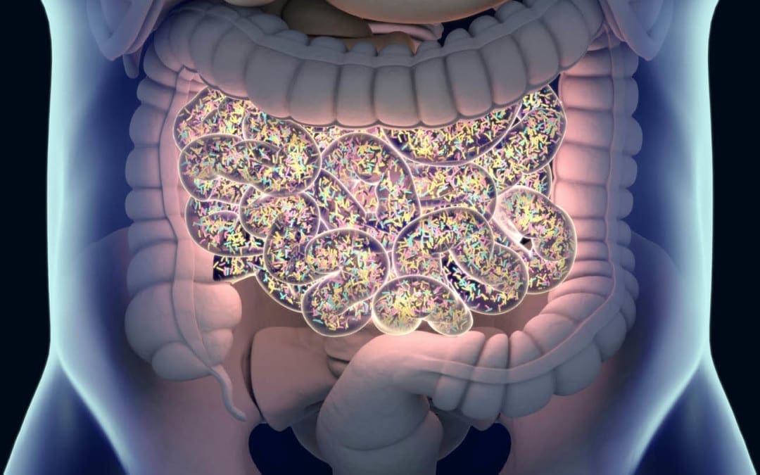
SIBO, or small intestinal bacterial overgrowth, is a severe health issue that ultimately affects the small intestine in the digestive system. This gastrointestinal (GI) tract condition happens when the bacteria that generally grows in several different regions of the gut begin to grow in the small intestine. SIBO can commonly cause pain, discomfort, and diarrhea, among other symptoms. It can also cause malnutrition as bacteria utilize the human body�s nutrients.�
What are the Symptoms of SIBO?
Small intestinal bacterial overgrowth, or SIBO, is a serious condition that includes symptoms which can commonly affect the gut. These can include: �
pain or discomfort in the stomach
gas
bloating
constipation
diarrhea
cramps
indigestion
a general feeling of fullness
weight loss
What are the Causes of SIBO?
Small intestinal bacterial overgrowth (SIBO) is a severe health issue that is unfortunately not yet fully understood by researchers and healthcare professionals. According to research studies and clinical trials, however, this gastrointestinal, or GI, tract condition can ultimately happen when the small intestine is anatomically abnormal, due to pH changes in the small intestine, when the human body’s immune system isn’t functioning accordingly, or due to malfunctions in the muscular activity of the small intestine which can commonly cause food and bacteria to remain and not be removed from the organ. �
SIBO, or small intestinal bacterial overgrowth, is also commonly associated with a variety of health issues. These can involve the following, including: �
a stomach bug, known as viral gastroenteritis
celiac disease
Crohn�s disease
low stomach acid levels, known as hypochlorhydria
IBS or irritable bowel syndrome
gastroparesis
portal hypertension
nerve damage
cirrhosis
several gastric bypass procedures
surgical interventions which cause strictures or adhesions
What are the Risk Factors of SIBO?
Moreover, researchers and healthcare professionals have determined that an underlying chronic health issue and a previous surgery or surgical intervention that affects the gastrointestinal (GI) tract can be several risk factors of SIBO. Other wellness problems which can ultimately cause SIBO include: �
diabetes
scleroderma
hypothyroidism
Parkinson’s disease
HIV
narcotics or drugs/medications which slow down the digestive system
What is the Diagnosis for SIBO?
If you’ve experienced any of the SIBO symptoms mentioned above, see your doctor immediately. The doctor will ask the patient about their symptoms and medical history. The doctor will also perform a physical examination which may include palpating or gently feeling the patient’s abdomen. A qualified and experienced healthcare professional may also order additional blood, fecal, and/or any other tests to diagnose small intestinal bacterial overgrowth. �
A breath test is another common test utilized for the diagnosis of SIBO. Excess bacteria in the small intestine can cause the release of hydrogen and methane, two common gases which can be identified through a breath test. This test is non-invasive and can be performed in a doctor�s office. Before a breath test, the patient will need to fast overnight. During a breath test, the patient will first breathe into a tube. Then, the patient will take a specialized sweet drink provided by the doctor and they will breathe into several other tubes at regular intervals for 2 to 3 hours after taking the specialized sweet drink. �
If common tests for SIBO are inconclusive, the doctor may need to sample the fluid from the patient’s small intestine to see what bacteria is growing there. �
What is the Treatment for SIBO?
Common treatment approaches for SIBO, or small intestinal bacterial growth, can ultimately include a combination of antibiotics and diet modifications. �
Antibiotics
Treatment for SIBO first involves getting the bacteria in the digestive system under control. This is generally achieved by utilizing antibiotics, such as ciprofloxacin (Cipro), metronidazole (Flagyl), or rifaximin (Xifaxan). Further treatment for SIBO may also require intravenous (IV) therapy for nutrition and fluids if the serious gastrointestinal (GI) tract condition has ultimately caused malnutrition or dehydration, among a variety of other symptoms. �
Although antibiotics may help reduce the amount of bacteria in the small intestine, however, these will not always help address the underlying chronic health issues that caused the wellness problem in the first place. If the qualified and experienced healthcare professional determines that the patient’s SIBO is due to an underlying chronic health issue, the patient will also need to begin treatment for that wellness problem. Diet modifications may also help treat SIBO. �
Diet Modifications
Further research studies and clinical trials are still required to demonstrate if diet can cause small intestinal bacterial overgrowth (SIBO) but, many people with SIBO have reported feeling relief from their symptoms after diet modifications. Talk to your doctor before making any modifications to your diet. �
Furthermore, people with SIBO or other chronic health issues may only need to make small diet modifications to treat their symptoms. These can include: �
Eating a balanced and nutritious diet
Consuming minimal meals more often to prevent having too much food sit in the stomach
Avoid eating gluten products, if you have celiac disease or any other similar chronic health issues
The doctor may also recommend the patient to try an elemental diet to help treat SIBO. An elemental diet replaces food and drinks with several liquid formulas throughout an extended period of time. In one small-scale research study and clinical trial, approximately 80 percent of participants with SIBO had a normal breath test result following an elemental diet for 15 days. The researchers ultimately determined that an elemental diet may be a highly effective treatment approach for SIBO. However, further evidence is still needed. Talk to your doctor before starting an elemental diet and follow their instructions. �
Taking probiotics may also help restore the gut bacteria. A 2010 research study and clinical trial demonstrated that probiotic treatment can be more safe and effective for SIBO than taking antibiotics. However, a 2016 review determined that further evidence for the efficiency of probiotics in SIBO treatment was ultimately inconclusive. The best treatment approach for a patient with SIBO is to follow a qualified and experienced healthcare professional’s advice. �
SIBO, or small intestinal bacterial overgrowth, is a well-known and often severe health issue that generally occurs because of an underlying chronic condition or disease. Common symptoms may ultimately determine the presence of SIBO. In addition, if the patient has a chronic health issue, such as celiac disease or Crohn’s disease, they should talk to a doctor to develop a long-term treatment plan. SIBO, or small intestinal bacterial overgrowth is treatable. If left untreated, this gastrointestinal (GI) tract problem can also cause dehydration and malnutrition. Patients should contact a doctor immediately if they suspect they have SIBO so that they can begin treatment right away. – Dr. Alex Jimenez D.C., C.C.S.T. Insight
Neurotransmitter Assessment Form
�
The following Neurotransmitter Assessment Form can be filled out and presented to Dr. Alex Jimenez. The following symptoms listed on this form are not intended to be utilized as a diagnosis of any type of disease, condition, or any other type of health issue. �
The scope of our information is limited to chiropractic, musculoskeletal, and nervous health issues or functional medicine articles, topics, and discussions. We use functional health protocols to treat injuries or disorders of the musculoskeletal system. Our office has made a reasonable attempt to provide supportive citations and has identified the relevant research study or studies supporting our posts. We also make copies of supporting research studies available to the board and or the public upon request. To further discuss the subject matter above, please feel free to ask Dr. Alex Jimenez or contact us at 915-850-0900.�
Curated by Dr. Alex Jimenez �
References:
Madormo, Carrie. �Everything You Should Know About Small Intestinal Bacterial Overgrowth (SIBO).� Edited by Suzanne Falck, Healthline, Healthline, 14 June 2017, www.healthline.com/health/sibo#symptoms.
Additional Topic Discussion: Chronic Pain
Sudden pain is a natural response of the nervous system which helps to demonstrate possible injury. By way of instance, pain signals travel from an injured region through the nerves and spinal cord to the brain. Pain is generally less severe as the injury heals, however, chronic pain is different than the average type of pain. With chronic pain, the human body will continue sending pain signals to the brain, regardless if the injury has healed. Chronic pain can last for several weeks to even several years. Chronic pain can tremendously affect a patient’s mobility and it can reduce flexibility, strength, and endurance. �
Neural Zoomer Plus for Neurological Disease
Dr. Alex Jimenez utilizes a series of tests to help evaluate neurological diseases. The Neural ZoomerTM Plus is an array of neurological autoantibodies which offers specific antibody-to-antigen recognition. The Vibrant Neural ZoomerTM Plus is designed to assess an individual�s reactivity to 48 neurological antigens with connections to a variety of neurologically related diseases. The Vibrant Neural ZoomerTM Plus aims to reduce neurological conditions by empowering patients and physicians with a vital resource for early risk detection and an enhanced focus on personalized primary prevention. �
Food Sensitivity for the IgG & IgA Immune Response
Dr. Alex Jimenez utilizes a series of tests to help evaluate health issues associated with food sensitivities. The Food Sensitivity ZoomerTM is an array of 180 commonly consumed food antigens that offers very specific antibody-to-antigen recognition. This panel measures an individual�s IgG and IgA sensitivity to food antigens. Being able to test IgA antibodies provides additional information to foods that may be causing mucosal damage. Additionally, this test is ideal for patients who might be suffering from delayed reactions to certain foods. Utilizing an antibody-based food sensitivity test can help prioritize the necessary foods to eliminate and create a customized diet plan around the patient�s specific needs. �
Formulas for Methylation Support
XYMOGEN�s Exclusive Professional Formulas are available through select licensed health care professionals. The internet sale and discounting of XYMOGEN formulas are strictly prohibited.
Proudly,�Dr. Alexander Jimenez makes XYMOGEN formulas available only to patients under our care.
Please call our office in order for us to assign a doctor consultation for immediate access.
If you are a patient of Injury Medical & Chiropractic�Clinic, you may inquire about XYMOGEN by calling 915-850-0900.
�
For your convenience and review of the XYMOGEN products please review the following link. *XYMOGEN-Catalog-Download �
* All of the above XYMOGEN policies remain strictly in force. �
If you are experiencing any of these situations, then you might have experienced some trouble losing weight.
Trying to lose weight is harder than it looks. While the secret of losing weight is more accessible said than done, people are always trying to live healthier lives by exercising and eating right. Some people can maintain a healthy weight throughout their lives effortlessly; however, for others, it is a struggle that starts from when they were a kid, and it gets harder when they start growing up. There have been books on how to lose weight, and people gaining weight when they are middle-aged, it is shown around their mid-section of their bodies. Although when a person is trying to lose weight due to health reasons or wanting to get better, it can be a long, arduous journey.
There are many reasons why individuals are having trouble losing weight. It might be due to being older and that the body changes along with getting older as well. Here are some of the reasons why it is difficult to lose weight, the older a person gets.
Losing Muscle Mass
When a person age, their metabolism changes with them. When they are younger, their metabolism can make a person exercise with high intensity. As they get older, their metabolism changes, and they will slow down a bit when they are exercising. Not only that, but a person can lose their muscle mass when they reach the age of 30.
Studies have shown that the amount of lean muscle mass can naturally decline 3 to 8 percent per decade when a person hits 30, and it will be much harder when they are at the age of 60. This is due to sarcopenia. Sarcopenia is a condition that is characterized by losing skeletal muscle mass and functioning. This condition is progressive, and some of the risk factors include age, gender, and the level of physical activity a person is doing. Since the strength and muscle mass decline in older adults mostly, it can lead to acute and chronic diseases that can harm the body.
There is a way to combat muscle loss for anyone that wants to have lean muscles and to lose weight, is to add weight training to their exercise regime. Research shows that lifting weights is perfect for anyone to make sure that the body stays toned and muscular while also preventing a metabolic slowdown. Since male and females bodies are different, doing weight lifting will help the muscles look lean and toned for females, while for males, their muscles look more prominent and bulker, depending on the weights they are using and how many reps they are doing.
Getting Overly Stressed
As we get older, the more stress we can get. Stress is made up of the hormone cortisol, which is released into the body. It can also be in two categories, which are short term and long term. With short term stress, it is effortless to manage since a person can be worried about a project for school, getting a job interview, or worrying about the little things, the cortisol hormone is short term and can be easily managed.
When it is long-term stress, it can lead to chronic illnesses if it is in a person for far too long. The pressures of work, having too many obligations, or stressing out due to deadlines for projects are bad for a person to have since the cortisol levels are building up in the body. Even having a sedentary job can cause the body to develop stress and weight gain.
There are ways to reduce stress in the body like finding hobbies to enjoy, exercising, always help the body release the tension that is pent up, even having a self-care day can do many wonders to a person and their body. Trying to de-stress the body is excellent and beneficial to anyone because being stress-free is essential for losing weight.
Major Lifestyle Changes
When it comes to significant lifestyle changes, it can be any one of these changes that can cause a person to gain weight. It does not always happen from the inside, but it can happen to people when they enter their thirties. Some changes can include starting a family, trying to find time out of a hectic schedule, distractions, or homework. Whatever the reasons are, these lifestyle changes require much attention. When that happens, then the pounds start to creep in, causing weight gain.
Medical Condition
Sometimes when a person gains weight, it is due to a medical condition they might have, and it makes it harder for them to lose weight as well. These medical conditions include PCOS (polycystic ovarian syndrome), sleep apnea, and hypothyroidism. When these conditions affect the targeted body systems, it can cause many health problems to the body by causing it to dysfunction.
“There are many reasons why losing weight is hard for anyone. There is a wide variety of reasons like eating junk foods, not getting enough sleep, staying hydrated, or a hectic lifestyle. If we take the time to change one thing to maintain a healthy lifestyle, then the weight will slowly but surely go away.” -Dr. Alex Jimenez D.C., C.C.S.T. Insight
Conclusion
Losing weight is hard for anyone, and it can be easy for some while difficult for others. Since everyone has a different body structure, trying to lose weight is one of the more laborious tasks if an individual has problems trying to shed off the weight. By changing some of the habits that are causing the weight gain, it may be beneficial to not only the person but to their body. Some products can help the body’s metabolism and support the function of sugars and amino acids that help support even the gastrointestinal lining, the endocrine system, and help maintain the blood sugar levels.
The scope of our information is limited to chiropractic, musculoskeletal, and nervous health issues or functional medicine articles, topics, and discussions. We use functional health protocols to treat injuries or disorders of the musculoskeletal system. Our office has made a reasonable attempt to provide supportive citations and has identified the relevant research study or studies supporting our posts. We also make copies of supporting research studies available to the board and or the public upon request. To further discuss the subject matter above, please feel free to ask Dr. Alex Jimenez or contact us at 915-850-0900.
References:
Dray, Tammy. �Why Is It Harder to Lose Weight as You Get Older?� LIVESTRONG.COM, Leaf Group, 2019, www.livestrong.com/article/417064-why-is-it-harder-to-lose-weight-as-you-get-older/.
Gunnars, Kris. �20 Common Reasons Why You’re Not Losing Weight.� Healthline, 20 Aug. 2018, www.healthline.com/nutrition/20-reasons-you-are-not-losing-weight.
Lawler, Moira. �5 Reasons It’s Harder to Lose Weight With Age.� EverydayHealth.com, 27 June, 2019, www.everydayhealth.com/weight/weight-gain-and-aging.aspx.
Santilli, Valter, et al. �Clinical Definition of Sarcopenia.� Clinical Cases in Mineral and Bone Metabolism: the Official Journal of the Italian Society of Osteoporosis, Mineral Metabolism, and Skeletal Diseases, CIC Edizioni Internazionali, Sept. 2014, www.ncbi.nlm.nih.gov/pmc/articles/PMC4269139/.
Walston, Jeremy D. �Sarcopenia in Older Adults.� Current Opinion in Rheumatology, U.S. National Library of Medicine, Nov. 2012, www.ncbi.nlm.nih.gov/pmc/articles/PMC4066461/.
Health coaches are becoming more and more crucial as modern and naturopathic medicine continues to improve. More than ever, the healthcare field is progressing at high speeds and professionals do not always have the time available that some patients desire. Here is where health coaches get involved. Basically, the position of a health coach was produced to fulfill the emptiness in several doctor offices. Many physicians contribute but don’t have the time or tools to help each individual and assist in constructing healthy habits on a day to day basis. But, health coaches are available to be a supportive mentor who guides and assists patients in making healthy lifestyle changes. Many patients who seek assistance to change their lifestyle are those afflicted by some kind of chronic pain, headaches, or joint swelling.
In the previous weeks, we have defined and explained what a health coach is and what they really do, as well as the first four steps a health coach might take with a patient. Throughout this article, the fifth and sixth steps will be broken down and analyzed.
Need a refresher? No problem!
Health Coaching in El Paso: Part 1 can be found by clicking�here
Health Coaching in El Paso: Part 2 can be found by clicking�here
Health coaching in El Paso: Part 3 can be found by clicking�here
Step 5: Visualizing Your Best Self
�
This step is extremely crucial. The reason being, without a vision of where an individual wants to be, they can easily get lost on their way to achieving a goal. A vision statement is not intended to be a specific sentence, but rather a loose description of what / who the patient is trying to become.
In order to create this statement, a health coach will work with the patient to clearly identify their skills, interests, and strengths. These are oftentimes similar to the items listed on the values sheet the patient filled out while the health coach was working with them back in�step 1. Other times, the health coach will assist the patient with their vision statement by asking things like:
What are you naturally good at?
What have you always wanted to see, do, or create?
What would help you feel more fulfilled?
In addition to these questions,� the health coach might encourage the individual by steering the conversation in a way that is related to their best self. With the help from a health coach, the patient can reflect and describe their best self as well as the emotions connected to their best self (thinking, feeling, and doing). A coach will also provide critical thinking questions related to a patient’s best self such as:
How do you know you’re there?
How do you know you’re not there?
How can you remember to be your best self and not slip back into the old ways of being?
Step 6: Creating A Plan For Resiliency
It is simply human nature that all people react to stressful situations differently.� However, one thing that is guaranteed is people will need a plan to get back on track. Undergoing life changes is not a simple task, but having a plan is.�An approach for building resilience must be tailored to the specific individual. A health coach will ensure the individuals that falling off track is natural, but how you get back on track is what counts. It starts with reflecting, seeking support, and making a plan to move forward.
When a patient is placed in a stressful situation, it is key they take a moment to recognize the situation and think about how they are feeling. During the moment, it may be difficult but with practice, reflection, and help from a health coach, the process becomes easier.
The best tips when it comes to addressing resiliency are to develop connections, set daily intentions, reflect on experiences, practice self-care, and be proactive.
A health coach may encourage a journal to help patients celebrate small victories and take responsibility for their own happiness. In addition to this, there are other resources available the patients may utilize such as books, self-help support groups, and asking themselves, “What do I typically find helpful in a stressful situation?”.
By utilizing a health coach and implementing these 6 steps into one’s life, the benefits are unbelievable. Identifying values, determining goals, building a plan for action, tracking progress and results, visualizing the best self, and creating a plan for resiliency will help individuals reach their health goals better than before.
By working with a health coach and remembering these exercises, individuals are extremely likely to be successful. Not only do they have someone for accountability, but they are learning ways to become more independent and thoughtful when it comes to their health. A positive community offers support that many individuals need to thrive. Naturopathic medicine and functional approaches are becoming more recognized for their ability to work on a variety of individuals. Take advantage of all the resources around that are there to help you.�– Kenna Vaughn, Senior Health Coach�
All information and resources for this post came from an Integrative Practioner article titled, “A Six-Step Approach To Health And Wellness Coaching: A Toolkit for Practice Implementation” and can be found by clicking�here; as well as listed below in the proper bibliography.
*The scope of our information is limited to chiropractic, musculoskeletal, and nervous health issues or functional medicine articles, topics, and discussions. We use functional health protocols to treat injuries or disorders of the musculoskeletal system. Our office has made a reasonable attempt to provide supportive citations and has identified the relevant research study or studies supporting our posts. We also make copies of supporting research studies available to the board and or the public upon request. To further discuss the subject matter above, please feel free to ask Dr. Alex Jimenez or contact us at�915-850-0900.
Miller, W. and Rose, G. (1991). Motivational Interviewing: Preparing People to Change Addictive Behavior. Guilford Publications.
Pecoraro, Wendy. �A Six-Step Approach to Health and Wellness Coaching: A Toolkit for Practice Implementation.��Official Media Integrative Practitioner, 17 Oct. 2019, www.integrativepractitioner.com/resources/e-books/a-six-step-approach-to-health-and-wellness-coaching-a-toolkit-for-practice-implementation.
Trzeciak, S. and Mazzarelli, A. (2019). Compassionomics. Studer Group.
If you are experiencing any of these situations, then you might want to consider seeing a chiropractor.
Transgender Discrimination
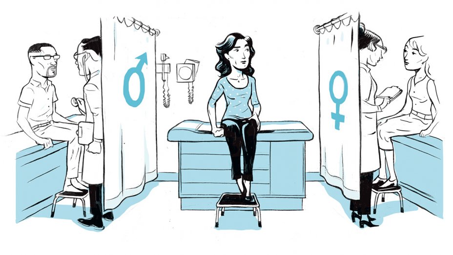
Going to get a routine checkup from either the doctor or the dentist is stressful enough for individuals. For transgender individuals, going to get a routine checkup is even more stressful for them as they are more often getting mistreated or even denied care that they needed. In a 2009 survey, around 70% of transgender and gender-nonconforming individuals have reported experience the following:
Refusal of health care
Healthcare professional refusing to touch or use precautions to individuals
Healthcare professionals using unnecessary abusive language
Blamed for their own health wellness
Healthcare professionals being abusive to the patients
Additional surveys also revealed that transgender health care is discriminated and has kept at least one-third of transgender individuals from seeking medical help for any illnesses or injuries that they may have encountered. It is especially startling that many transgender patients have educated their doctors about transgender health.
Transgender is defined as �an individual who feels that their gender identity does not match their physical body and is different from the gender they are born in.� A 2016 data analysis by The Williams Institute, found out that about 1.4 million American individuals that identify as transgender.
Transgender individuals are beginning to speak about their problems and issues. They talk about the concerns about how office staff are feeling betrayed during their transitional status, all the way to others discriminating in the health care they are receiving. Transgender individuals are providing medical professionals what they can and should do to make sure that they feel safe in the medical professional’s care. Without awareness and education, the healthcare providers are not doing; these issues are more likely to escalate with the growing transgender population.
What Practitioners Need To Do
In March 2019, two individuals Emma Vosicky and Jaime Pagano, addressed to the students and faculty at the National University of Health Sciences (NUHS) Pride Medical Alliance (PMA) club about the challenges they faced. These two individuals were worried about how they would be treated differently by the office staff and were in fear of those who would betray them by sharing their information. These transgender speakers went ahead and discussed the difficult challenges that went beyond the physical transformations that they and many others have faced when seeking medical care.
Vosicky went ahead and discussed how necessary it was to “out” herself to a medical professional when they were asking her about the medication she was taking or discussing the previous medical history that did not match her appearance. Both speakers suggested that healthcare providers should consider different ways that they can let their patients know that they are non-discriminatory to them.
Pagano discussed how he felt much safer when he sees an intake form that included different sex options in a doctor’s lobby. Terms that are in the intake form includes gender non-conforming, non-binary, trans-female, and trans male, alongside with male and female. He said that this feels helpful to the provider he was seeing to be aware and cognizant that everyone is not living in a male/female only world. Pagano also mentioned that he feels more confident that his provider will be more clinically aware of his needs.
The NUHS faculty member Jamine Blesoff, ND, has worked with transgender youth and stressed that it is vital for physicians to ensure that their patients’ care is essential throughout each stage of their transitions.� Dr. Blesoff noted that health practitioners still must provide a PAP test for men who are transitioning from being female as well as a prostate exam for pre-surgical women. Dr. Blesoff expresses concern that some doctors will not provide any type of service to transgender patients.
“It is a universal requirement that healthcare practitioners adhere to HIPAA laws and to make sure that their transgender patients are treated with dignity, respect, and above all else, ensuring that they receive the needed medical care that they deserve like everyone else.”-Dr. Alex Jimenez D.C., C.C.S.T. Insight
Being Gender Neutral
Healthcare professionals should always build trust with any patients that walks through the door. Healthcare providers have to make sure that the intake forms provide space for patient�s gender identity. In addition to the patient’s physical status, transgender patients can indicate their preferred identity, and healthcare professionals can ask what pronouns the patients preferred like he/she/they and uses them throughout their visit.
Speak Respectfully
Doctors should consider using their patient�s chosen name instead of their �real� name if it does not show up in their records and ask the patient if a different name is listed. Healthcare professionals should politely apologize if they use the individual’s wrong name or identity. Even though it may be a bit challenging for long-time patients, but as long as healthcare professionals are making an effort, it will become a way of demonstrating respect not only to the patient but to the doctors as well.
“I always believe that intent matters more than words,” Sam Brinton said, who is the Head of Advocacy and Government Affairs at The Trevor Project. Brinton also mentioned, “There is a difference between ‘I cannot’ and ‘I am trying.’ If you intend to hurt me by not using my pronouns, that matters more than any words you say.”
Recognize the Physical Discomfort
For transgender patients to feel safe and that they are getting the medical care that they need, doctors should take care of them and be respectful to their patient�s needs. For transgender patients, it is already stressful enough for them to get a routine check. When doctors are respectful of their patient’s needs and not to continue procedures to them, that will cause them shame and physical discomfort.
Treat the Ailments Only
Healthcare professionals should consider what kind of information or examination that they are giving to their patients the care they needed. So providing necessary medical care like back pains, stomach problems, immune disorders, or a general checkup is essential.
Educate the Staff
All medical staff that interacts with patients must educate themselves on how to provide comfort and care when they are dealing with transgender patients. Medical providers and medical staff must apply the knowledge of interacting with patients on a day to day basis.
Conclusion
Transgender healthcare is a necessity for these individuals that are trying to get the same benefits that everyone else is getting. Healthcare professionals must be respectful and provide the best care to offer for patients with different identities and backgrounds. Educating and being aware of what the patient is going through is part of the doctor’s job to assist not only themselves better but also inform the patient a solution while making them feel comfortable. Some products are here to support anyone’s ailments and provide support to the intestines, gastrointestinal function, and muscular system.
The scope of our information is limited to chiropractic, musculoskeletal, and nervous health issues or functional medicine articles, topics, and discussions. We use functional health protocols to treat injuries or disorders of the musculoskeletal system. Our office has made a reasonable attempt to provide supportive citations and has identified the relevant research study or studies supporting our posts. We also make copies of supporting research studies available to the board and or the public upon request. To further discuss the subject matter above, please feel free to ask Dr. Alex Jimenez or contact us at 915-850-0900.
References:
Flores, Andrew R., et al. �How Many Adults Identify As Transgender In The United States?� The William Institute, June 2016, williamsinstitute.law.ucla.edu/wp-content/uploads/How-Many-Adults-Identify-as-Transgender-in-the-United-States.pdf.
Marshall, Tari. �Transgender Health Care: How to Meet Their Needs.� Transgender Health Care: How to Meet Their Needs, 20 Nov. 2019, blog.nuhs.edu/the-future-of-integrative-health/transgender-health-care-how-to-meet-their-needs.
Marshall, Tari. �When He/She May Be They/Them.� LinkedIn, 13 Feb. 2018, www.linkedin.com/pulse/when-heshe-may-theythem-tari-marshall?trk=portfolio_article-card_title.
Team, Lambda. �Lambda Legal Releases Health Care Discrimination Survey Results; More Than Half of LGBT and HIV Positive Respondents Report Discrimination.� Lambda Legal, 4 Feb. 2010, www.lambdalegal.org/news/ny_20100204_lambda-releases-health.
Team, NUHS. �Pride Club Program Addresses Transgender Experiences with Medical Professionals: National University of Health Sciences Illinois & Florida.� Earn a Degree Chiropractic, Naturopathy, and Acupuncture Medicine |�National University of Health Sciences, 13 Mar. 2019, www.nuhs.edu/news/2019/3/pride-club-program-addresses-transgender-experiences-with-medical-professionals/.
Team, The Trevor Project. �Saving Young LGBTQ Lives.� The Trevor Project, 2019, www.thetrevorproject.org/#sm.00013irq131dh2e6qpejz1qoa103y.
If you are experiencing any of these situations, then you might have a magnesium deficiency.
Good health is one of the things to be thankful for. Unfortunately, 84 million adults in the U.S. are living with prediabetes, while another 27 to 28 million adults are affected with type 2 diabetes, so good health is not a given for everyone. According to the National Osteoporosis Foundation, 10 million Americans have osteoporosis, and another 44 million have low bone density, putting them at an increased risk. From the body to the brain, psychological and mood issues like depression and anxiety plague people. There is something that may be beneficial for all of these issues and is a workhorse nutrient that does not get its share of the spotlight. It has been regulated to the shadows behind the flashier and more buzzworthy compounds that get recognition than this nutrient. Magnesium is the critically essential, time-tested, go-to reliable nutrient that everybody needs.
The human body contains about 25 grams of magnesium, which is needed for over 300 enzymes to react. The data from the NHANES (National Health and Nutrition Examination Survey) indicated that the majority of Americans from all ages consume less that than their respective EARs (estimated average requirements) on magnesium. It is a massive problem because magnesium deficiency plays a role in hypertension, cardiovascular diseases, type 2 diabetes, osteoporosis, and migraine headaches.
Magnesium and Glucose Levels
Magnesium is required for several enzymes in glycolysis, which is the first process in glucose metabolism in the body, and it may explain why it is such an essential factor for blood sugar regulation in the body. Epidemiological evidence indicates that magnesium intake is inversely correlated with the risk of type 2 diabetes. Studies have shown that higher magnesium intakes may help reduce the risk of type 2 diabetes as much as 17%, and 48% of people with type 2 diabetes may have hypomagnesemia.
The inverse correlations have been observed between circulating magnesium levels, fasting blood glucose, and insulin level. There is even a response to an OGTT (oral glucose tolerance test) for those with type 2 diabetes. Research shows that higher magnesium intakes are also associated with reducing the risk for cardiovascular mortality, particularly in women as it is estimated that 100 mg/day increase in dietary magnesium may confer as much as 25% reduction in the risk of cardiovascular mortality. Researchers have called subclinical magnesium deficiency “principal dicer of cardiovascular disease and a public health crisis,” so naturopathic practitioners suggesting adding magnesium-rich foods to a person’s diet is beneficial to prevent magnesium deficiency from happening.
Magnesium and Mental Health
In regards to mental health, evidence has suggested that magnesium deficiency may play a role in the etiology of depression and that high-dose supplementation of magnesium may improve this condition. Studies found that other issues that have responded favorably to magnesium supplementation include irritability, insomnia, postpartum depression, and substance abuse in the body. There is some suggestive but inconclusive evidence that indicates that magnesium supplementation may be beneficial for individuals with mild anxiety and possibly owing to its role as a natural relaxing agent.
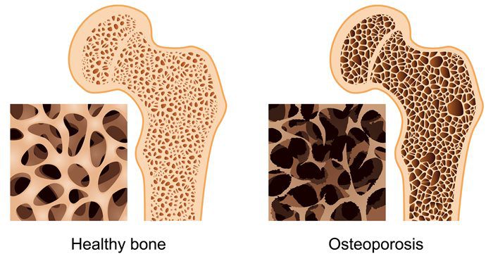
Magnesium and Osteoporosis
For osteoporosis, calcium gets all the attention when it comes to bone mineral density; however, magnesium is an essential component for the physical structure of bone density as well. There is about 60% of the body’s magnesium stored in the bones, and considering the high prevalence of suboptimal magnesium intake in North America, the concurrent high prevalence of osteoporosis is unsurprising. Concerning bone health, low magnesium status may interfere with the efficacy of vitamin D supplementation. In the Journal of the American Osteopathic Association, a review was covered in which researchers affirmed that vitamin D could not be metabolized without the sufficient levels of magnesium.
Adding Magnesium-rich Food To Your Feast
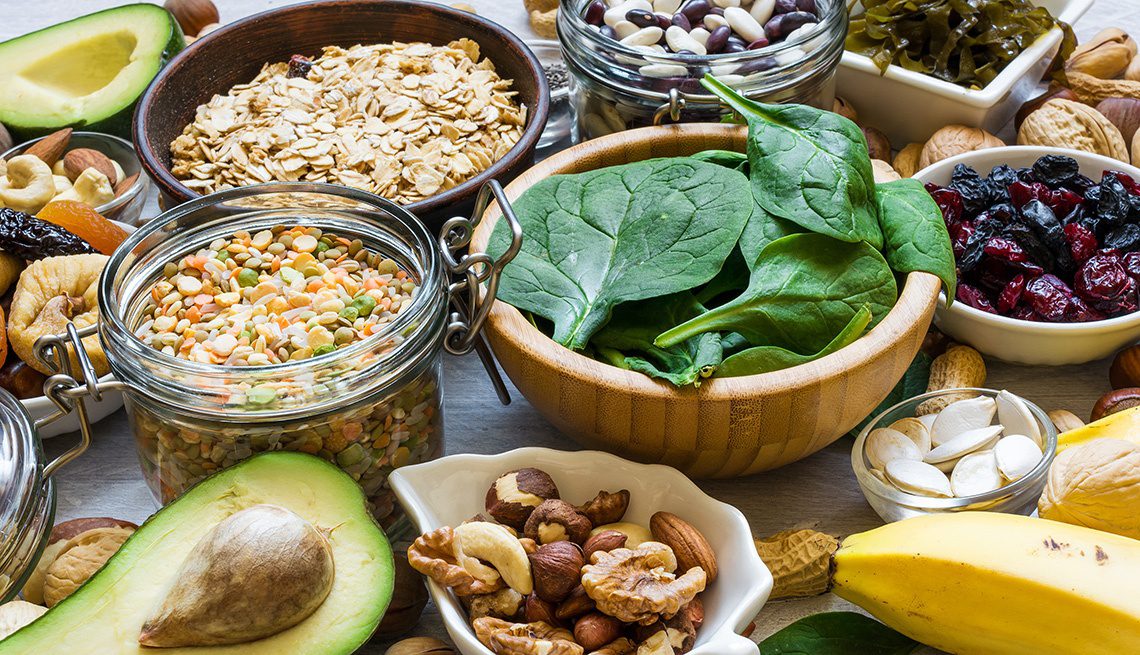
With Thanksgiving coming around the corner, there is a way to bring magnesium to the holiday table. The good news is that this crucial mineral fits perfectly into Thanksgiving entertainment. People can serve mixed nuts as part of appetizers or hors d’oeuvres while their guests are socializing. Mixed nuts can provide a substantial amount of magnesium. They can be an excellent addition to turkey stuffing/dressing or a whole grain salad, which can provide even more magnesium that the body needs. Serving leafy greens like chard and spinach are reliable sources of magnesium, as well as certain beans like black beans and kidney beans are filled with magnesium.
Since nuts, seeds, and beans are high in phytic acid, which is a compound that binds to the minerals. So in order to increase the bioavailability of magnesium in these foods, soaking nuts, seeds, and beans is a traditional preparation method to neutralize some of this problematic molecule.
For dessert, adding chocolate is an excellent way to get magnesium in the body. Since the cocoa powder is a rich source of magnesium, research has been speculating that the chocolate cravings might be the body’s way of crying for magnesium. Not to mention, when foods are much higher in magnesium, they are not the usual subjects for intense cravings like chocolate.
“So for Thanksgiving, adding magnesium-rich foods can help cut back the sodium and carb intake of the holiday feast can be beneficial to your body to function correctly and good for your health.”-Dr. Alex Jimenez D.C., C.C.S.T. Insight
Conclusion
Magnesium is an excellent and beneficial nutrient for anyone to add to their Thanksgiving dinner. The nutrient plays many roles in the body like regulating blood sugar, improving mental health as a natural relaxing agent, and preventing osteoporosis from occurring. Adding this nutrient and some products can help the body metabolize and stable the blood sugar levels to their normal range for beneficial results.
The scope of our information is limited to chiropractic, musculoskeletal, and nervous health issues or functional medicine articles, topics, and discussions. We use functional health protocols to treat injuries or disorders of the musculoskeletal system. Our office has made a reasonable attempt to provide supportive citations and has identified the relevant research study or studies supporting our posts. We also make copies of supporting research studies available to the board and or the public upon request. To further discuss the subject matter above, please feel free to ask Dr. Alex Jimenez or contact us at 915-850-0900.
References:
Boyle, Neil Bernard, et al. �The Effects of Magnesium Supplementation on Subjective Anxiety and Stress-A Systematic Review.� Nutrients, MDPI, 26 Apr. 2017, www.ncbi.nlm.nih.gov/pmc/articles/PMC5452159/.
Bruinsma, K, and DL Taren. “Chocolate: Food or Drug?” Journal of the American Dietetic Association, U.S. National Library of Medicine, Oct. 1999, www.ncbi.nlm.nih.gov/pubmed/10524390.
Castiglioni, Sara, et al. �Magnesium and Osteoporosis: Current State of Knowledge and Future Research Directions.� Nutrients, MDPI, 31 July 2013, www.ncbi.nlm.nih.gov/pmc/articles/PMC3775240/.
DiNicolantonio, James J, et al. �Subclinical Magnesium Deficiency: a Principal Driver of Cardiovascular Disease and a Public Health Crisis.� Open Heart, BMJ Publishing Group, 13 Jan. 2018, www.ncbi.nlm.nih.gov/pmc/articles/PMC5786912/.
Eby, George A, and Karen L Eby. �Rapid Recovery from Major Depression Using Magnesium Treatment.� Medical Hypotheses, U.S. National Library of Medicine, 2006, www.ncbi.nlm.nih.gov/pubmed/16542786.
Fang, Xin, et al. �Dose-Response Relationship between Dietary Magnesium Intake and Cardiovascular Mortality: A Systematic Review and Dose-Based Meta-Regression Analysis of Prospective Studies.� Journal of Trace Elements in Medicine and Biology: Organ of the Society for Minerals and Trace Elements (GMS), U.S. National Library of Medicine, Dec. 2016, www.ncbi.nlm.nih.gov/pubmed/27053099.
Fang, Xin, et al. �Dose-Response Relationship between Dietary Magnesium Intake and Risk of Type 2 Diabetes Mellitus: A Systematic Review and Meta-Regression Analysis of Prospective Cohort Studies.� Nutrients, MDPI, 19 Nov. 2016, www.ncbi.nlm.nih.gov/pmc/articles/PMC5133122/.
Higdon, Jane. �Magnesium.� Linus Pauling Institute, 14 Oct. 2019, lpi.oregonstate.edu/mic/minerals/magnesium#structural-roles.
Serefko, Anna, et al. �Magnesium and Depression.� Magnesium Research, U.S. National Library of Medicine, 1 Mar. 2016, www.ncbi.nlm.nih.gov/pubmed/27910808.
Spiga, Rosangela, et al. �Are Circulating Mg2+ Levels Associated with Glucose Tolerance Profiles and Incident Type 2 Diabetes?� Nutrients, U.S. National Library of Medicine, 14 Oct. 2019, www.ncbi.nlm.nih.gov/pubmed/31615167.
Team, DFH. �Preparing Beans and Legumes � What to Know.� Designs for Health, 9 Oct. 2018, blog.designsforhealth.com/preparing-beans-and-legumes.
Team, DFH. �Put Magnesium on the Menu at Thanksgiving.� Designs for Health, 19 Nov. 2019, blog.designsforhealth.com/node/1151.
Team, NOF. �Https://Cdn.nof.org/Wp-Content/Uploads/2015/12/Osteoporosis-Fast-Facts.pdf.� National Osteoporosis Foundation, 2015.
Unknown, Unknown. �Diabetes Statistics.� National Institute of Diabetes and Digestive and Kidney Diseases, U.S. Department of Health and Human Services, 1 Sept. 2017, www.niddk.nih.gov/health-information/health-statistics/diabetes-statistics.
Unknown, Unknown. �Office of Dietary Supplements – Magnesium.� NIH Office of Dietary Supplements, U.S. Department of Health and Human Services, 11 Oct. 2019, ods.od.nih.gov/factsheets/Magnesium-HealthProfessional/#h4.
Unknown, Unknown. �Office of Dietary Supplements – Magnesium.� NIH Office of Dietary Supplements, U.S. Department of Health and Human Services, 11 Oct. 2019, ods.od.nih.gov/factsheets/Magnesium-HealthProfessional/#h7.
Uwitonze, Anne Marie, and Mohammed S. Razzaque. �Role of Magnesium in Vitamin D Activation and Function.� The Journal of the American Osteopathic Association, American Osteopathic Association, 1 Mar. 2018, jaoa.org/article.aspx?articleid=2673882.
Waanders, Femke, et al. �Hypomagnesaemia and Its Determinants in a Contemporary Primary Care Cohort of Persons with Type 2 Diabetes.� Endocrine, U.S. National Library of Medicine, 24 Oct. 2019, www.ncbi.nlm.nih.gov/pubmed/31650393.
Yanovski, Susan. �Sugar and Fat: Cravings and Aversions.� OUP Academic, Oxford University Press, 1 Mar. 2003, academic.oup.com/jn/article/133/3/835S/4688015.
If you are experiencing any of these situations, why not try a HIIT workout to relieve these symptoms.
Everyone can agree that they do not have enough time to exercise. When asked why people will not work out, one of the reasons is that due to their hectic lifestyle, the lack of time comes out on top of it all. The U.S. Department of Health and Human Services recommends that adults should get between 150 and 300 minutes per of moderate-intensity. There is a way to cut that time commitment in half by opting for high-intensity workouts instead. With high-intensity interval training or HIIT, it is one of the proven ways to reap all the benefits of exercise in less time. Research shows that spending less time doing HIIT may even be better than spending more time doing less intense exercises for individuals.
What is HIIT?
High-intensity interval training or HIIT alternates explosive bursts of full-throttle efforts with periods of recovery. It can either be rest or a lower-intensity exercise. In many fitness centers and gyms, HIIT workouts often include both cardio and resistance training; however, HIIT workouts can be done as a strictly cardio routine.
During intense burst in a HIIT workout, a person is working out at around 80 percent of their max heart rate for 15 seconds to a few minutes. Between each of those periods, a person is either slowing down or resting completely to let their heart rate come back down to around 50 percent.
A person can calculate different target heart rates by using an online calculator. During a workout, a person can wear a heart rate monitor to keep track on much they are exerting themselves. For a lower-tech option, Denver-based certified personal trainer Lindsay Kelly recommends the “talk test.” The way the “talk test” is when a person is doing their target intensity heart rate like sprinting; for example, it should be hard to speak more than two words without taking a breath. Then when they are in the recovery period, the reverse factor is real.
Why HIIT Works
HIIT is so effective because it allows a person to exercise at a higher intensity for such a short period. The exertion gets the heart working and the blood pumping better than any moderate-intensity exercise can bring with their prolonged periods of rest.
The Importance of Rest
While a person might not realize it, the rest periods are built into the HIIT workout and are a critical part of the routine. They force the body to adjust to a very different state of activity, which is excellent for cardiovascular conditioning.
Feel The Afterburn
Another benefit of a HIIT workout is that even after a person is finished with their HIIT workout, it keeps on working for them. Research shows that when individuals keep on burning calories after their HIIT workout at a higher than they would after a continuous exertion workout. It is commonly known as the “afterburn effect,” and it helps people extend the benefits of their efforts.
The Benefits of HIIT
Researchers have been studying HIIT extensively, and the results are precise: HIIT workouts are better than continuous exercise when it comes to improving health in a variety of ways. One of the health benefits of a HIIT workout is that it improves cardiorespiratory fitness, which is the health of the heart and breathing. This matters to a person who is trying to get in as much exercise as possible with little time because cardiorespiratory fitness is a primary factor in the risk of diseases and death. Studies have shown that HIIT workouts can increase cardiorespiratory fitness at twice the rate of continuous exercises.
The health benefits of HIIT does not stop there, as other research studies have shown that HIIT can help with the following areas of the body.
Endurance
By improving cardiorespiratory fitness, HIIT can improve a person’s stamina. What it does is that it enhances the body’s ability to consume and use oxygen. One study has compared a regular endurance training to HIIT by looking at how they affect maximal oxygen consumption known as VO2max. The research found out that HIIT was superior to endurance training by improving VO2max in healthy young to middle-aged adults. Once a person starts to build their endurance, they can increase the length or the intensity of the HIIT working periods and enjoy the significant health benefits it provides.
Heart Health
One of the significant contributors to cardiovascular disease and death is high blood pressure, and one of the best ways to keep it in check is through regular exercise. The traditional recommendation for blood pressure modulating has been to exercise at moderate intensity for at least 30 minutes on most or all day so that way high blood pressure will not transform into hypertension. Several studies have suggested that HIIT may be an even better option, and one study shows that while both continuous exercise and HIIT helps with blood pressure control, HIIT is the only workout to help reduce arterial stiffness. Arterial stiffness is a predictor of cardiovascular disease in people with high blood pressure.
Brain Function
When a person feels that that mental clarity after a good workout, it is not their imagination. The brain and mental health benefits of exercise are well documented. Research shows that HIIT helps explicitly improve the cognitive function, including short-term memory, verbal memory, attention, and processing speed in the brain. HIIT also increases the amount of oxygen that the brain gets from the blood.
Diabetes Management
Since exercise is an essential part of diabetes management, research shows that HIIT may be a wise exercise choice for anyone who has type 2 diabetes. Studies have shown that HIIT workouts can improve endothelial function, insulin sensitivity, glucose control, and other health effects of diabetes that are better than continuous exercise.
Conclusion
HIIT workouts are perfect for anyone who does not have enough time out of their busy schedule. With the alternating burst of exercises and periods of recovery, HIIT workouts are beneficial to anyone with a short amount of time to complete them. HIIT includes both cardio and resistance training and works with the entire body. Some products are excellent in countering the metabolic effects of temporary stress and supporting the body�s system.
The scope of our information is limited to chiropractic, musculoskeletal, and nervous health issues or functional medicine articles, topics, and discussions. We use functional health protocols to treat injuries or disorders of the musculoskeletal system. Our office has made a reasonable attempt to provide supportive citations and has identified the relevant research study or studies supporting our posts. We also make copies of supporting research studies available to the board and or the public upon request. To further discuss the subject matter above, please feel free to ask Dr. Alex Jimenez or contact us at 915-850-0900.
References:
Chobanian, Aram V., et al. �Seventh Report of the Joint National Committee on Prevention, Detection, Evaluation, and Treatment of High Blood Pressure.� AHA Journals, 1 Dec. 2003, www.ahajournals.org/doi/full/10.1161/01.hyp.0000107251.49515.c2.
Council on Sports, HHS Office. �Physical Activity Guidelines for Americans.� HHS.gov, US Department of Health and Human Services, 1 Feb. 2019, www.hhs.gov/fitness/be-active/physical-activity-guidelines-for-americans/index.html.
Dupuy, Oliver, et al. �Effect of Interval Training on Cognitive Functioning and Cerebral Oxygenation in Obese Patients: A Pilot Study.� Latest TOC RSS, Medical Journals Limited, 1 Nov. 2014, www.ingentaconnect.com/content/mjl/sreh/2014/00000046/00000010/art00016.
Francois, Monique E, and Jonathan P Little. �Effectiveness and Safety of High-Intensity Interval Training in Patients with Type 2 Diabetes.� Diabetes Spectrum: a Publication of the American Diabetes Association, American Diabetes Association, Jan. 2015, www.ncbi.nlm.nih.gov/pmc/articles/PMC4334091/.
Gillen, Jenna B., and Martin J. Gibala. �Is High-Intensity Interval Training a Time-Efficient Exercise Strategy to Improve Health and Fitness?� Applied Physiology, Nutrition, and Metabolism, 27 Sept. 2013, www.nrcresearchpress.com/doi/10.1139/apnm-2013-0187#.XdQT5y2ZP1J.
Guimar�es, Guilherme Veiga, et al. �Effects of Continuous vs. Interval Exercise Training on Blood Pressure and Arterial Stiffness in Treated Hypertension.� Hypertension Research: Official Journal of the Japanese Society of Hypertension, U.S. National Library of Medicine, June 2010, www.ncbi.nlm.nih.gov/pubmed/20379194.
Milanovi?, Zoran, et al. �Effectiveness of High-Intensity Interval Training (HIT) and Continuous Endurance Training for VO2max Improvements: A Systematic Review and Meta-Analysis of Controlled Trials.� SpringerLink, Springer International Publishing, 5 Aug. 2015, link.springer.com/article/10.1007/s40279-015-0365-0.
Pescatello, Linda S, et al. �American College of Sports Medicine Position Stand. Exercise and Hypertension.� Medicine and Science in Sports and Exercise, U.S. National Library of Medicine, Mar. 2004, www.ncbi.nlm.nih.gov/pubmed/15076798.
Unknown, Unknown. “Is High-Intensity Interval Training Right for You?” Fullscript, 12 Nov. 2019, fullscript.com/blog/high-intensity-interval-training.
Weston, Kassia S, et al. �High-Intensity Interval Training in Patients with Lifestyle-Induced Cardiometabolic Disease: a Systematic Review and Meta-Analysis.� British Journal of Sports Medicine, BMJ Publishing Group Ltd and British Association of Sport and Exercise Medicine, 1 Aug. 2014, bjsm.bmj.com/content/48/16/1227.short.
Stomach pain, burning, or aching 1-4 hours after eating?
Agitated, easily upset, nervous?
Lightheaded if meals are missed?
Digestive problems subside with rest and relaxation?
If you are experiencing any of these situations, then you might be experiencing a histamine attack on your immune system.
During an allergic response, the body’s immune system starts to react by releasing various immune compounds to protect itself from foreign substances that identify as harmful. One of these immune compounds, known as histamine, is commonly present in a variety of foods. When histamine is elevated in the body, it is due to a high dietary intake or an inability to break it down, so individuals may experience allergic symptoms from a histamine reaction.
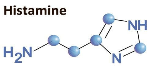
What is Histamine?
Histamine is a compound that is formed through the metabolism of specific amino acids in the immune system. There are a variety of levels of histamine that is found naturally in the foods that people consume. It is also produced by the body where it is in specific immune cells, including mast cells and basophils. During an allergic and other immune response, histamine is released from these cells, and consuming large quantities of histamine that is over 100 mg may result in a mild adverse reaction. Studies have shown that if histamine is consumed in a higher amount that is over 1000 mg, it can lead to histamine intoxication or histamine poisoning.
What is Histamine Intolerance?
Under normal conditions, histamine is released in the body or ingested through food, and it is broken down by two enzymes: HNMT (histamine-N-methyltransferase) and DAO (diamine oxidase). High levels of histamine can occur in individuals that have reduced activity of these enzymes. When histamine levels are increased, or the ability to break down histamine is impaired, individuals may experience histamine intolerance, which will generally present itself as an allergic reaction to the immune system.
What Causes Histamine Intolerance?
Specific individuals may have an increased sensitivity to biogenic amines like histamine. Some factors have been associated with an increased risk of histamine intolerance, including:
Certain health conditions (coronary heart disease, hypertension, respiratory diseases)
Vitamin B12 deficiency
Certain medications that inhibit the activity of histamine-degrading enzymes (acetylcysteine, metamizole, metoclopramide, metronidazole, verapamil)
Mast cell conditions can increase the secretion of histamine. Since mast cells are found throughout the body, they are involved with the innate immune response as well as being the primary source of histamine in the intestines. Studies show that when specific immune receptors detect a foreign substance in the body, the mast cells secrete inflammatory compounds like histamine as a protective response. Mast cell activation is characterized by increasing plasma and urine histamine levels as well as an increased histamine metabolite in the urine. Several conditions are associated with mast cell activation, including:
Allergies that are mediated by IgE (immunoglobulin-E) and other hypersensitivities
Symptoms of rhinoconjunctivitis (nasal congestion, post-nasal drip, sneezing)
Pruritus (itchy skin)
Urticaria (hives)
Histamine-free Diet
With dietary support for histamine intolerance, it may involve a histamine-free diet. Studies have been examining the effects of four-week histamine-free diet intervention on 22 individuals that have CU (chronic urticaria). Chronic urticaria is a common skin condition that is characterized by episodes of red marks and swelling that last longer than six weeks on the body.
A study found that when it is being compared to baseline, plasma histamine levels were significantly reduced when following the diet. Additionally, USS (urticaria severity score) and (UAS), both decreased following the intervention. It means that a histamine-free diet may help improve symptoms that are associated with dietary histamine intake or histamine intolerance like chronic urticaria in the body.
Research also shows that when individuals follow a histamine-reduced diet, it may increase the levels of the DAO enzyme. Research also demonstrated that when individuals followed the histamine-reduced diet for an average of 13 months, the increased levels of DAO were correlated with the degree of compliance to the diet. When high-histamine and histamine-liberated foods are eliminated, the individual�s tolerance levels can be determined by slowly reintroducing foods to test for potential reactions.
Certain foods may increase histamine levels by providing a dietary source or by liberating histamine in the body. Studies have found that it is essential to note that inconsistent levels of histamine are found in various foods and that the levels may fluctuate based on the maturity, storage, and processing of the food.
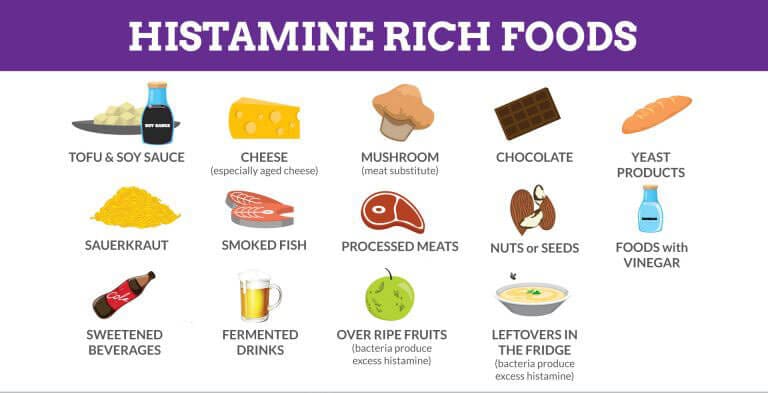
Here are the high-histamine foods to avoid on a histamine-free diet. They are:
Aged cheese (cheddar, gouda, parmesan)
Alcohol (beer, champagne, wine)
Certain produce (avocado, eggplant, spinach, tomato)
Cured meats (fermented sausage, salami)
Fermented vegetables (sauerkraut)
Fish products (dried anchovies, fish sauce)
Here are the histamine-liberating foods to avoid on a histamine-free diet as well. They are:
Certain fruits (citrus, pineapple, banana, strawberries, papaya)
With methods surrounding food preparations that should be considered, researchers have suggested that people should consume food that is fresh as possible and boiled rather than frying or grilling food may help reduce the intake of biogenic amines like histamine. Studies have shown that spoiled foods have been found to have high levels of histamine, so it is essential to be mindful when consuming leftovers, especially leftover fish. There are some individuals with histamine intolerance that may benefit from taking antihistamine medication or a DAO supplement. There are also certain nutrients, including copper, vitamin B6, and vitamin C that can help support histamine degradation.
Conclusion
When the body is suffering from an allergic response, its’ immune system starts to react by sending out various immune compounds that attack harmful foreign substances. Histamine is one of the immune compounds that is produced and broken down to HNMT and DAO. Histamine can also trigger an asthma attack on individuals, while certain foods can contain high-histamine and histamine-liberating properties that can be harmful in the body. Some products use an advanced formula that helps support the immune system, targets amino acids, and supports antioxidant processes to make sure that the body is functioning correctly.
The scope of our information is limited to chiropractic, musculoskeletal, and nervous health issues or functional medicine articles, topics, and discussions. We use functional health protocols to treat injuries or disorders of the musculoskeletal system. Our office has made a reasonable attempt to provide supportive citations and has identified the relevant research study or studies supporting our posts. We also make copies of supporting research studies available to the board and or the public upon request. To further discuss the subject matter above, please feel free to ask Dr. Alex Jimenez or contact us at 915-850-0900.
References:
Martin, San Mauro, et al. �Histamine Intolerance and Dietary Management: A Complete Review.� Adrianaduelo, 31 Aug. 2016, www.adrianaduelo.com/wp-content/uploads/2016/09/2016_Histamine-intolerance-and-dietary-management.pdf.
Chung, Bo Young, et al. �Effect of Different Cooking Methods on Histamine Levels in Selected Foods.� Annals of Dermatology, The Korean Dermatological Association; The Korean Society for Investigative Dermatology, Dec. 2017, www.ncbi.nlm.nih.gov/pmc/articles/PMC5705351/.
Dougherty, Joseph M. �Allergy.� StatPearls [Internet]., U.S. National Library of Medicine, 28 July 2019, www.ncbi.nlm.nih.gov/books/NBK545237/.
Fong, Michael. �Histology, Mast Cells.� StatPearls [Internet]., U.S. National Library of Medicine, 20 Sept. 2019, www.ncbi.nlm.nih.gov/books/NBK499904/.
Lackner, Sonja, et al. �Histamine-Reduced Diet and Increase of Serum Diamine Oxidase Correlating to Diet Compliance in Histamine Intolerance.� European Journal of Clinical Nutrition, U.S. National Library of Medicine, Jan. 2019, www.ncbi.nlm.nih.gov/pubmed/30022117.
Maintz, Laura, and Natalija Novak. �Histamine and Histamine Intolerance.� OUP Academic, Oxford University Press, 1 May 2007, academic.oup.com/ajcn/article/85/5/1185/4633007.
Reese, Imke, et al. �German Guideline for the Management of Adverse Reactions to Ingested Histamine: Guideline of the German Society for Allergology and Clinical Immunology (DGAKI), the German Society for Pediatric Allergology and Environmental Medicine (GPA), the German Association of Allergologists (AeDA), and the Swiss Society for Allergology and Immunology (SGAI).� Allergo Journal International, Springer Medizin, 2017, www.ncbi.nlm.nih.gov/pmc/articles/PMC5346110/.
Son, Jee Hee, et al. �A Histamine-Free Diet Is Helpful for Treatment of Adult Patients with Chronic Spontaneous Urticaria.� Annals of Dermatology, The Korean Dermatological Association; The Korean Society for Investigative Dermatology, Apr. 2018, www.ncbi.nlm.nih.gov/pmc/articles/PMC5839887/.
S�nchez-P�rez, S�nia, et al. �Biogenic Amines in Plant-Origin Foods: Are They Frequently Underestimated in Low-Histamine Diets?� Foods (Basel, Switzerland), MDPI, 14 Dec. 2018, www.ncbi.nlm.nih.gov/pmc/articles/PMC6306728/.
Unknown, Unknown. �Histamine Intolerance & Diet: What You Should Know.� Fullscript, 11 Nov. 2019, fullscript.com/blog/histamine-intolerance.
IFM's Find A Practitioner tool is the largest referral network in Functional Medicine, created to help patients locate Functional Medicine practitioners anywhere in the world. IFM Certified Practitioners are listed first in the search results, given their extensive education in Functional Medicine