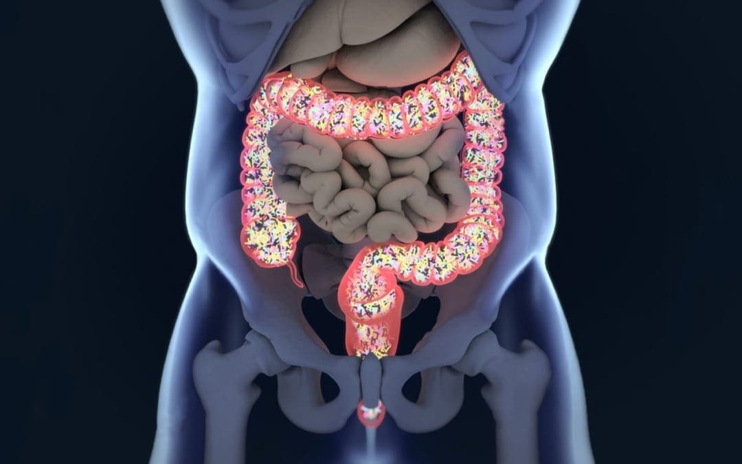
Children are not born with a fully developed microbiome, and a baby’s diet has a large impact on the foundation set for a healthy guts future (Biotics Education Team, 1).� Setting up a child to have healthy gut flora from early stages can help them:
In the TEDDY study published in Nature Medicine, it shows that a child’s microbiome goes through 3 transitional phases:
Developmental phase (3�14 months)
Transitional phase (15�30 months)
Stable phase (31�46 months)(Stewart et al., 3)
Throughout the developmental stage, those with a higher breastfeeding rate were associated with increased levels of Bifidobacterium.� “However, once the infants were weaned, there was a rapid loss of the�Bifidobacterium spp.,�and a quick turnover occurred in the microbiome, which featured a higher population of bacteria within the�Firmicutes�phylaphase (Biotics Education Team, 1)”.� Once infants begin to wean off milk, it is helpful to start providing them with probiotic powders.
Prebiotics are the dietary fiber that the live organisms in the probiotics need to eat in order to flourish.
Some foods that include prebiotics are:
Vegetables
Fruits
Legumes
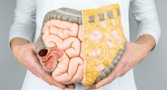
It is great to start toddlers on prebiotics and probiotics because it can help them to continue to have a healthy gut. A healthy gut can help prevent many issues that adults face later on in life (Veereman-Wauters, 4) Having a healthy gut can help to protect the gut from harmful bacteria and fungi, it can aid in sending signals to the immune system, regulate inflammation, create a supportive barrier in the cell lining of the colon and reduce the risk of cancer (Lewis, 2)�
Probiotics are safe for most children and can reduce the risk of upper respiratory tract infections and well as helping to reduce their risk of allergies. It is beneficial to have toddlers on probiotics and prebiotics so they do not develop a “leaky gut”. By starting children on probiotics and prebiotics young, it can aid their overall health for life.
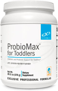
ProbioMax� for Toddlers
Prebiotic and Probiotic Support for Toddlers*
�Overall, it is best to start building the child’s microbiota through the maternal diet in pregnancy, expose them to environments, and talk with their pediatrician about starting them on probiotics. It’s better to start young and build a healthy foundation than to be diagnosed in their 20’s with leaky gut from something that could have been prevented. – Insight from Kenna Vaughn, Health Coach�
NCBI Resources:
Our knowledge of microbiota is rapidly developing and changing. A relatively young field, the science of gut bacteria has been quickly taken up by industry. Most drugstores sell probiotics in some form or another, and yogurt and other fermented foods are frequently hailed as healthy for the gut because they contain live bacteria. Probiotics are food or supplements that contain living microbes intended to support or improve your microbiome’s health. If your favorite yogurt contains �live and active cultures,� you are getting a dose of probiotics along with your breakfast. These microbes are thought to bolster or replace the bacteria communities in the gut of people.
�Cites:
Biotics Education Team. �Impact of Diet on Baby’s Microbiome.� Biotics Research Blog, blog.bioticsresearch.com/impact-of-diet-on-babys-microbiome.
Lewis, Sarah. �Probiotics and Prebiotics: What’s the Difference?� Healthline, Healthline Media, 3 June 2017, www.healthline.com/nutrition/probiotics-and-prebiotics.
Stewart, Christopher J., et al. �Temporal Development of the Gut Microbiome in Early Childhood from the TEDDY Study.� Nature News, Nature Publishing Group, 24 Oct. 2018, www.nature.com/articles/s41586-018-0617-x.
Veereman-Wauters, Gigi. �Application of Prebiotics in Infant Foods.� The British Journal of Nutrition, U.S. National Library of Medicine, Apr. 2005, www.ncbi.nlm.nih.gov/pubmed/15877896.
The phrase, �You are what you eat� implies that the way we are defines us as the food we all consumed. However, our gut tells us otherwise as the food we eat, may in fact be leading us to obesity. Our gut plays a role in our overall health, when we eat good food our gut is happy and when we eat bad food our gut will tell us by fighting off the bad food. A recent study showed us that the bacteria in our gut produce amyloid and lipopolysaccharides. These two microbiomes seem to show us that together, with proper dieting that these microbiomes can prevent Alzheimer�s Disease.
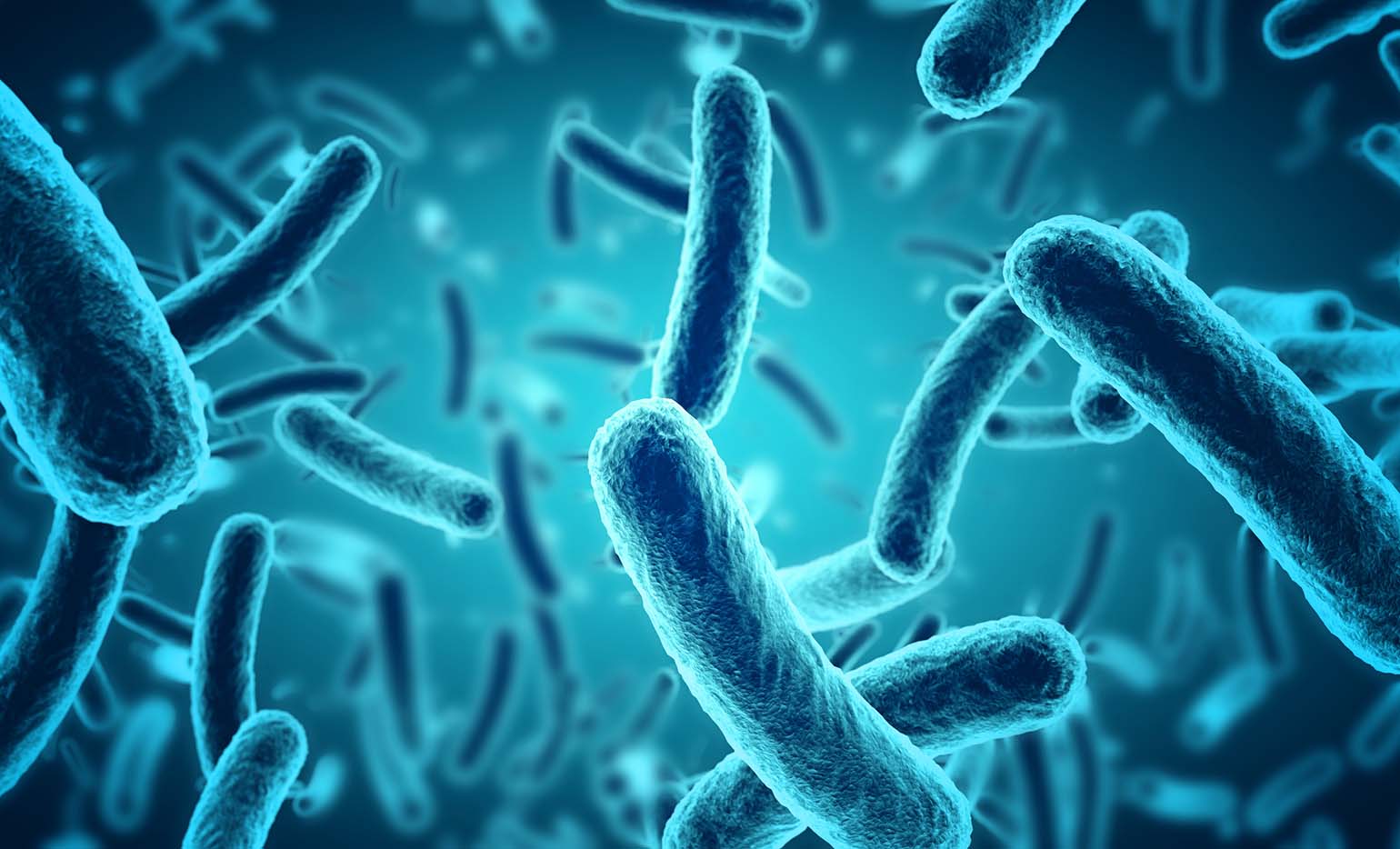
As the microbiomes and the bacteria that co-exist in our gut, there are the two most predominant groups that have also played a key role in our lifestyle: gram-positive Firmicutes and gram-negative Bacteroidetes- both play a huge role in obesity. Firmicutes are bad bacteria that lead us to obesity. When we eat processed food and sugars, our body starts to crave it more, thus leading us to be overweight.
Junk Food
When we eat junk food, all that sugar and fat are feeding the Firmicutes. Firmicutes thrive on sugar since our bodies need it and it can be both good or bad While Bacteroidetes are the good bacteria that leads us to a healthy gut. Bacteroidetes are in the stomach regions as well as the Firmicutes. These two predominant bacteria groups tell us that the food we eat can actually affect our bodies when we eat bad foods or good foods.
However, Dr. Kristen Senella mentioned that we all have a different balance of Firmicutes and Bacteroidetes since we are all different shapes and sizes. Depending on our health and food lifestyle, we can have either a low Firmicutes and a high Bacteroidetes or a high Firmicutes and a low Bacteroidetes. Plus, having either a high or low count of Firmicutes can lead to weight gain or weight loss; depending on which healthy lifestyle and exercise regime you are following.
Gram-Positive
Gram-positive bacteria will appear blue or violet, while gram-negative bacteria will appear red or pink under the microscope. When studying the gut and the bacteria groups that it is hosting, scientist use mice to study how their guts react to different diets they are put through so that way we, as humans, can take either pills to help our bodies maintain a healthy lifestyle or to read and do our own research. One group is fed in a healthy lifestyle and doesn�t experience diseases or ailments that we face. And the other group is fed with a bad lifestyle where they are prone to many of the diseases and fatigue as their life span is shortened very quickly. In order for us to actually maintain a healthy lifestyle and importantly feel good is to make sure our Firmicutes are not too dangerously low, but we can control it with probiotics.
Probiotics
Probiotics can vary from yogurt, fermented vegetables, kombucha, and miso. But there are certain companies that also reign supreme in the probiotic market. Activia yogurt and Yakult are two of the most well-known companies that use the live microorganisms to help us maintain a healthy lifestyle as well as keeping our gut�s microbiome in check. When we have some sort of probiotic foods in our system, we are preventing certain ailments and diseases going out of control. Like our cholesterol, blood pressure, being lactose intolerant, or recurring abdominal pains.
When we mix probiotics into our food when we are trying to maintain a healthy lifestyle, we can see a vast improvement in how we have more energy, we feel full that we don�t have to overeat or mindless snacking, and overall we feel good in our gut as we go through our daily routine. From 2007 to now, roughly 3.9 million Americans use probiotics to maintain a healthy gut, however, those probiotics are just a fraction of what the six types of foods that can help maintain a healthy gut microbiome to help support a healthy lifestyle.
Healthy Lifestyle
For instance, a good healthy lifestyle is eating your basic food groups; whether it be plant-based or omnivorous, as well as, exercising a couple of times out of the year. A bad healthy lifestyle is eating processed food and not exercising, which leads to obesity and cardiac arrest. Depending on the person and the efforts that they are willing to maintain a healthy lifestyle, they can achieve longevity by taking care of their gut first and foremost.
Family In Kitchen Making Morning Breakfast Together
Protein
Let�s start with protein. Protein can vary with lean meats like chicken and beef or plant-based like beans, legumes or tofu. Any of these types of protein can help our bodies by making us make our muscles grow, but also control the bacteria in our guts. Next up is fats. Fats can vary like good and bad bacteria. There are good fats like fish, nuts, olive oil, and avocado; as well as, bad fats like butter, lard, and fatty foods. Granted that we can overindulge on the trans fats as there are many fast-food chains, but we can moderate ourselves to not eat out at fast food joints all the time.
Yes, they are cheap and easy to access, however now and days, we as humans are now cooking more in our homes and meal prepping our meals to be healthier. Digestible and Non-Digestible Carbohydrates are mostly starch, sugars, and fibers. These two food groups can make our gut feel happy or upset depending on the food we consume. Sugars, starches, and fibers help our bodies by feeling full with the starches, the fibers help our bowel movements in case our gut feels bloated, and the sugars gives us microburst of energy for our fast-paced lives.
Fermented & Polyphenols
The last two food groups are fermented food and Polyphenols. Both of these food groups have amazing properties since we see them everywhere in the food market, hiding in plain sight. Fermented foods like yogurt and kimchi are a few examples of ways of keeping our guts happy and stopping many diseases. Polyphenols are antioxidant foods like dark chocolate, berries, dark greens, and certain fruits. These help our gut curb that sugar hunger and all in all taste really good.
All in all, our gut microbiomes are important to us and our overall health as we all try to maintain and achieve a healthy lifestyle. The phrase �we are what we eat� still implies to all of us, however, it is up to us to actually put in the work and constantly try out different foods to make sure that our gut is still functioning properly. No matter which diet you choose, pick one that will work with your body and your gut since we all are made differently. But our gut should be the first thing that we should listen to.
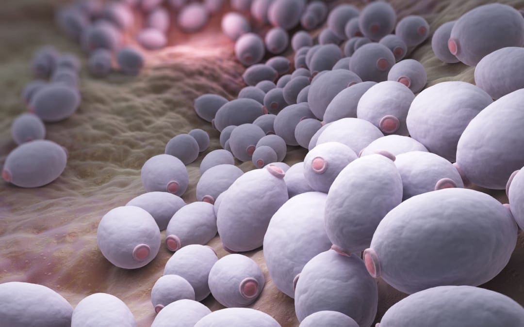
Candida is a yeast that grows naturally in the human mouth and the intestines.
Small amounts aid in nutrient digestion and absorption.
However, overgrowth of candida can damage the intestinal lining and release toxic byproducts directly into the circulatory system.
If not addressed Candida overgrowth can turn chronic and lead to:
Fungal skin infections
Chronic fatigue syndrome
Fibromyalgia
Autoimmune disease
Brain fog
Mood swings
Vaginal infections
Seasonal allergies
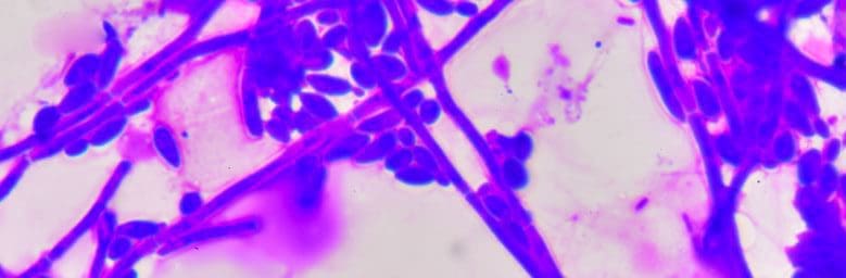
The most damaging fact is that it can puncture holes through the intestinal lining.
Candida grows roots as it spreads.
The roots can tear through the intestinal wall as they search for sustenance that can result in leaky gut.
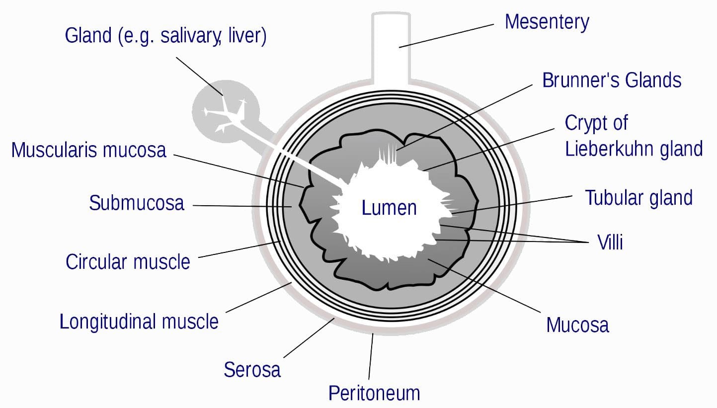
Leaky gut releases endotoxins from the intestinal lumen, like lipopolysaccharide (LPS), that can enter into circulation, triggering an innate immune response that often results in low-grade inflammation.
These gut bacteria lyse, release LPS into the intestinal lumen, where no damage can be done to a healthy gut.
But if the intestinal lining is damaged, LPS can enter directly into circulation, and trigger off a low-grade inflammation anywhere in the body.
Prevention
One way to protect the microbiome against Candida and LPS is with a combination of probiotic spores and yeast.
This combination has the power to control:
Pathogenic infections
Repair intestinal damage
Strengthen the immune system for future infections
However, most probiotics don’t effectively survive digestion to colonize the large intestine.
But probiotic spores and yeasts come equipped to survive the harsh gastric passage and�safely enter the intestines.
PROBIOTICS
Probiotic spores are likely the most promising therapy for metabolic endotoxemia, while currently there are no other probiotics or compounds that have demonstrated the same effect.
There are many types of probiotics that offer different types of beneficial bacteria to help for the proper functioning of the body. Here are 7 types.
Lactobacillus Acidophilus
Lactobacillus Reuteri
Lactobacillus Bulgaricus
Streptococcus Thermophilus
Bifidobacterium Bifidum
Saccharomyces Boulardii
Bacillus Subtilis
Candida Control
When probiotics are not enough to contain a chronic�Candida overgrowth it may be helpful to incorporate natural compounds like
Propolis is a resinous material that has built-in antifungal properties that are used by honeybees to protect the inside lining of the hive.
Bee propolis supports the immune system and fights infections without having to call on the immune system.
This spares energy, avoids activation of immune cells/responses and prevents inflammatory reactions that cause irritation and pain discomfort.
Undecylenic acid can also control fungal overgrowths in the gut.
It is a monounsaturated fatty acid with antifungal properties.
It can be used as a topical ointment that is safe for the most sensitive places like:
Skin
Mouth
Vaginal cavity
Undecylenic acid has also been found to be highly effective in treating Tinea pedis, better known as, athletes foot.
A total body system approach to Candida overgrowth is ideal
Address gut health as the source of Candida overgrowth
Restore the intestinal barrier
Utilize natural compounds (undecylenic acid and bee propolis), to balance intestinal cultures of Candida
This combination is a natural and effective way to improve gut barrier function and control harmful gut infections.
6 Day *DETOX DIET* Treatment | El Paso, TX (2019)
Fred Foreman is a basketball coach who depends on his well-being to be able to participate in his everyday tasks and responsibilities. That’s when coach Foreman started the 6 Day Detox Program from Xymogen, what was developed to help renew and enhance the human body’s natural cleansing and detoxification capabilities. Fred Foreman discusses his experience with the 6 Day Detox Program, describing the benefits he experienced as well as the effort he had to make, to support his overall well-being through the detox. Fred Foreman feels a great sense of fulfillment with the 6 Day Detox Program and he encourages other people, who also wish to improve their overall health and wellness, to detox their body. Coach Foreman highly recommends the 6 Day Detox Program as an alternative treatment choice for overall health and wellness.
NCBI Resources:
Probiotics are the good bacteria (or friendly bacteria) that line your gut and help in the absorption of nutrients from the food and thus boost up your immune system. Digestive disorders, candida, frequent attack of cold and flu, autoimmune disease, skin problems, etc. are some side effects we will experience due to lack of enough probiotics. In this world, due to unhealthy agricultural practices (little or no probiotics in food) and the intake of antibiotics for every health problem (kill the existing good bacteria). So, we have to include more probiotic-rich foods in our diet.
Functional neurology primarily focuses on the fundamentals of neuron health and it is mainly based on neuroplasticity theories. It’s believed that the brain and the nervous system are capable of changing, and can become malleable, due to a reaction to certain stimulation. The brain can be shaped by sensory, motor, cognitive, or emotional experiences. �
The creation of synapses in the nervous system depends on the stimulation they receive. Neurons which receive too much stimulation are the ones which become stronger and those which don’t receive stimulation become weaker and eventually diminish. It is believed that it is possible to create new neurons even after there has been damage to the nervous system. �
The Role of Functional Neurology
Functional neurology evaluates changes in the nervous system before these become severe health issues. The practice of functional neurology has been adopted by several modalities of practice, such as chiropractic, psychology, occupational therapy and even by conventional healthcare professionals. Functional neurology is commonly practiced by chiropractors. �
The practice of neurology involves applying neuroscience research from laboratory studies to determine how it can be practically applied in health care. The brain is protected by supporting the nervous system. The ultimate goal of functional neurology is to treat brain and nervous system health issues without the utilization of drugs or together with conventional treatment approaches. Functional neurologists can help treat a wide variety of neurological health issues, including: �
Neurodegenerative disorders: Alzheimer�s disease, Parkinson�s disease, dementia, and multiple system atrophy.
Demyelinating conditions: Multiple sclerosis, transverse myelitis, and leukodystrophies.
Trauma and brain injuries: Concussions and whiplash-associated disorders.
Vestibular conditions: Motion sickness, dizziness/disequilibrium, labyrinthitis, vertigo, and Meniere’s disease.
Movement disorders: Tics, restless leg syndrome, myoclonus, and dystonia.
Neuro-developmental conditions: Autism spectrum disorders, ADHD, Asperger’s syndrome, Tourette syndrome, dyslexia, processing disorders, and global developmental delay.
Headaches and pain syndromes: Cluster headaches, complex regional pain syndrome, migraines, and fibromyalgia
Functional neurological disorders which are best referred to as a group of physical, sensory and cognitive symptoms which do not seem to have an identifiable organic etiology.
Functional Neurology Treatment
The primary goal of functional neurology is to promote, support, and restore the optimal function of the brain and the nervous system, as opposed to the absence of pathology. Sometimes it’s not always possible to determine the natural source of a person’s neurological disease and its symptoms. Functional neurology can be particularly beneficial in these instances. �
The patient’s medical history and a non-invasive evaluation are required for diagnosis. Treatment is determined based on the patient’s current and targeted well-being. Any blood tests, x-rays, MRIs and/or other tests are also evaluated. During the evaluation, the healthcare professional will observe all aspects of the patient, including eye movements and posture, which can demonstrate the function of the brain and the nervous system. Blood pressure, pulse, and reflexes are also evaluated. �
Neuro-developmental conditions and behavioral disorders are generally treated with functional neurology. Anxiety is commonly increased in patients with these type of health issues, therefore, it is recommended that the non-invasive evaluation is performed in a way which does not trigger anxiety in the patient. Functional neurology treatment is individualized and every part of the treatment approach is customized to the individual’s treatment requirements. �
Functional neurology emphasizes on encouraging patients to practice self-care so that face-to-face treatment with a healthcare professional does not continue for months or years without end. Home exercise programs are developed to treat the associated health issues, meaning that functional neurology treatment is incorporated into the patient’s daily activities. �
Biochemistry and Nutrition in Functional Neurology
Functional neurology treatment focuses on retraining the brain. Neurons need energy and stimulation to survive and thrive, therefore, functional neurology treatment may involve exercises, such as eye exercises, cognitive activities, balancing activities, and joint adjustments. Different stimulation can affect different regions and pathways in the human brain. �
Moreover, functional neurology treatment may also involve a nutritional and biochemical approach by eliminating several factors which may potentially affect neurons. These can ultimately include toxins, chemicals, and infection, among other factors. Dietary modifications and supplementation may also be included to provide optimal energy for neurons. �
An individualized treatment approach is applied to each individual otherwise there exists the risk of over-stimulating and exceeding the capacity of a patient’s nervous system. The goal of functional neurology treatment is to improve brain and nervous system health, neural processing, communication, and all signaling involving the brain and the entire human body. �
Functional neurology focuses on the diagnosis and treatment of the human brain and the nervous system utilizing sensory and cognitive based treatment methods and techniques to promote, support, and restore neuroplasticity, integrity, and functional optimization. Functional neurology can be utilized to help improve a variety of neurological diseases and health issues, including Alzheimer’s disease. Functional neurology is frequently practiced by chiropractors. – Dr. Alex Jimenez D.C., C.C.S.T. Insight
The purpose of the article above is to discuss the purpose of functional neurology in the treatment of neurological disease. Neurological diseases are associated with the brain, the spine, and the nerves. The scope of our information is limited to chiropractic, musculoskeletal and nervous health issues as well as functional medicine articles, topics, and discussions. To further discuss the subject matter above, please feel free to ask Dr. Alex Jimenez or contact us at 915-850-0900 . �
Curated by Dr. Alex Jimenez �
Additional Topic Discussion: Chronic Pain
Sudden pain is a natural response of the nervous system which helps to demonstrate possible injury. By way of instance, pain signals travel from an injured region through the nerves and spinal cord to the brain. Pain is generally less severe as the injury heals, however, chronic pain is different than the average type of pain. With chronic pain, the human body will continue sending pain signals to the brain, regardless if the injury has healed. Chronic pain can last for several weeks to even several years. Chronic pain can tremendously affect a patient’s mobility and it can reduce flexibility, strength, and endurance.
Formulas for Methylation Support
XYMOGEN�s Exclusive Professional Formulas are available through select licensed health care professionals. The internet sale and discounting of XYMOGEN formulas are strictly prohibited.
Proudly,�Dr. Alexander Jimenez makes XYMOGEN formulas available only to patients under our care.
Please call our office in order for us to assign a doctor consultation for immediate access.
If you are a patient of Injury Medical & Chiropractic�Clinic, you may inquire about XYMOGEN by calling 915-850-0900.
�
For your convenience and review of the XYMOGEN products please review the following link.*XYMOGEN-Catalog-Download �
* All of the above XYMOGEN policies remain strictly in force. �
Magnesium is an essential mineral, yet it tends to be overlooked as a health concern. To illustrate, a study shows that up to 75% of individuals are not intaking the recommended daily amount (2). This stems from the fact that most foods have steadily decreased the amount of magnesium they provide. Thus leaving many consumers with the need to supplement. Different magnesium supplements have been shown to facilitate the body in different ways.� Magnesium citrate can help to lower blood pressure whereas Magnesium L-Threonate can help with memory loss. So, which magnesium supplement should you be taking?��� �
Magnesium Citrate Overview
Magnesium Citrate is one of the most bioavailable forms of magnesium out there, meaning it’s ready and easily absorbed by the body’s digestive tract (5). In addition to this, Magnesium Citrate has been shown to improve arterial stiffness, lower blood pressure, and in turn, help to prevent cardiovascular disease in patients who are slightly obese (3). However, be aware that magnesium citrate is also used to treat constipation, therefore it can act as a laxative by helping to absorb water in the intestines (1). �
Magnesium L-Threonate Overview
Magnesium L-Threonate is a newer form of magnesium, therefore there are fewer studies showing its full capabilities. However, Magnesium L-Threonate is the only form of magnesium to cross the blood-brain barrier more readily. The blood-brain barrier is extremely selective in order to reduce the amount of toxins entered into the CNS (central nervous system). Due to this, it has been proven to increase synapse density and aid in memory loss, cognitive decline, as well as help to improve short term memory (4). In a long term study of 17 years performed with Magnesium L-Threonate, the results manifested that subjects were 37% less likely to get dementia (4). �
If you are looking for something to assist in relaxation, sleep, and memory; Magnesium L-Threonate is the supplement for you. Although, if you are looking for something to relieve occasional constipation and something to aid in blood pressure that mixes well with water and has little to no taste, magnesium citrate is the route you should consider. Getting started on the right magnesium supplement today could set you up with a better tomorrow. – Kenna Vaughn, Health Coach Insight
The scope of our information is limited to chiropractic, musculoskeletal and nervous health issues as well as functional medicine articles, topics, and discussions. To further discuss the subject matter above, please feel free to ask Dr. Alex Jimenez or contact us at 915-850-0900 . �
References
(1)Cisar�, Fabio, et al. �Bowel Preparation for Gastrointestinal Endoscopic Procedures With Sodium Picosulphate-Magnesium Citrate Is an Effective, Safe, and Well-Tolerated Option in Pediatric Patients: A Single-Center Experience.� Gastroenterology Nursing : the Official Journal of the Society of Gastroenterology Nurses and Associates, U.S. National Library of Medicine, 2018, www.ncbi.nlm.nih.gov/pubmed/30063687.
(2)Guerrera, Mary P, et al. �Therapeutic Uses of Magnesium.� American Family Physician, U.S. National Library of Medicine, 15 July 2009, www.ncbi.nlm.nih.gov/pubmed/19621856.
(3)Schutten, Jo�lle C, et al. �Effects of Magnesium Citrate, Magnesium Oxide and Magnesium Sulfate Supplementation on Arterial Stiffness in Healthy Overweight Individuals: a Study Protocol for a Randomized Controlled Trial.� Trials, BioMed Central, 28 May 2019, www.ncbi.nlm.nih.gov/pubmed/31138315.
(4)”Science Review: Magnesium L-Threonate.” Metagenics Institute. 01 Aug. 2019 .
(5)Walker, Ann F, et al. �Mg Citrate Found More Bioavailable than Other Mg Preparations in a Randomised, Double-Blind Study.� Magnesium Research, U.S. National Library of Medicine, Sept. 2003, www.ncbi.nlm.nih.gov/pubmed/14596323. �
Orthotics Treat Other Areas Than the Feet
�
�
Additional Topic Discussion: Xymogen� Supplements
The average adult in the United States takes one or more dietary supplements on a regular basis. Dietary supplements include vitamins, minerals, herbals and botanicals, amino acids, enzymes, and a variety of other products. Several of the most common dietary supplements include vitamin D and vitamin E, minerals such as calcium and iron, herbs such as echinacea and garlic, and products such as glucosamine, probiotics, and fish oils. For people who don’t have balanced nutrition, taking dietary supplements can help them get necessary amounts of essential nutrients and can help improve overall well-being.
Formulas for Methylation Support
�
XYMOGEN�s Exclusive Professional Formulas are available through select licensed health care professionals. The internet sale and discounting of XYMOGEN formulas are strictly prohibited.
� Proudly,�Dr. Alexander Jimenez makes XYMOGEN formulas available only to patients under our care.
� Please call our office in order for us to assign a doctor consultation for immediate access.
� If you are a patient of Injury Medical & Chiropractic�Clinic, you may inquire about XYMOGEN by calling 915-850-0900.
For your convenience and review of the XYMOGEN products please review the following link.*XYMOGEN-Catalog-Download � * All of the above XYMOGEN policies remain strictly in force. �
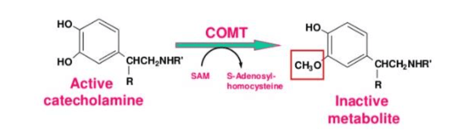
� Methylation, commonly referred to as the “one-carbon metabolism”, is the transfer or formation of methyl (CH3) groups, such as MTHFR, COMT, and DNMT. Methylation frequently utilizes SAMe as a methyl donor. Methylation is fundamental for a variety of bodily structures and functions, including cell division, gene expression, early CNS development, immune cell differentiation, neurotransmitter biosynthesis and/or metabolism, histamine clearance, detoxification, hormone biotransformation, cellular energy metabolism, phospholipid synthesis, and the myelination of peripheral nerves, among other structures and functions. � � Among one of it’s most essential functions on structures, DNA methylation highly regulates a variety of epigenetic marks, making them substantially stable, such as the covalent bonds of X and Y chromosomes in men and women. Methylation occurs at CpG sites with DNMT enzymes. Moreover, DNA methylation can help regulate and/or manage other bodily structures and functions. Histone methylation, RNA methylation, and mitochondrial DNA methylation (miDNMT) can help induce or inhibit gene expression. Recent research studies have also demonstrated that these same enzymes may additionally be involved in demethylation. �
Methylation and Fetal Programming
� According to research studies, while there are a variety of epigenetic marks which remain seemingly unchanged through inheritance, there are numerous other epigenetic marks which can change due to external factors, known as metastable epialleles. These changes can also change throughout inheritance as well as remain seemingly unchanged through inheritance. Metastable epialleles provide individuals with considerable differences and can be passed down to offspring during critical stages of fetal development. According to many more research studies, methylation can also affect epigenetic marks outside fetal programming windows. �
DNA methylation was originally considered to be stable and irreversible, however, external factors can also affect epigenetic methylation. The purpose of the following article is to discuss the importance of methylation for a variety of bodily structures and functions. Furthermore, it’s fundamental to understand how nutrition, fitness, lifestyle, supplements, and medicines, can improve DNA methylation and overall health and wellness. – Dr. Alex Jimenez D.C., C.C.S.T. Insight
Smoothies and Juices for Methylation Support
�
While many healthcare professionals can recommend diet and lifestyle changes to improve methylation support, there are several options you can try yourself at home. As described above, methylation support supplementation should be determined by a healthcare professional. Smoothies and juices are a fast and easy way to include all the necessary nutrients you need for methylation support without any side-effects. The smoothies and juices below are part of the Methylation Diet Food Plan.
Sea Green Smoothie Servings: 1 Cook time: 5-10 minutes
1/2 cup cantaloupe, cubed
1/2 banana
1 handful of kale or spinach
1 handful of Swiss chard
1/4 avocado
2 teaspoons spirulina powder
1 cup of water
3 or more ice cubes
Blend all ingredients in a high-speed blender until completely smooth and enjoy!
1/2 cup blueberries (fresh or frozen, preferably wild)
1 medium carrot, roughly chopped
1 tablespoon ground flaxseed or chia seed
1 tablespoons almonds
Water (to desired consistency)
Ice cubes (optional, may omit if using frozen blueberries)
Blend all ingredients in a high-speed blender until smooth and creamy. Best served immediately!
Sweet and Spicy Juice Servings: 1 Cook time: 5-10 minutes
1 cup honeydew melons
3 cups spinach, rinsed
3 cups Swiss chard, rinsed
1 bunch cilantro (leaves and stems), rinsed
1-inch knob of ginger, rinsed, peeled and chopped
2-3 knobs whole turmeric root (optional), rinsed, peeled and chopped
Juice all ingredients in a high-quality juicer. Best served immediately!
Ginger Greens Juice Servings: 1 Cook time: 5-10 minutes
1 cup pineapple cubes
1 apple, sliced
1-inch knob of ginger, rinsed, peeled and chopped
3 cups kale, rinsed and roughly chopped or ripped
5 cups Swiss chard, rinsed and roughly chopped or ripped
Juice all ingredients in a high-quality juicer. Best served immediately!
Zesty Beet Juice Servings: 1 Cook time: 5-10 minutes
1 grapefruit, peeled and sliced
1 apple, washed and sliced
1 whole beet, and leaves if you have them, washed and sliced
1-inch knob of ginger, rinsed, peeled and chopped
Juice all ingredients in a high-quality juicer. Best served immediately!
Protein Power Smoothie Serving: 1 Cook time: 5 minutes
1 scoop protein powder
1 tablespoon ground flaxseed
1/2 banana
1 kiwi, peeled
1/2 teaspoon cinnamon
Pinch of cardamom
Non-dairy milk or water, enough to achieve desired consistency
Blend all ingredients in a high-powered blender until completely smooth. Best served immediately!
ProLon� Fasting Mimicking Diet
Balanced methylation support can be achieved through proper nutrition. The ProLon� fasting mimicking diet offers a 5-day meal program which has been individually packed and labeled to serve the foods you need for the FMD in precise quantities and combinations. The meal program is made up of ready-to-eat or easy-to-prepare, plant-based foods, including bars, soups, snacks, supplements, a drink concentrate, and teas. The products are scientifically formulated and great tasting. Before starting the ProLon� fasting mimicking diet, 5-day meal program, please make sure to talk to a healthcare professional to find out if the FMD is right for you. The ProLon� fasting mimicking diet can help promote methylation support, among a variety of other healthy benefits.
�
Many doctors and functional medicine practitioners can recommend nutritional advice and/or guidelines to help improve DNA methylation. Proper diet and lifestyle changes can ultimately help improve DNA methylation. Understanding how methylation can affect an individual’s epigenetics throughout the early stages of life can help promote health and wellness. The scope of our information is limited to chiropractic, musculoskeletal and nervous health issues as well as functional medicine articles, topics, and discussions. To further discuss the subject matter above, please feel free to ask Dr. Alex Jimenez or contact us at 915-850-0900�.
Curated by Dr. Alex Jimenez
Additional Topic Discussion:�Acute Back Pain
Back pain�is one of the most prevalent causes of disability and missed days at work worldwide. Back pain attributes to the second most common reason for doctor office visits, outnumbered only by upper-respiratory infections. Approximately 80 percent of the population will experience back pain at least once throughout their life. Your spine is a complex structure made up of bones, joints, ligaments, and muscles, among other soft tissues. Injuries and/or aggravated conditions, such as�herniated discs, can eventually lead to symptoms of back pain. Sports injuries or automobile accident injuries are often the most frequent cause of back pain, however, sometimes the simplest of movements can have painful results. Fortunately, alternative treatment options, such as chiropractic care, can help ease back pain through the use of spinal adjustments and manual manipulations, ultimately improving pain relief.
Formulas for Methylation Support
�
XYMOGEN�s Exclusive Professional Formulas are available through select licensed health care professionals. The internet sale and discounting of XYMOGEN formulas are strictly prohibited.
Proudly,�Dr. Alexander Jimenez makes XYMOGEN formulas available only to patients under our care.
Please call our office in order for us to assign a doctor consultation for immediate access.
If you are a patient of Injury Medical & Chiropractic�Clinic, you may inquire about XYMOGEN by calling 915-850-0900.
�
For your convenience and review of the XYMOGEN products please review the following link.*XYMOGEN-Catalog-Download
* All the above XYMOGEN policies remain strictly in force.
To promote methylation support through diet and lifestyle changes, many healthcare professionals recommend following a diet food plan which is nutritionally replete and rich in nutrients for DNA methylation status and activity, anti-inflammatory, low in Glycemic index, rich in antioxidants and phytonutrients as enzyme modulators, optimal in hydration, and supportive of detoxification. �
Moreover, to promote methylation support, a diet plan should avoid excess calories, folic acid-fortified foods, alcohol, minimize AGE formation, added sugars, foods from animals raised with the use of antibiotics and hormones, high-mercury content fish like tuna, king mackerel, swordfish, and shark as well as utilizing plastic food and beverage containers. According to a research study on diet and aging, calorie restriction can slow down or reverse the decline in age-related DNA methylation associated with diseases. As mentioned previously, alcohol consumption can tremendously affect DNA status and activity which may interfere with SAMe levels and may ultimately impact folate metabolism by inhibiting MTR and MAT enzymes, among other structures and functions. �
Furthermore, many doctors and functional medicine practitioners also recommend following intermittent fasting, a diet plan which cycles between fasting and non-fasting, alongside a low carbohydrate diet. According to several research studies, an extended nighttime fast can stimulate a decreased ketone production which may help decrease inflammation and protect the epigenome. �
Healthcare professionals can provide patients with nutrition advice and recommendations for lifestyle changes to help promote methylation support. While potential side effects of supplements have been discussed in previous articles, doctors and functional medicine practitioners can recommend supplementation alongside nutrition and lifestyle changes. Below, we will demonstrate a variety of superfoods, foods, and microbes which can be utilized to help promote DNA methylation status and activity.
B. adolescentis � appears to be the highest producer of 5mTHF
B. pseudocatenulatum
Nutrition and lifestyle changes can help promote methylation support without experiencing the side effects of supplements. A wide array of superfoods, foods, and microbes can help promote methylation support, alongside a diet food plan, supplementation, and intermittent fasting. The purpose of the following article is to discuss how nutrition and lifestyle modifications can help promote methylation support. It’s fundamental to understand how nutrition, fitness, lifestyle, supplements, and medicines, can improve DNA methylation and overall health and wellness. – Dr. Alex Jimenez D.C., C.C.S.T. Insight
Smoothies and Juices for Methylation Support
�
While many healthcare professionals can recommend diet and lifestyle changes to improve methylation support, there are several options you can try yourself at home. As described above, methylation support supplementation should be determined by a healthcare professional. Smoothies and juices are a fast and easy way to include all the necessary nutrients you need for methylation support without any side-effects. The smoothies and juices below are part of the Methylation Diet Food Plan.
Sea Green Smoothie Servings: 1 Cook time: 5-10 minutes
1/2 cup cantaloupe, cubed
1/2 banana
1 handful of kale or spinach
1 handful of Swiss chard
1/4 avocado
2 teaspoons spirulina powder
1 cup of water
3 or more ice cubes
Blend all ingredients in a high-speed blender until completely smooth and enjoy!
1/2 cup blueberries (fresh or frozen, preferably wild)
1 medium carrot, roughly chopped
1 tablespoon ground flaxseed or chia seed
1 tablespoons almonds
Water (to desired consistency)
Ice cubes (optional, may omit if using frozen blueberries)
Blend all ingredients in a high-speed blender until smooth and creamy. Best served immediately!
Sweet and Spicy Juice Servings: 1 Cook time: 5-10 minutes
1 cup honeydew melons
3 cups spinach, rinsed
3 cups Swiss chard, rinsed
1 bunch cilantro (leaves and stems), rinsed
1-inch knob of ginger, rinsed, peeled and chopped
2-3 knobs whole turmeric root (optional), rinsed, peeled and chopped
Juice all ingredients in a high-quality juicer. Best served immediately!
Ginger Greens Juice Servings: 1 Cook time: 5-10 minutes
1 cup pineapple cubes
1 apple, sliced
1-inch knob of ginger, rinsed, peeled and chopped
3 cups kale, rinsed and roughly chopped or ripped
5 cups Swiss chard, rinsed and roughly chopped or ripped
Juice all ingredients in a high-quality juicer. Best served immediately!
Zesty Beet Juice Servings: 1 Cook time: 5-10 minutes
1 grapefruit, peeled and sliced
1 apple, washed and sliced
1 whole beet, and leaves if you have them, washed and sliced
1-inch knob of ginger, rinsed, peeled and chopped
Juice all ingredients in a high-quality juicer. Best served immediately!
Protein Power Smoothie Serving: 1 Cook time: 5 minutes
1 scoop protein powder
1 tablespoon ground flaxseed
1/2 banana
1 kiwi, peeled
1/2 teaspoon cinnamon
Pinch of cardamom
Non-dairy milk or water, enough to achieve desired consistency
Blend all ingredients in a high-powered blender until completely smooth. Best served immediately!
ProLon� Fasting Mimicking Diet
Balanced methylation support can be achieved through proper nutrition. The ProLon� fasting mimicking diet offers a 5-day meal program which has been individually packed and labeled to serve the foods you need for the FMD in precise quantities and combinations. The meal program is made up of ready-to-eat or easy-to-prepare, plant-based foods, including bars, soups, snacks, supplements, a drink concentrate, and teas. The products are scientifically formulated and great tasting. Before starting the ProLon� fasting mimicking diet, 5-day meal program, please make sure to talk to a healthcare professional to find out if the FMD is right for you. The ProLon� fasting mimicking diet can help promote methylation support, among a variety of other healthy benefits.
�
Many doctors and functional medicine practitioners can recommend nutritional advice and/or guidelines to help improve DNA methylation. Proper diet and lifestyle changes can ultimately help improve DNA methylation. Nutrition and lifestyle modifications, including methylation superfoods, foods, and microbes, can ultimately help promote methylation support. The scope of our information is limited to chiropractic, musculoskeletal and nervous health issues as well as functional medicine articles, topics, and discussions. To further discuss the subject matter above, please feel free to ask Dr. Alex Jimenez or contact us at 915-850-0900�.
Curated by Dr. Alex Jimenez
Additional Topic Discussion:�Acute Back Pain
Back pain�is one of the most prevalent causes of disability and missed days at work worldwide. Back pain attributes to the second most common reason for doctor office visits, outnumbered only by upper-respiratory infections. Approximately 80 percent of the population will experience back pain at least once throughout their life. Your spine is a complex structure made up of bones, joints, ligaments, and muscles, among other soft tissues. Injuries and/or aggravated conditions, such as�herniated discs, can eventually lead to symptoms of back pain. Sports injuries or automobile accident injuries are often the most frequent cause of back pain, however, sometimes the simplest of movements can have painful results. Fortunately, alternative treatment options, such as chiropractic care, can help ease back pain through the use of spinal adjustments and manual manipulations, ultimately improving pain relief.
Formulas for Methylation Support
�
XYMOGEN�s Exclusive Professional Formulas are available through select licensed health care professionals. The internet sale and discounting of XYMOGEN formulas are strictly prohibited.
Proudly,�Dr. Alexander Jimenez makes XYMOGEN formulas available only to patients under our care.
Please call our office in order for us to assign a doctor consultation for immediate access.
If you are a patient of Injury Medical & Chiropractic�Clinic, you may inquire about XYMOGEN by calling 915-850-0900.
�
For your convenience and review of the XYMOGEN products please review the following link.*XYMOGEN-Catalog-Download
* All the above XYMOGEN policies remain strictly in force.
IFM's Find A Practitioner tool is the largest referral network in Functional Medicine, created to help patients locate Functional Medicine practitioners anywhere in the world. IFM Certified Practitioners are listed first in the search results, given their extensive education in Functional Medicine


 � Among one of it’s most essential functions on structures, DNA methylation highly regulates a variety of epigenetic marks, making them substantially stable, such as the covalent bonds of X and Y chromosomes in men and women. Methylation occurs at CpG sites with DNMT enzymes. Moreover, DNA methylation can help regulate and/or manage other bodily structures and functions. Histone methylation, RNA methylation, and mitochondrial DNA methylation (miDNMT) can help induce or inhibit gene expression. Recent research studies have also demonstrated that these same enzymes may additionally be involved in demethylation. �