The human body has numerous musculoskeletal muscles that allow the host to do various movements without pain or discomfort. Each muscle group has tendons, muscles, ligaments, and connective tissues surrounding the skeletal joint and protecting the skeletal structure. Each muscle group in the body allows different functions, from turning the neck from side to side to enabling the legs to provide motion when walking. Now naturally, the body ages over time, which can lead to muscle weakness in the muscle groups and affect the connective tissues, or various disruptors can develop in a healthy body that can also affect the muscles and connective tissues. Fortunately, the multiple muscle groups and connective tissues are affected by overlapping risk profiles. In that case, there are many treatments and techniques that many pain specialists utilize to restore the body and relieve pain-like symptoms associated with musculoskeletal disorders. Today’s article examines connective tissues, how conditions can affect the connective tissues, and how the MET technique stretches or strengthens the body’s connective tissue. We provide information about our patients to certified medical providers that offer available therapy techniques like MET (muscle energy techniques) for individuals dealing with chronic conditions associated with disorders affecting the body’s connective tissues that can correlate and develop with overlapping pain profiles. We encourage each patient appropriately by referring them to our associated medical providers based on their diagnosis results. We accept that education is a spectacular way when asking our providers the most crucial questions at the patient’s acknowledgment. Dr. Alex Jimenez, D.C., assesses this information as an educational service. Disclaimer
What Are Connective Tissues?
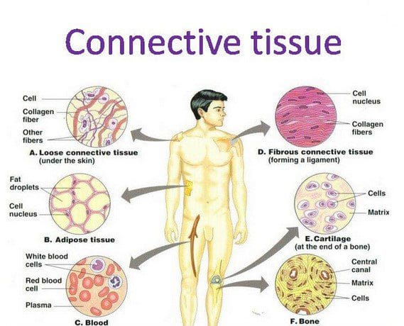
The human body is a multiplex machine that is composed of many tissues that surround the skeletal joints and vital organs with the basic functions that the body produces. Research studies reveal that, as the name has implied, connective tissues in the body refer to the several different body tissues that connect and support the other tissues by binding them to the body. Now there are three different categories that connective tissue can be broken down into:
Loose connective tissue
Dense connective tissue
Specialized connective tissues
These three different connective tissue categories have functions that allow the body to perform properly and provide support to the rest of the musculoskeletal system. The dense connective tissues make up the body’s tendons and ligaments that move the hands and feet while having a higher collagen fiber density. The loose connective tissues help keep the vital organs in place. And finally, the specialized connective tissues are composed of adipose tissues, cartilage, lymphoid tissues, etc. When the body begins to age naturally or is dealing with issues affecting the connective tissues, it can develop musculoskeletal disorders associated with the connective tissue.
Disorders Affecting The Connective Tissues
Have you been experiencing muscle pain or weakness in your body? Do your hands or feet feel tired? Or do you feel stiffness and pain in your joints? Many pain-like symptoms are associated with musculoskeletal disorders affecting the body’s connective tissues. As stated earlier, when the body begins to age naturally, the various muscles in the body can develop into musculoskeletal disorders associated with the connective tissues. Aging can affect connective tissue function as the cartilage from the specialized connective tissues has less elasticity and changes the proteoglycan both quantitatively and qualitatively, according to the book, “Clinical Applications of Neuromuscular Techniques,” written by Leon Chaitow, N.D., D.O., and Judith Walker DeLany, L.M.T. Additional research studies have revealed that environmental factors associated with the body’s immune system can affect the connective tissues. This is known as connective tissue disorder, and it can be comprised of numerous conditions that can affect the immune system and cause overlapping symptoms in the musculoskeletal system. This includes some of the following:
Inflammation in the joints causes them to lock up
Muscle weakness where myofascial entrapment affects the muscle fibers
Fatigue
Vitamin deficiency
An Introduction To MET- Video
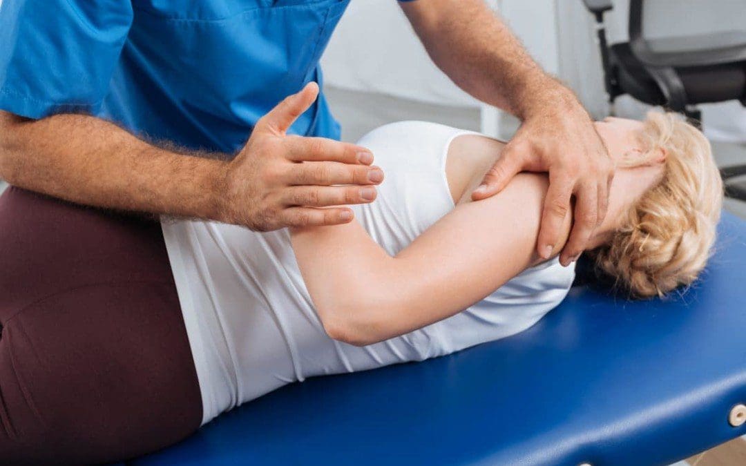
Have you been feeling stiffness in your muscles or joints? Does it hurt when you are bending over and lifting heavy objects? Or are you feeling tired constantly? When the body deals with these issues, it can affect more than the muscles and connective tissues. This can lead to symptoms of stiffness and aches in the joints while restricting the range of motion to the muscles. When this happens to the body, many pain specialists utilize MET (muscle energy technique) and relieve those symptoms. Studies reveal that MET is a manual treatment for soft tissue, helping mobilize the joints and stretch tight muscles and fascia to improve circulation to the connective tissues and drain the lymphatic system. The video above introduces how MET is used on the body.
The MET Technique On Connective Tissues
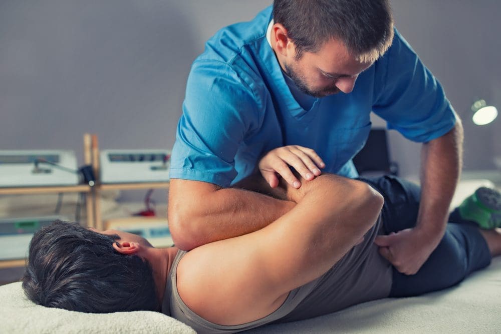
Research studies reveal that since the muscles and joints are being held together by connective tissues, using the MET technique allows pain specialists to stretch the muscles and joints to release tension and other symptoms associated with pain. When pain specialists use the MET technique on the body, it can help strengthen the weaker muscles while paying attention to how short the muscles are affecting the body. While the MET technique can help support the muscles with combined physical therapy, it can help stretch the tight muscles and overworked connective tissues. This allows the body to be restored and get back to normal. Many pain specialists like chiropractic care allow the MET technique to stretch the trapped connective tissues and free the body’s structures to correct postural imbalances.
Conclusion
The body’s connective tissues support each muscle, organ, and skeletal structure. When issues affect the body, the various muscle groups, and connective tissues start to develop overlapping symptoms associated with pain. When pain-like symptoms affect the body, many people will go to a pain specialist and be treated using the MET technique to restore the muscles and body and return to normal.
References
Chaitow, Leon, and Judith Walker DeLany. Clinical Applications of Neuromuscular Techniques. Churchill Livingstone, 2003.
Kamrani, Payvand, et al. “Anatomy, Connective Tissue.” In: StatPearls [Internet]. Treasure Island (FL), StatPearls Publishing, 24 Jan. 2022, https://www.ncbi.nlm.nih.gov/books/NBK538534/.
Page, Phil. “Current Concepts in Muscle Stretching for Exercise and Rehabilitation.” International Journal of Sports Physical Therapy, U.S. National Library of Medicine, Feb. 2012, https://www.ncbi.nlm.nih.gov/pmc/articles/PMC3273886/.
Rao, Vijay, and Simon Bowman. “Latest Advances in Connective Tissue Disorders.” Therapeutic Advances in Musculoskeletal Disease, U.S. National Library of Medicine, Aug. 2013, https://www.ncbi.nlm.nih.gov/pmc/articles/PMC3728978/.
Thomas, Ewan, et al. “The Efficacy of Muscle Energy Techniques in Symptomatic and Asymptomatic Subjects: A Systematic Review.” Chiropractic & Manual Therapies, U.S. National Library of Medicine, 27 Aug. 2019, https://www.ncbi.nlm.nih.gov/pmc/articles/PMC6710873/.
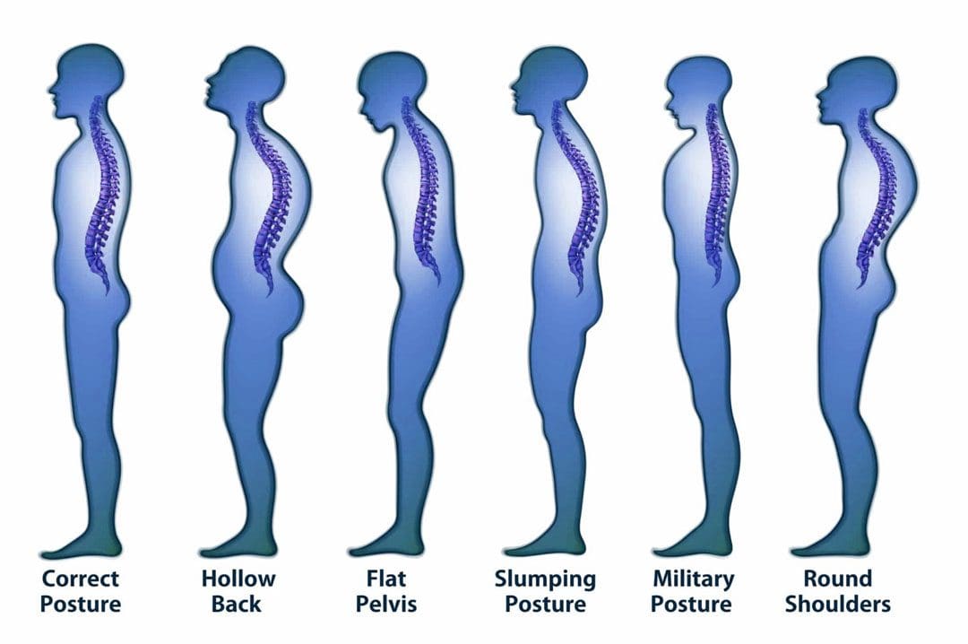
Every day, the body is in constant rest or active motion when needed, from working to exercising and getting adequate rest to repeat the cycle. However, as the body is in this dynamic/rest motion, unintentionally, many individuals will be hunched forward, causing their posture to be slouched for long periods. To that point, it can cause the surrounding neck, shoulder, and back muscles to be pulled and overly stretched, causing pain when the individual gets out of the reclined position. When a person is constantly being hunched over, the action alone could lead to poor posture, which can cause misalignment to the spine and be associated with many chronic conditions that affect their way of life. Fortunately, various treatments can help alleviate poor posture and its associated symptoms. Today’s article examines what defines good posture, the influences that can affect body posture, and how treatment techniques like MET (muscle energy technique) can help improve posture. We mention our patients to certified medical providers that provide available therapy treatments like MET (muscle energy techniques) for individuals suffering from chronic conditions associated with poor posture that can correlate with overlapping risk profiles. We encourage each patient when it is appropriate by referring them to associated medical providers based on their diagnosis or needs. We understand and accept that education is a marvelous way when asking our providers crucial questions at the patient’s request and acknowledgment. Dr. Alex Jimenez, D.C., uses this information as an educational service. Disclaimer
What Defines Good Posture?
Have you been experiencing referred pain in your neck, shoulders, or lower back? Do you feel pain when stretching after being hunched over throughout the day? Or have you noticed that your neck is slanted, which causes your head to poke in front of your shoulders? Many of these issues are correlated with poor posture. Many of us have heard the saying from our parents, “Stand up straight!” And this is a reminder that having good posture correlates with good spinal health. The book, “Clinical Applications of Neuromuscular Techniques,” written by Leon Chaitow, N.D., D.O, and Judith Walker DeLany, L.M.T, mentions that posture is used to describe the static state of the spine. There are two different types of posture: static and dynamic. Static posture is when the body is in motion, while dynamic posture is when the body is resting. So good posture allows the spine to naturally curve with minimal pain affecting the cervical, thoracic, and lumbar regions.
Influences That Affect Body Posture
As stated early, many of us unintentionally hunch our bodies over time. This is one of the issues as we constantly look down on our phones, and as we get older, it can affect our ability to balance ourselves. Research studies reveal that improper posture can affect static and dynamic balance as we age. This means that when we are constantly hunched over as older adults, we are more prone to the risk of falling and causing long-term disability to our bodies. Additional research studies also mentioned that chronic conditions like forward head posture (which correlates to constantly looking down at the phone) could cause a persistent and abnormal contraction of the neck and shoulder muscles to become dysfunctional. To that point, it can cause pressure on the muscles, fascia, and nerves in the cervical-thoracic regions of the body. When bad posture affects the body over time, it can develop into musculoskeletal disorders if not treated immediately.
5 Way To Improve Posture- Video
Have you felt muscle strain on your neck, shoulders, and back? Have you felt relief when you stretch after being hunched over? Do you feel unstable when walking? These issues could be correlated with your posture if you have been experiencing these issues. When it comes to the body, it is important to make sure that maintaining good posture is not just to please your parents but to have a healthy spine. When we are constantly hunched over, it can cause the muscles and connective tissues to have gravitational strain and shorten the length of the muscles. However, realizing that you have poor posture early on can be treated. The video above shows the five best ways to improve your posture and how to strengthen the back, neck, and shoulder muscles from developing chronic conditions. Exercise alone can not be the only solution; combining it with chiropractic therapy allows the body to be fully restored with various techniques to reduce pain-like symptoms.
How The Met Technique Helps Improve Posture
So how would chiropractic care help with improving posture? Many chiropractors use techniques like MET (muscle energy technique) and spinal manipulation to help restore the body to realignment. Studies reveal that the combinations of MET and stretching can help lengthen the short muscles and restore range of motion to the body. Chiropractors use their hands and various tools to help realign the spine from subluxation and return the body to normal while freeing the tense muscles. Chiropractic care decreases the body’s risk of back injuries while reducing wear and tear on the muscles and joints, contributing to poor posture.
Conclusion
Overall, it is important to maintain good posture to prevent unwanted chronic issues from causing pain-like symptoms to the body. Recognizing the problems contributing to poor posture, treatment, and exercise can help stretch and strengthen the back muscles from hunching over. Maintaining good posture allows the body to be pain-free and prevents many unwanted symptoms from developing.
References
Chaitow, Leon, and Judith Walker DeLany. Clinical Applications of Neuromuscular Techniques. Churchill Livingstone, 2003.
Cohen, Rajal G, et al. “Lighten up! Postural Instructions Affect Static and Dynamic Balance in Healthy Older Adults.” Innovation in Aging, U.S. National Library of Medicine, 24 Mar. 2020, https://www.ncbi.nlm.nih.gov/pmc/articles/PMC7092748/.
Lee, Joon-Hee. “Effects of Forward Head Posture on Static and Dynamic Balance Control.” Journal of Physical Therapy Science, U.S. National Library of Medicine, Jan. 2016, https://www.ncbi.nlm.nih.gov/pmc/articles/PMC4756019/.
Phadke, Apoorva, et al. “Effect of Muscle Energy Technique and Static Stretching on Pain and Functional Disability in Patients with Mechanical Neck Pain: A Randomized Controlled Trial.” Hong Kong Physiotherapy Journal : Official Publication of the Hong Kong Physiotherapy Association Limited = Wu Li Chih Liao, U.S. National Library of Medicine, 14 Apr. 2016, https://www.ncbi.nlm.nih.gov/pmc/articles/PMC6385145/.
Environmental factors can affect the body and lead to chronic conditions involving the musculoskeletal system. When issues like stress, physical inactivity, and traumatic events affect the muscle groups in the upper and lower extremities, it causes the various muscles to tense up and be succumbed to multiple injuries that could potentially develop trigger points. Now trigger points can cause overlapping risk profiles and pain-like issues that can affect a person’s mobility and stability. However, many ways can alleviate the pain-like symptoms associated with trigger points affecting the musculoskeletal system. Many pain specialists use techniques to stretch the tense muscle and release the trigger point nodule in the muscle fibers. Today we will look at how myofascial trigger point formation affects the body, how MET (muscle energy techniques) are used to relieve trigger point formation, and how chiropractic care uses the MET technique on trigger points. We mention our patients to certified medical providers that provide available therapy treatments like MET (muscle energy techniques) for individuals suffering from chronic conditions associated with trigger point formation on the musculoskeletal system. We encourage each patient when it is appropriate by referring them to associated medical providers based on their diagnosis or needs. We understand and accept that education is a marvelous way when asking our providers crucial questions at the patient’s request and acknowledgment. Dr. Alex Jimenez, D.C., uses this information as an educational service. Disclaimer
Myofascial Trigger Points Affecting The Body
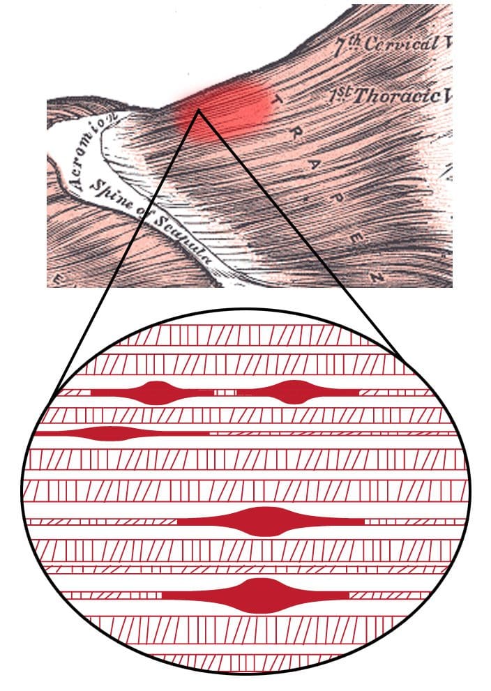
Have you been dealing with pain in different locations in your body? Do you feel that your muscles feel tight or tensed constantly? Or do you feel muscle strain when lifting or carrying heavy objects? Many of these pain-like issues correlate with myofascial trigger points affecting the body. According to research studies, myofascial pain syndrome or trigger points are hard palpable nodules discrete along the taut skeletal muscle band that can be painful when active or compressed. Now trigger points can cause the affected muscles to be hypersensitive, which to that point, can spread pain when being touched, known as referred pain. A great example would be if tense shoulder muscles have a cluster of trigger points and, when touched, send pain to the neck.
Trigger points in the musculoskeletal system can be present in soft tissues that can cause dysfunction and promote pain in the affected muscle area. Trigger points are developed in any scenario, from trauma like an auto accident to repetitive motions for extended periods. Two features can cause trigger point formation that can create these nodules: active and latent trigger points. Active trigger points, according to “Clinical Applications of Neuromuscular Techniques,” written by Leon Chaitow, N.D, D.O, and Judith Walker DeLany, L.M.T, mentioned that when pressure is applied to active trigger points it can cause referred pain associated with symptoms of painful sensations to the affected muscle. While latent trigger points, when pressure is applied to them, can cause referred pain that a person experienced in the past and occurs recently. Latent trigger points can also develop into active trigger points correlating to overlapping risk profiles. The book also stated that when the fascia and connective muscle tissues have been overused or strained, it can lead to trigger point formation development.
MET Trigger Point Therapy-Video
Have you been dealing with referred pain in different areas of your body? Do you feel that your muscles are tense and aching? Or do you feel muscle strain when lifting or carrying heavy objects? If you have been dealing with these issues, they are correlated to trigger point formation in your musculoskeletal system. Why not try MET or muscle energy technique therapy? Studies reveal that muscle energy techniques were developed originally to treat soft tissue, stretch tight muscles and fascia, and mobilize joints while improving blood circulation and draining the lymphatic system. So how do trigger point formation can be treated with MET techniques? Well, since trigger points can cause tight, hypersensitive spots that can be located in various taut muscle bands, MET techniques from pain specialists can help stretch and break up the tight nodules in the muscles to achieve muscle restoration at full resting length. The video above demonstrates how MET is used as trigger point therapy.
MET Techniques On Trigger Point Formation
So how do MET techniques work on trigger point formation in the musculoskeletal system? According to research studies, MET techniques utilize soft tissue manipulation to improve the myofascial system’s and joints’ functional parameters. Many pain specialists, like chiropractors, use this technique and other tools to help restore the body’s natural range of motion in the joints while providing a pain-reducing effect to the numerous musculoskeletal disorders. Additional research studies also mentioned that MET/NET (neuro-emotional) techniques could help relieve pain sensitivity from the affected muscle area.
How Chiropractic Care Uses MET Techniques On Trigger Points
So how would chiropractic care utilize MET techniques on an individual with trigger points? Due to its effectiveness and drug-free approach, chiropractic care can help smooth out the muscle and fascia by applying pressure with their hands or special tools to relieve trigger point pain. With MET techniques, chiropractors can help release muscle stiffness, tightness, and shortness to restore the body and re-align the spine. With continued chiropractic treatment, the body can reduce the future formation of trigger points in the muscle fibers while preventing further issues from developing.
Conclusion
Trigger point formation can occur in different muscle areas in the body, leading to overlapping risk profiles associated with pain. When the body is dealing with referred pain caused by trigger points, it can cause numerous issues affecting a person’s daily activity. Luckily, pain specialists like chiropractic care can incorporate techniques like MET and spinal manipulation to re-align the body, stretch out the stiff muscles, and promote a restored range of motion back to the musculoskeletal system. By going through daily treatments, the body can begin to heal naturally and prevent future injuries.
References
Bablis, Peter, et al. “Neuro Emotional Technique for the Treatment of Trigger Point Sensitivity in Chronic Neck Pain Sufferers: A Controlled Clinical Trial.” Chiropractic & Osteopathy, U.S. National Library of Medicine, 21 May 2008, https://www.ncbi.nlm.nih.gov/pmc/articles/PMC2427032/.
Chaitow, Leon, and Judith Walker DeLany. Clinical Applications of Neuromuscular Techniques. Churchill Livingstone, 2003.
Shah, Jay P, et al. “Myofascial Trigger Points Then and Now: A Historical and Scientific Perspective.” PM & R : the Journal of Injury, Function, and Rehabilitation, U.S. National Library of Medicine, July 2015, https://www.ncbi.nlm.nih.gov/pmc/articles/PMC4508225/.
Thomas, Ewan, et al. “The Efficacy of Muscle Energy Techniques in Symptomatic and Asymptomatic Subjects: A Systematic Review.” Chiropractic & Manual Therapies, U.S. National Library of Medicine, 27 Aug. 2019, https://www.ncbi.nlm.nih.gov/pmc/articles/PMC6710873/.
Wendt, Michał, and Małgorzata Waszak. “Evaluation of the Combination of Muscle Energy Technique and Trigger Point Therapy in Asymptomatic Individuals with a Latent Trigger Point.” International Journal of Environmental Research and Public Health, U.S. National Library of Medicine, 14 Nov. 2020, https://www.ncbi.nlm.nih.gov/pmc/articles/PMC7696776/.
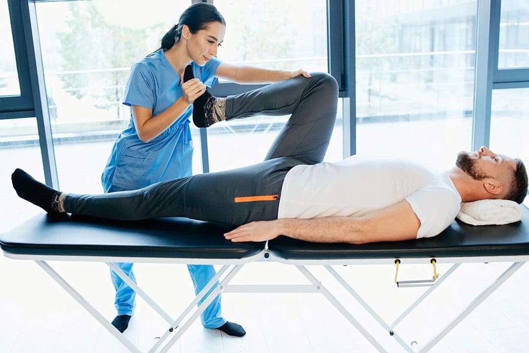
The various muscles, tendons, and ligaments inside the body surround the skeletal joint to provide movement and multiple actions to allow the host to be mobile. The body also has various muscle groups, with soft tissues surrounding the vital organs to help support the body. Since the human body is mobile, many factors can cause issues to the body’s host and lead to chronic overlapping risk profiles that can correlate with pain in the joints and muscle tissues. When these factors are causing pain in the musculoskeletal system, various treatment techniques can help reduce the pain-like symptoms and help restore the body. MET, or muscle energy technique, is one of the different treatment techniques used by pain specialists like chiropractors, massage therapists, physical therapists, and occupational therapists on many individuals with musculoskeletal pain. Today’s article looks at the musculoskeletal system, how the issues affect the muscles, and how muscle energy technique is utilized to reduce muscle pain associated with the musculoskeletal system. We mention our patients to certified medical providers that provide available therapy treatments like MET (muscle energy techniques) for individuals suffering from chronic conditions associated with the musculoskeletal system. We encourage each patient when it is appropriate by referring them to associated medical providers based on their diagnosis or needs. We understand and accept that education is a marvelous way when asking our providers crucial questions at the patient’s request and acknowledgment. Dr. Alex Jimenez, D.C., uses this information as an educational service. Disclaimer
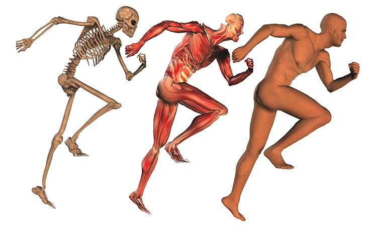
An Overview Of The Musculoskeletal System
The musculoskeletal system plays a huge role in the body, consisting of numerous muscle groups, tissues, ligaments, joints, and organs controlled by the central nervous system. The central nervous system provides the motor-sensory function to the musculoskeletal system, allowing the body to rest and move around. What the central nervous system does to the musculoskeletal system, according to research studies, it is revealed that these two systems have a relationship with each other as they are interconnected. Besides the various muscle groups that help surround the skeletal joints and provide mobility to the body, we will look at the connective tissue associated with the facial system and how muscle activity is affected by chronic issues.
Connective Tissue & The Fascial System
Regarding the musculoskeletal system, the connective tissue is one of the single abundant materials that allow each muscle group to be connected to its specific body region. The connective tissue comprises the body’s bones, muscles, blood vessels, and lymph nodes while embracing all the soft tissues and organs. The body’s connective tissue also works with the fascial system, giving the body the fundamental requirements. The fascial system is the structural form of the body since the fascial system is composed of connective tissues. With these two systems connecting and working together, it allows the muscles in the body to respond to various actions thrown at in different environments. The fascia web allows all muscle tissues to exist in isolation and interwoven with other structures to provide mobility.
Muscle Activity
Everything from the connective tissues to the fascia is involved in muscle activity in the musculoskeletal system. When the various muscles start to work with the body’s most movement, it is combined with one or more muscles acting as the prime mover or antagonist, allowing synergistic muscles to assist and contract simultaneously. The various muscle groups in the musculoskeletal system allow different actions, often repeated, to become stabilizing or antagonizing muscles. A great example is looking at the upper and lower extremities of the body. The upper extremities allow the arms, neck, head, and shoulders to have mobility when it comes to bending, twisting, and turning. While the lower extremities allow the hips, low back, legs, and feet to allow, stability and flexion to make the body move. However, these muscle groups can be affected by multiple factors that can affect muscle activity and lead to overlapping soft tissue pain profiles.
Issues Affect Muscle Activity
Since the body is a complex machine, different environmental factors can affect muscle groups in various ways and cause numerous pain issues. Now when it comes to environmental factors, many negative influences do play a role in affecting the musculoskeletal system in three categories:
Biomechanical: trauma, overusing the muscles, congenital, etc.
Biochemical: endocrine imbalances, inflammation, ischemia, nutritional deficiency, etc.
Psychosocial: anxiety, depression, chronic stress, etc.
These influences can cause the muscles to tense up and restrict blood flow, causing pain and trigger points to form in the muscle fibers and making a person feel miserable. Fortunately, therapeutic techniques allow the muscles to relax and release the tension that the person is feeling.
MET(Muscle Energy Technique)-Video
What Is Muscle Energy Technique?
When people feel stressed, and their muscles become tight, they can develop pain-like symptoms that correlate with chronic issues. Fortunately, a revolution has taken place that many pain specialists like chiropractors and massage therapists take place when it comes to manipulative therapy through a technique known as MET or muscle energy technique. According to research studies, MET is an osteopathic manipulative medicine designed to improve the body’s musculoskeletal function. This technique helps target soft tissues and contributes to joint mobilization. The muscle energy technique allows the tight muscles and fascia to be stretched, improving circulation and lymphatic flow since chiropractors or doctors of chiropractic care utilize spinal manipulation to realign the body and restore joint function.
Additional studies also reveal that MET combined with chiropractic care allows pain reduction in the muscles and can increase the body’s range of motion. This technique is essential for chronic and acute low back pain, trigger point pain, and other musculoskeletal dysfunctions associated with environmental factors.
The Various Stretching Techniques Of MET
The main objective of MET is to induce relaxation of hypertonic musculature, which also stretches the muscles to reduce pain-like symptoms. Now many treatments like chiropractic care can combine different techniques to reduce pain and restore mobility to the individual. With MET, various stretching techniques can allow chiropractors to stretch the tense muscles while restoring the range of motion. Some of the stretching techniques that pain specialists use include:
Facilitated stretching: Allows chiropractors and massage therapists to use strong/light isometric contractions to treat the muscles and be actively stretched. Reduces muscle cramps, tissue damage, or pain to the affected muscle group while utilizing breathing techniques and producing sufficient post-isometric relaxation.
Active-isolated stretching: Allows chiropractors and massage therapists to stretch the affected muscle actively while using precise localization to allow the affected muscle to receive a specific extension. This allows the muscles to relax through a short repetitive contraction and retraction to increase oxygenated blood flow. This stretching technique prevents the activation of the myotatic stretch reflex on the affected muscle.
Static stretching: In yoga, the individual can maintain a position for a few minutes to allow deep breathing and slowly release contracted and tensed muscle tissues to relax. This stretch also releases myofascial trigger points from the affected muscle groups.
Ballistic stretching: This stretch provides a series of rapid, bouncing movements that allow the short muscles in the body to be lengthened rapidly.
Conclusion
When the body encounters environmental factors that can cause pain-like symptoms to the host, it can develop into pain and other chronic conditions affecting a person’s life. Many techniques like MET (muscle energy technique) allow the musculoskeletal system to stretch out tense muscles and help restore mobility to the body. Pain specialists like chiropractors can incorporate various MET stretching techniques combined with spinal manipulation to restore the body to its original state.
References
Chaitow, Leon, and Judith Walker DeLany. Clinical Applications of Neuromuscular Techniques. Churchill Livingstone, 2003.
Murphy, Andrew C, et al. “Structure, Function, and Control of the Human Musculoskeletal Network.” PLoS Biology, U.S. National Library of Medicine, 18 Jan. 2018, https://www.ncbi.nlm.nih.gov/pmc/articles/PMC5773011/.
Thomas, Ewan, et al. “The Efficacy of Muscle Energy Techniques in Symptomatic and Asymptomatic Subjects: A Systematic Review.” Chiropractic & Manual Therapies, U.S. National Library of Medicine, 27 Aug. 2019, https://www.ncbi.nlm.nih.gov/pmc/articles/PMC6710873/.
Waxenbaum, Joshua A, and Myro Lu. “Physiology, Muscle Energy – StatPearls – NCBI Bookshelf.” In: StatPearls [Internet]. Treasure Island (FL), StatPearls Publishing, 25 July 2022, https://www.ncbi.nlm.nih.gov/books/NBK559029/.
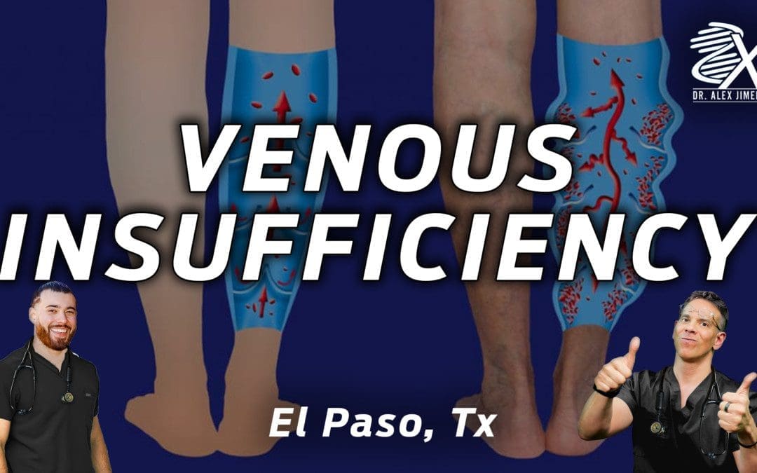
Dr. Jimenez, D.C., presents what you need to know about venous insufficiency. Many factors and lifestyle habits cause an effect on our bodies, which can lead to chronic disorders that can impact our musculoskeletal system and potentially lead to pain-like symptoms associated with chronic conditions. In this presentation, we will look at what venous insufficiency is, the symptoms, and how to prevent venous insufficiency from affecting the lower extremities. We mention our patients to certified medical providers that provide available therapy treatments for individuals suffering from chronic conditions associated with Lyme disease. We encourage each patient when it is appropriate by referring them to associated medical providers based on their diagnosis or needs. We understand and accept that education is a marvelous way when asking our providers crucial questions at the patient’s request and acknowledgment. Dr. Alex Jimenez, D.C., uses this information as an educational service. Disclaimer
What Is The Venous System?
Dr. Alex Jimenez, D.C., presents: So we will go over tackling common cardiovascular problems and venous insufficiency. So let’s discuss this common complication in our practices: venous insufficiency and the functional medicine approach. So if you look at venous or blood flow, you look at the heart. The heart will pump blood to the arteries and the arterials, the arteries and arterials will pump to capillary beds, and venules will go to veins. Veins will then move the blood to the subclavian vein, and the lymph ducts will also drain in the subclavian vein.
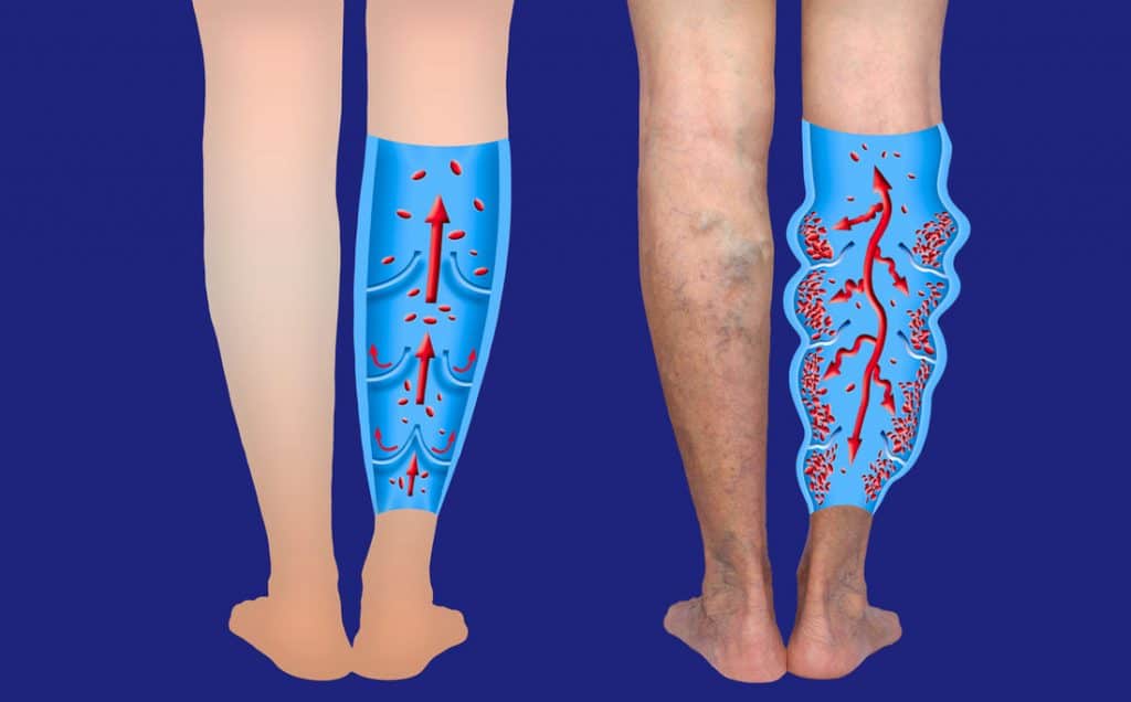
The subclavian vein will then go into the heart, and in the process, it continues and circulates. The big difference between veins and arteries is that arteries have muscles within them, and the muscles will contract, regulate blood pressure, and help keep the blood flowing. But veins do not have that luxury. Veins will depend on our skeletal muscles around them; if we contract them a lot, we’re helping with circulation. So, being active, moving around, and flexing our muscles will keep the pressure in the superficial system to about 20 to 30. And then, when it starts going to the deeper system with the valves, what happens is that the valves will stop blood from flowing back. So the blood can only go in one direction.
And that is basically to have a healthy venous system. You want to be often exercising, and you want to have that higher venous pressure and flow. So what is the pathophysiology of chronic venous insufficiency? You have incompetent valves, or you can have incompetent valves, you can have thrombosis, and you can have obstruction. And that can lead to elevated venous pressure. High venous pressure can lead to vein dilation, skin changes, and ulceration, but also elevated venous pressure can worsen incompetent valves, thrombosis, and obstruction. And then you get this vicious cycle, and usually, it’s the lower extremities; they get worse and worse. So if you want to look at the contributing factors, look at the functional medicine matrix. Venous insufficiency pathogenesis hits many places on the functional medicine matrix, multiple places we can look at in the lower body extremities.
Venous Insufficiency & Its Signs
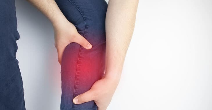
Dr. Alex Jimenez, D.C., presents: So what are the clinical manifestations of venous insufficiency? The symptoms are limb itching, heaviness, fatigue, particularly in the legs, pain in the legs, swelling, and tightness. The skin can get dry and become irritated. You might not be dealing with autoimmunity if you have this dry, irritated skin. You might be dealing with venous insufficiency. They can get muscle cramps. So your muscle cramps might not be a magnesium deficiency. Your muscle cramps might be venous insufficiency pain worse when standing or seated with their legs dangling. So when you’re sitting, the legs are dangling, and the pain improves when you elevate your legs and walk. And that actually can differentiate from arterial insufficiency. Remember, you get claudication in peripheral artery disease and arterial insufficiency. That’s when you walk and exert yourself. And because the blood vessels going to the muscles and the legs are tighter because of atherosclerosis, you get pain from walking.
Whereas venous insufficiency is the other side of the system, you walk and start feeling better. Why? Because those muscles are pumping the veins and moving blood through instead of the blood just being stagnant and sitting there. So edema you can get, which is swelling. Stasis dermatitis, which is dermatitis, red and swelling, and inflamed varicose veins, can be seen in this picture. Now the diagnosis is usually made by clinical signs and symptoms. So the clinical signs, what are the signs to look out for? For this part, go to your favorite search engine and look up every one of these symptoms that we mentioned so you know what it looks like. We are sure you’ve seen it before, but remind yourself what these things look like so that it can help you; it can help you when you’re diagnosing and looking at your patients.
Lymphodematosclerosis
Dr. Alex Jimenez, D.C., presents: Suppose a person has varicose veins. You can have lymphodematosclerosis, which is the champagne bottle sign. When you search that, look at that and see how the leg will look like an upside-down champagne bottle. Why? Because there’s a lot of fibrosis and hard tissue, and that tissue is holding that blood. You can’t get much edema, and you can’t get much swelling because it’s so tight, the blood can’t move in there. So look up the champagne bottle, not just the regular one, but look up a champagne bottle or lymphodematosclerosis, and you will remember that image when you see it. Then you will remember that image. You can get ulcerations because there’s decreased blood movement. So you get ulcers, and you can get hyperpigmentation. We see this often when you have a darkened skin color in the lower extremities from the constant fluid or blood leaking.
That’s hemosiderin deposits or iron deposits from popping blood cells. And you can get skin atrophy. So by typing these clinical signs on the internet that correlate with venous insufficiency, you have a good visual of what these things look like. So what is the functional medicine treatment plan? We’re going to look at the risk factors of chronic venous insufficiency, and we’re going to look at the adaptable ones, and based on that, we can give patients recommendations and plans. So obesity works on decreasing fat, sedentary life, being active, checking estrogen and hormone levels, and reducing estrogen and increasing progesterone. If you have to get out of that estrogen dominance, we want to look at those risk factors, see which ones are adjustable, and start working with them.
Ways To Reduce Venous Insufficiency
Dr. Alex Jimenez, D.C., presents: So you have this person with venous insufficiency. Check on their obesity levels, so you work on lowering their body fat and see if they have a sedentary lifestyle and getting them moving high. Check their hormone levels and see where their estrogen levels are regulated. If you check the IFM hormone module, check it out because it has some really good information on how to balance hormones in a functional medicine way. Make sure that they’re standing for a short period. At least occasionally, have them walk around, and you can have them set a timer. So every so often, every 20, 30 minutes, they walk around to keep their legs and blood flow moving. Work on decreasing smoking. And mentioning these risk factors to the patient can make them aware that this can worsen their venous insufficiency. Other conservative therapies include leg elevation. So have them lay down by putting their legs up to allow gravity to help push the blood down. Compression therapy. So have them wear compression stockings and stasis dermatitis; sometimes, you must use topical dermatologic steroids and some of those agents, which can be helpful there.
You may consider earthing. There was a research study that showed that if you put your feet on the ground outside barefoot, not in the insulated houses, then what can happen is, is the viscosity of your red blood cells will decrease. So the red blood cells will clump less, and you can have better movement and circulation. Pharmacological therapies and supplementations to target venous insufficiency. So what can we do while we’re looking at doing two things? We want the venous tone to be improved. So you want to tighten those veins up. On the arteries, you want to loosen them up. Usually, when an individual has hypertension, we want the veins to tighten those bad boys up so that circulation can happen. And then you want to improve the flow. You want the blood to be able to flow through the veins better.
Supplements For Venous Tone
Dr. Alex Jimenez, D.C., presents: So let’s take a look at the venous tone. This is one of the places where we’re ahead of the game in functional and integrative medicine because if you look at the conventional literature, even up-to-date research, many people are using up-to-date now to see how often they diagnose weak venous tone. So we can take a look at that. But if you look at what you can do for venous tone? It has two supplements. Regarding venous tone and increasing venous tone, two supplements can support the venous system: horse-chestnut seed extract (Escin) and diosmin.
So those are the two things that are mentioned. And we, in functional and integrative medicine, are more prepared to deal with this because we know about pharmacy grade; we learn about giving them a good product that is third-party tested and doesn’t have those toxic fillers and whatnot. The second way of treating venous insufficiency from a medical point of view is by improving venous flow. You want blood viscosity to be thinner. You don’t want the blood not to be as prone to clotting so the blood can flow easier. So here are some agents you can use. You can use aspirin; you can use pentoxifying; you can use nattokinase, which can help lower fibrinogen. Regarding venous insufficiency, it can cause the body to have high fibrinogen. So nattokinase can help lower elevated fibrinogen.
Conclusion
Dr. Alex Jimenez, D.C., presents: If they’re not on aspirin or any blood thinners and have high fibrinogen and venous insufficiency, it might also be a good one to put somebody on omega-3s. We are trying to get their omega-3 levels up, and they are useful when optimizing help with venous flow. You’re going to have people to come and see you, and you’re going to be treating them for other things. And because you’re functional medicine, you’re part of the cool club; what’s going to happen is they’re not even going to tell you about their venous insufficiency, and it’s going to get better just because of the treatments that you’re doing. And it will be epic. And if all else fails, you refer to associated medical specialists to help your patient. So, in conclusion, take care of your veins and look for the signs to prevent venous insufficiency from causing more issues in the lower extremities, and utilize vitamins and supplements to reduce the pain and inflammation in the muscles and joints.
Dr. Jimenez, D.C., presents how implementing different strategies for patients to incorporate exercise in their health and wellness journey in this 2-part series. Many factors and lifestyle habits tend to take over our daily lives, leading to chronic disorders that can impact our bodies and cause many unwanted symptoms. In this presentation, we will look at different strategies and options to incorporate into our patients regarding health and wellness. Part 1 looks at how to implement exercise in a clinical setting. We mention our patients to certified medical providers that provide available therapy treatments for individuals suffering from chronic conditions associated with Lyme disease. We encourage each patient when it is appropriate by referring them to associated medical providers based on their diagnosis or needs. We understand and accept that education is a marvelous way when asking our providers’ crucial questions at the patient’s request and acknowledgment. Dr. Alex Jimenez, D.C., uses this information as an educational service. Disclaimer
Different Strategies For Patients
Part 1 in the last presentation mentioned what to do when examining patients. We said how to implement different strategies to incorporate exercise into a daily routine for many individuals who want to kickstart their health and wellness journey. By coming up with a plan, many doctors can help their patients develop a personalized plan to cater to the individual; it can allow both the patient and doctor to see what works and what doesn’t. Part 1 also explains how to delegate with the patients to help ease them into implementing exercise as part of their daily routine. Delegation is described as a transfer of responsibility for the performance of the patient’s care while retaining accountability for the outcomes. The main point here is you are delegating the educational process related to the exercise prescription. You can use it for the diet prescription, or you can use it for anything that tends to be educational and formatted for your patients.
Based on the documentation complexity, we would ensure a face-to-face encounter with the patient to meet the legal requirement for insurance to bill it as a 99-213 or a 99-214. So what we do with our health coaches is we also want to have them do other cross-trained roles in our office because we’re a small little practice. So, our health coaches are involved with our patients and know how to assess if an interested new patient would be a good candidate for our services. They are great at using the technology we do with some of our new patients, whether it’s a BIA or if we prescribe heart math. So they are great with technology and with education around nutrition, exercise, whatever you can train your health coach to do, then you can create a way to delegate for her to do it, whether it’s through insurance or cash.
Okay, now last but certainly not least, it is so important to know, and you know this if you have children or you know if you have a family member, which we know you do that what you say and what you do are two different things. So there are studies that show an association that if a provider is exercising or implementing a journey of improving their exercise and diet, it shows up more in their recommendations. And when a provider talks about it authentically during a motivational interviewing process with a patient, it’s obvious to the patient that it’s important to the provider because they’re not just talking the talk; they’re walking the walk, which is important for all of us. We are patients as well. To consider that one of the best ways to start an exercise prescription program and your office is to do one for yourself.
Creating a Workout Environement
Walk yourself through it and see the little bumps and aspects of the journey so you can speak authentically and start that office workout challenge in your own office. And we did that in our office, and we noticed that people would be coming in, and some people would be doing desk pushups, and they were like, “What are you doing?” and we would respond, “We’re just getting our desk pushups in. Hold on for a second; I’ll be right with you.” Or somebody comes in, and we’re doing squats and conversing about a patient. It sounds humorous, but they know that we mean business when we say let’s do an exercise prescription. So remember that for patients learning things is lovely, but it doesn’t change outcomes; doing things changes results and your behavior matters.
We hope you have found this portion of our day-to-day useful. We are excited to see that knowing that exercise is an underutilized tool in our armamentarium for optimizing our patients’ lives. So we will continue discussing our strategies for implementing activity in our practices. How do we incorporate exercise into our patients?
It can start as simple as asking them about their movement, seeing what they enjoy doing when it comes to exercise, and creating something slow. Just five to 10 minutes commit, saying, “Okay, well, if you like walking, could you walk for 10 minutes daily? Please ensure you track and return in two to three weeks, and we’ll review that?” And then, from there, sometimes, the providers will give them a cardiovascular prescription. We’ll provide them with resistance training and a stretch prescription. But the cool thing is that we can reiterate it by saying. “You should see one of our health coaches and one of our educators in two to three weeks so they can go over a stretch program, a resistance program, or figure out what exercise would be best for you.” We’ll use some of our tools and do the bioimpedance test to check the percent fat, percent water, and connective muscle tissue that looks at the phase angle. The phase angle is how strong the cell’s repellent electricity and the higher their phase angle, the better they would do with chronic diseases and cancer. We encourage improving this phase angle, improving hydration, and showing them the difference between weight and fat. There’s a big difference between the two.
Delegating & Functional Medicine
We also delegate with the health coaches as we develop a personalized treatment plan for the patients, and we can do it in two different ways. So one option is to bill for chronic care management. What this means is that, say, if the patient has a chronic disorder affecting their daily activities? Our health coaches can call them on their phones and discuss their plans. The second option is an office visit, allowing the patient to converse with the health coach and review their personalized program.
So incorporating these two options into your patients allows many doctors to gather all the information, assess the situation, and discuss the plan with the patients to improve or kickstart their health and wellness journey. When it comes to implementing exercise as part of the health and wellness journey for the patients, we are the leverage group to incorporate exercise as part of the treatment. Working with health coaches, nutritionists, personal trainers, and physical therapists who deliver different exercise routines to the patient’s needs is part of the journey. How does this apply to individuals with joint and mobility issues associated with autoimmune disorders like arthritic diseases?
So anybody with arthritic diseases or a chronic illness, we prefer them very actively a physical therapist who has a whole program for people with autoimmune disease and its correlating symptoms that have overlapping risk profiles. We also have a referral program for water aerobics and low-impact programs to reduce pain-like symptoms. So getting people up and moving is key. Movement is key.
Another strategy is implementing functional medicine combined with exercise. Functional medicine allows doctors and patients to determine where the problem is in the body. Functional medicine also works with associated referred medical providers to develop a treatment plan for the patient and help create a relationship between both the doctor and the patient. So making these nice little allies on the outside for the things you don’t want to or can’t do is an amazing tool with exercise. Or it could be with nutrition, or it could be with stress management. It’s the same thing with lifestyle. Do either do it in-house or out? The choice is up to you.
And so, what are these static things that we often think are static that we do every day that we can begin to incorporate stretching to activate our parasympathetic nervous system? Incorporating non-exercise activity thermogenesis into your life. And that’s something all of us in a stressful life could use a little more. And when you integrate it into your life, it’s top of mind so that you’re sitting there with your patient and thinking, “How can I encourage them?” By relating to the patient, you can show them tips or tricks to incorporate into their personalized treatment plan.
Motivational Interviewing
The goal is to use motivational interviewing and the aspects of motivational interviewing not to convince them to exercise but to understand their resistance to roll with it. Many individuals work two jobs, so telling them to exercise will not make them stop everything and start working out by relating and asking for the right questions like, “So you’re trying to get off of this blood pressure medication, and I love that you’re committed to that. So what other things can you see, or is there any part of the exercise or physical activity that you could consider that could keep you moving towards your goal of getting off this medicine?”
Helping people see that they have this time limitation. We acknowledge and roll with their resistance but then give them the discrimination to say, “Yeah, and you’re here because you want to get healthy. And I must tell you, exercise is one of the big levers. So if you do nothing, you will keep getting what you’re getting. So what can we do? Does anything else come to your mind as a solution?” We can’t tell you how much it improves things when you have the patient be the person who comes up with the idea of what to do next versus feeling the burden of having to be the one who psychically knows what this patient’s going to do. Plus, it gets exhausting trying to anticipate the right answer for the patient.
By letting the patients be accountable for their actions and their treatment, it is important to have that communication with them and see how they keep themselves motivated through their exercise regime, whether they are eating the right amount of healthy foods, going to therapy treatments, and are they taking their supplements? You will go back and forth with their choices and offer suggestions because it doesn’t apply to exercise, but exercise is the one that people will sometimes completely believe in but will resist. They’re more likely to take on a diet sometimes than they are to take on exercise. So you can apply these principles to anything like taking supplements, taking a shake, taking the diet, whatever happens, to be their resistance point in a functional medicine treatment plan. You can use these things. Sometimes, we have to consider that that might help a patient.
Conclusion
These are your go-to suggestions, but the patients get to pick a time and are in the control seat instead of you telling them because this will provide resistance to their treatment plans and cause them to not commit to their health and wellness journey. But relating to them, offering suggestions, and constantly communicating with them allows the individual to try different things that will work with them and can show massive positive results in their health and wellness journey.
Dr. Jimenez, D.C., presents how to implement exercise as part of your daily routine. Many factors and lifestyle habits tend to take over our daily lives, and in this 2-part series, we will look at how to implement exercise in a clinical setting. Part 2 will continue the presentation. We mention our patients to certified medical providers that provide available therapy treatments for individuals suffering from chronic conditions associated with Lyme disease. We encourage each patient when it is appropriate by referring them to associated medical providers based on their diagnosis or needs. We understand and accept that education is a marvelous way when asking our providers’ crucial questions at the patient’s request and acknowledgment. Dr. Alex Jimenez, D.C., uses this information as an educational service. Disclaimer
How To Implement Strategies?
Dr. Alex Jimenez, D.C., presents: Today we will discuss how to implement strategies using exercise as a prescription. Remember, just like we talked about how a healthy diet full of nutritious, whole foods can be used as a prescription, we want this science to make it to the patient and create outcomes because otherwise, this is just a bunch of things you know and not something that you know how to put into practice. So we’ve listened; we know that’s what you’re up to, so let’s get started. We will discuss some general aspects of implementing exercise as a prescription and some ideas we use in our practice. And then, of course, share the brilliant ideas with some of the other colleagues who also are figuring out ways to make this work in their practice. The first thing we want to share with you is when you’re approaching a patient with an exercise prescription, assuming the patient’s interested, you should ascertain first how this person is motivated.
Because it always makes sense to ride their motivation wave than to come from the standpoint that this is what I want from you, and this is why you need to do it. The first thing we want to put out there is that you want to ensure that this patient has a reason to want to exercise. So it’s less about a doctor’s orders or a provider’s recommendation, and you want to partner with our patients therapeutically, which means understanding their motivation. So for most people, there are two ways we can reinforce the outcome of a positive implementation of the exercise. First, we want to optimize those factors related to one-on-one communication with our patients. And then, number two, optimize the environment in our practice for success. Okay, so we’ll go over these things in detail now.
It only sometimes works if we give them a prescription and assume they want to do it. So if Joan Rivers was your patient in the past, this might have been her reason for not wanting to exercise, and you must be able to roll with it. Let’s talk about how we can do that. This works with patients, spouses, and children; it is wise to persuade people to do things and make them think it is their idea. So, with much bigger goals in mind, Nelson Mandela used the same principle. So we want you to think about who you are working with and who you are partnering with; these are some common functional medicine personas that you may come across, especially if you’re in more of a private practice, whether it’s cash or membership type of practice, you might see this persona in people.
Look For The Personas
Dr. Alex Jimenez, D.C., presents: Are these all personas the same? Not necessarily, as people have different reasons to exercise. For example, say you have a chronically ill individual who needs their hands to be held or have individuals who read many fitness magazines following these leaders through a whole lifestyle lens. And the way you engage with each of these personas is based on their goal for exercise. So, the unwell individual may have different goals, challenges, or limitations than the lifestyle lens individual. So make sure you know who you are working with, and if you need more clarification, have a conversation with them to find out.
Let’s say you’ve gotten through that step, and now you’re in the actual conversation of, “Hey, let’s figure out how to get this exercise thing to create benefits in your life.” As you’re having the conversation, you might learn to use some aspects of motivational interviewing. So rolling with resistance, for example, sometimes people say, “Nope, I don’t want to exercise.” So in this example, you might say, “Okay, if you don’t want to exercise at a gym, what other options have you heard of that you might want to consider?” Let’s say that’s how you opened it up and remember that there’s always a way to roll with the resistance, and it’s focused on acknowledging the patient’s input. You’re responding to them by saying, “Okay, fine. You don’t want to work at a gym. I get that,” while expressing empathy. Many individuals have tried to work at a gym, and the machines tend to injure them when used incorrectly, intimidate them, or the equipment is not made for their size structure.
Emphasize With Your Patients
Dr. Alex Jimenez, D.C., presents: Many people want to avoid exercising; this is one of the many frustrating things because you feel the equipment needs to be made for you. So notice that you can empathize without judging and then roll with resistance and ensure they understand that you acknowledge their input about the situation. These things are common sense to you. Many of us may not employ these to the fullest potential to motivate our patients to implement exercise as part of their daily routines. The important and obvious thing is to refrain from arguing with your patient. Because all that will go to create for most people is more resistance, so if they say, “Hey, I don’t want to exercise right now,” you can say, “Would you be willing to talk about exercising as a goal in the future?”
And if they say, “Yeah, I need to make it through December,” you can reply with, “Okay, great, let’s have you follow up with me in January. Does that work for you?” So again, avoiding arguing and expressing empathy can put people’s minds at ease and prevent resistance. Another factor that many people often do when it comes to implementing exercise as part of their routine is by developing discrepancy. So sometimes, people say things that conflict with the daily habits that they already follow. So they might say, “Yeah, I want to exercise because I don’t want to take a statin medication, but I don’t have time to exercise.” So this is where you help them understand like you recognize that exercise is one of the key ways to reduce your need for a statin medication. And you get that if we leave this cholesterol the way it is, it will cause more risks for your patients. But at the same time, time is a factor. So you come up with some ideas to benefit your patients and incorporate exercise as a routine.
Develop A Plan
Dr. Alex Jimenez, D.C., presents: Remember that you don’t have to solve everything for someone. You could put things out like developing discrepancies for the patient and then let the patient generate solutions that work. So also support self-efficacy. This means that we are not going to change the behavior. The patient is the one who has to change the behavior, and their understanding of their capacity to change their behavior is essential. So whatever you can do to point out the positives, acknowledge whatever they’ve done, even if it’s like, “Hey, it’s wonderful that you bought sneakers. I understand that you didn’t do anything we discussed; life happened. I want to acknowledge you for getting the sneakers because that makes it much easier to start the plan now.” So support self-efficacy whenever possible. Now other more tangible obstacles keep someone from wanting to implement exercise.
Many times it’s either on a mental or physical plane. So here are some solutions that we’ve listed for some of the common mental obstacles we’ve seen. Some people don’t want to be out in public because of concerns about body image. So, they can often go to a special kind of gym if they want to go to a gym, or they can do at-home videos or a personal trainer. Sometimes it gets boring, and they would often moan and groan about it when they are exercising; however, if they are doing fun exercises like dancing or swimming, they will become more motivated and start to change their exercise regime throughout the week. You could do these things despite needing more knowledge or confidence about doing it correctly or on time.
Incorporate A Trainer Or A Health Coach
Dr. Alex Jimenez, D.C., presents: That’s when you might want to bring in a health coach or personal trainer, and with physical obstacles which may be related to a person hasn’t been exercising for a long time and assuming that you’ve cleared them to be able to initiate an exercise plan, maybe there are ways that you can say, “Okay listen, I want you to walk at a low intensity to start with, and you know, over the next month I’d like you to build up two 5,000 steps a day.” This can be a routine set for three days a week, four days a week, or whatever you decide with them and does that work for the patient. That might be one way to work on physical or perceived physical limitations. And then there may be people who have real-time constraints. So the two ways to handle this; is to optimize NEAT or HIIT workouts.
These can be simple activities we do throughout the day, like taking the stairs, parking further away, walking during your lunch break, and having walking appointments and meetings. While watching TV in the evening, you could pump some free weights in your bedroom or your living room. Or if they are more avid exercisers and are open to taking on some HIIT training, that could be a way to get some concentrated cardio and strength training signals in the body. Next, we want to discuss the different scenarios we may have regarding our office structures that support implementing exercise. A common scenario would be that you need a dedicated person in-house to help people implement the exercise prescription.
Use Resources
Dr. Alex Jimenez, D.C., presents: Okay, so if you are the provider, health coach, and personal trainer, we want you to consider using resources. You must recognize your boundaries in terms of not being able to be everything to everyone but using your resources effectively. Because we can’t create boundaries that are so tight that you’re not making the type of office that you want, meaning one that incorporates exercise prescriptions. So we’re going to talk about an office workout and exercise grid and how we will work with the local community, personal trainers, and gyms to refer out. And we have trained them to look at our exercise prescription as a guideline even though we are not legally partnered with them. They use these prescriptions as a way of communicating what our goals are. Here are some tools that we use that we are going to share out.
And then, especially in certain times like we’re having right now, we also referred to online resources. So this office workout prescription was created by our team, and we handed out this resource to our patients. We encourage them to find a buddy in their office or home because it is generally more fun. There are data to suggest that when you exercise in a social format, Like participating in team sports, it creates more benefits than doing an individual sport or being at the gym with your AirPods centered only on yourself. So there is this association where having a social element to your exercise regimen increases the benefits. Set up reminders on your phone when you’re at the office to do these hourly five-minute exercises.
And then we also have an online link where our trainers and health coaches demonstrate proper form and modifications for these office workouts. And then, of course, once you give any resource, whether it’s this office workout prescription or any other help, determine with the patient what we want to do about this. We don’t want to give out this prescription and say we hope it works. The main question is that do you want to have accountability? “Hey, can you come back to see us in a month, and let’s see where you are with it?” Or, “Hey, can you consider taking it to this next level after a month if you feel good and come back to see us in two months?” Or, “Hey, once you’re done with this, why don’t we talk in two months to recheck your lipids and know if you made a bump in your LDL particle number so that we can lower the dose of your statin or get you off the statin.”
So we don’t recommend just doing the exercise prescription and leaving it open-ended in terms of follow-up; make it like any other prescription; if you were to put someone on a statin, you would follow up with them. So just like that, you would follow up with someone you prescribe an exercise prescription. Again, it’s really practical. It can be done whether you work in an office, a home office, or you don’t work at an office but work in the house. So it’s in your IFM toolkit. And it has a Monday through Friday, an eight-to-five grid of what you do throughout the week. So it diversifies exercises and makes it, so all your muscle groups are incorporated using the stuff you have in an office or a typical home.
Delegate With Your Patients
Dr. Alex Jimenez, D.C., presents: So it is beautiful for the “I don’t know what to do” people, and it’s a great start for sedentary people. Then you can also consider any technology that is of interest to you. Here are some that our health coach and personal trainer have suggested based on what the patient’s goals are. They may be trying to run a 5k, then find an app that might work for them there. Or they may incorporate yoga to work on their mind-body access or flexibility. You can personalize it to the type of workout if they’re interested in HIIT, yoga, or Pilates. Again, find technologies you enjoy, and check them out yourself. Or you can make a little cheat sheet that can be given out or put as a template. Here’s something important that we want you to consider if you still need to do it.
It’s called delegation. This can not be done alone; this is a group effort to allow the individual to have a team to back them up and help improve their health and wellness journey. Now, this is done in healthcare all over the place. For respiratory therapists, many people will do delegated work from the healthcare provider. So it’s just a transfer of responsibility for the performance of patient care. Now, remember that it’s still done under the provider’s responsibility. You should consider that different states and insurance contracts may have little nuances on how they would want you to do delegation. Still, we know habits have changed, and we need help to keep up with them to meet the requirement.
So how would we delegate a patient? We would go through a thorough examination, like taking their BMIS/BIAs with the Inbody Machine, and then go through a series of functional medicine tests to determine what issues or overlapping risk profiles are affecting them. Then the doctor and their associated medical providers will develop a personalized treatment plan for that patient that incorporates a healthy diet and exercise regime for them to follow.
Conclusion
Dr. Alex Jimenez, D.C., presents: Making these small changes is beneficial in the long haul regarding a person’s health and wellness journey. It may take a while to get accustomed to the routine, and sometimes it can be frustrating. However, finding what works and doesn’t work with the patient and making these changes can result in a better solution that benefits the person.
IFM's Find A Practitioner tool is the largest referral network in Functional Medicine, created to help patients locate Functional Medicine practitioners anywhere in the world. IFM Certified Practitioners are listed first in the search results, given their extensive education in Functional Medicine