El Paso, TX. Chiropractor, Dr. Alexander Jimenez discusses the anatomy of nerve fibers, receptors, spinal tracts and brain pathways. Regions of the Central Nervous System (CNS) coordinate various somatic processes using sensory inputs and motor outputs of peripheral nerves. Important areas of the CNS that play a role in somatic processes are separated in the spinal cord brain stem. Sensory pathways that carry peripheral sensations to the brain are referred to an ascending pathway, or tract. Various sensory modalities follow specific pathways through the CNS. Somatosensory stimuli activate receptors in the skin, muscles, tendons, and joints throughout the entire body. The somatosensory pathways are divided into two separate systems based on the location of the receptor neurons. Somatosensory stimuli from below the neck run along the sensory pathways of the spinal cord, and the somatosensory stimuli from the head and neck travel through cranial nerves.
ANATOMY OF RECEPTORS, NERVE FIBERS, SPINAL CORD TRACTS AND BRAINSTEM PATHWAYS
RECEPTORS AND RECEPTOR BASED THERAPY
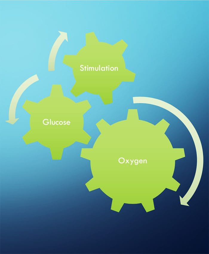
NEURONS NEED THREE THINGS TO SURVIVE!
FUNCTIONAL NEUROLOGY KEY CONCEPTS
The cell needs three things to survive.
Oxygen, glucose and stimulation.
Stimulation = Chiropractic, exercise, etc.
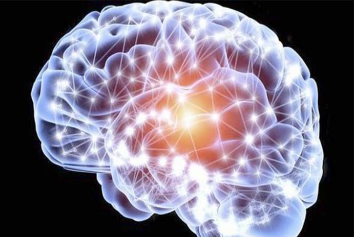
Stimulation leads to neuronal growth
Neuronal growth leads to plasticity
Subluxations alter the frequency of firing of neurons
Activation of one side will stimulate ipsilateral cerebellum and contralateral cortex (usually)
Proper stimulation CAN reduce pain.
CHIROPRACTIC IS RECEPTOR-BASED THERAPY
INTRODUCTION
The ongoing activity and output of the CNS are greatly influenced, and sometimes more or less determined, by incoming sensory information.
The basis of this incoming sensory information is an array of sensory receptors, cells that detect various stimuli and produce receptor potentials in response, often with astonishing effectiveness.
The health of the neuron, however, plays a huge role in how neurons can produce receptor potentials, the endurance of the neuron and the ability to create plasticity.
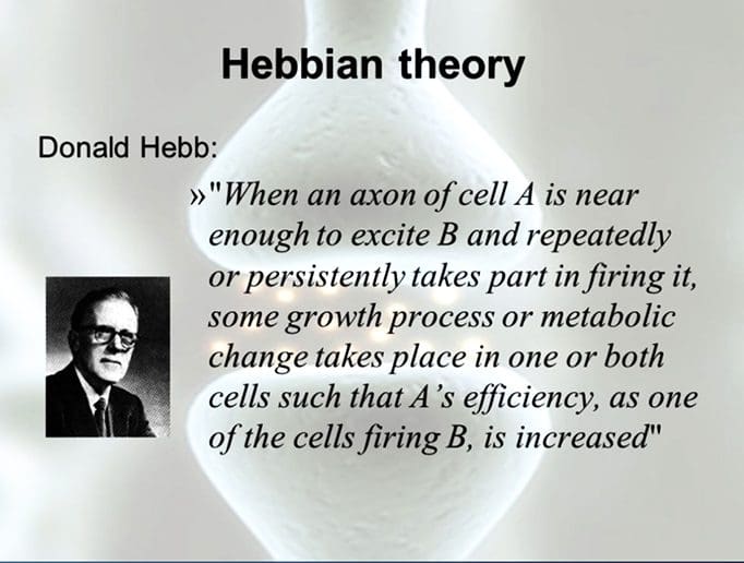
�Neurons that fire together, wire together.� Hebbian Theory
TYPES OF RECEPTORS
Chemoreceptors
Smell, taste, interoceptors
Thermoreceptors
Temperature
Mechanoreceptors
Cutaneous receptors for touch, auditory, vestibular, proprioceptors
Nociceptors
Pain
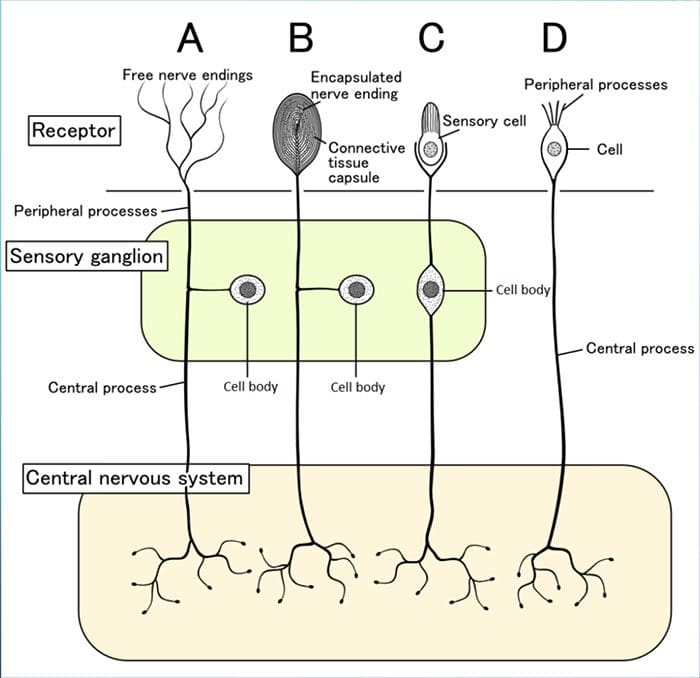
PARTS OF RECEPTORS
Although their morphologies vary widely, all receptors have three general parts:
1. Receptive Area 2. Area Rich In Mitochondria
Health of the neurons within the receptors will determine its response to stimulation
3. Synaptic Area To Pass Messages To The CNS
RECEPTIVE FIELDS
These are particular areas in the periphery where application of an adequate stimulus causes the receptors to respond.
Neurons in successive levels of sensory pathways (second- order neurons, thalamic and cortical neurons-also have receptive fields, although they may be considerably more elaborate than those of the receptors.
TRANSDUCTION
Sensory receptors use ionotropic and metabotropic mechanisms to produce receptor potentials
Sensory receptors transduce some physical stimulus into an electrical signal � a receptor potential � that the nervous system can understand.
Sensory receptors are similar to postsynaptic membranes as their adequate stimuli are analogous to neurotransmitters.
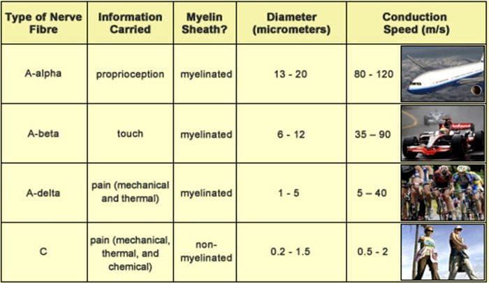
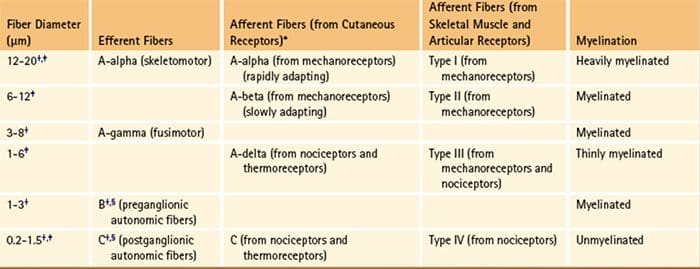
THE DIAMETER OF A NERVE FIBER IS CORRELATED WITH ITS FUNCTION
BIGGER = FASTER
Larger fibers conduct action potentials faster than do smaller fibers.
A? fibers are the largest and most rapidly conducting myelinated fibers.
The slowest conducting fibers of the body are the C fibers
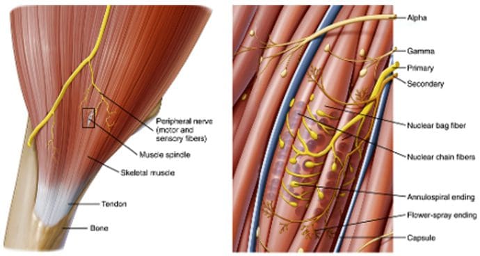
RECEPTORS IN MUSCLES AND JOINTS DETECT MUSCLE STATUS AND LIMB POSITION
MUSCLE SPINDLES
Muscle spindles (Fig. 9-14) are long, thin stretch receptors scattered throughout virtually every striated muscle in the body.
These muscle spindles sense muscle length and proprioception (�one�s own� perception).
They are quite simple in principle, consisting of a few small muscle fibers with a capsule surrounding the middle third of the fibers.
These fibers are called intrafusal muscle fibers (fusus is Latin for �spindle,� so intrafusal means �inside the spindle�), incontrast to the ordinary extrafusal muscle fibers (�outside the spindle�).
The ends of the intrafusal fibers are attached to extrafusal fibers, so whenever the muscle is stretched, the intrafusal fibers are also stretched.
The central region of each intrafusal fiber has few myofilaments and is noncontractile, but it does have one or more sensory endings applied to it.
When the muscle is stretched, the central part of the intrafusal fiber is stretched, mechanically sensitive channels are distorted, the resulting receptor potential spreads to a nearby trigger zone, and a train of impulses ensues at each sensory ending.
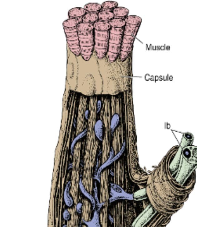
GOLGI TENDON ORGANS
Golgi tendon organs are spindle-shaped receptors found at the�junctions between muscles and tendons. They are similar to Ruffini endings in their basic organization, consisting of interwoven collagen bundles surrounded by a thin capsule (Fig. 9-16).
Large sensory fibers enter the capsule and branch into fine processes that are inserted among the collagen bundles. Tension on the capsule along its long axis squeezes these fine processes, and the resulting distortion stimulates them.
If tension is generated in a tendon by making its attached muscle contract, tendon organs are found to be much more�sensitive and can actually respond to the contraction of just a few muscle fibers.
Thus Golgi tendon organs very specifically monitor the tension generated by muscle contraction and come into play whe
n fine adjustments in muscle tension need to be made (e.g., when handling a raw egg).
�
Thus the mode of action of Golgi tendon organs is quite different from that of muscle spindles (Fig. 9-17). If a muscle�contracts isometrically, tension is generated across its tendons, and the tendon organs signal this; however, the muscle spindles signal nothing because muscle length has not changed (assuming that the activity of the gamma motor neurons remains unchanged).
In contrast, a relaxed muscle can be stretched easily, and the muscle spindles fire; the tendon organs, however, experience little tension and remain silent. A muscle, by virtue of these two types of receptors, can have its length and tension monitored simultaneously.
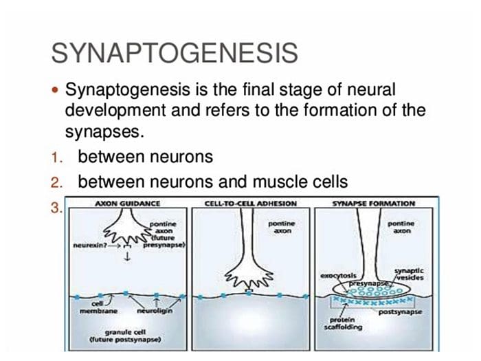
Neurons are believed to establish neuronal connections through innate predetermined programs during the developmental process of the brain. It’s also believed that neurons gravitate to areas of attraction and move away from areas of repulsion in a theory known as the chemoaffinity hypothesis. The Chemoaffinity hypothesis claims that neurons first make connections with their targets based on interactions with specific molecular markers and, therefore, that the first wiring diagram of an organism is indirectly determined by its genotype.
These markers are created during cellular differentiation and aid not just with synaptogenesis, but also act as guidance cues for their individual axon. The development of the mature nervous system formations demands axons to navigate to their correct targets in order to establish neuronal connections or synaptic connections. Growing axons create highly motile structures, known as growth cones, which direct the axon to its goal. They do it by responding to specific guidance molecules that either attract or repel the growth cone.
The Theory of Neuronal Connections
The concept that axons are directed principally by molecular determinants, rather than mechanical determinants, such as cells, extracellular material and other neurons, was established by Roger Wolcott Sperry, a neuropsychologist and neurobiologist, in 1963. However, it was not until the discovery of guidance molecules including netrins, semaphorins, ephrins and Slits, that Sperry’s chemoaffinity hypothesis became widely recognized as a prevalent mechanism for guidance of not only axons, but of all cells.
In 1981, Roger Sperry received the Nobel Prize for Physiology or Medicine for his discoveries concerning the functional specialization of the cerebral hemispheres. He performed studies on patients with epilepsy in whom the corpus callosum, or the bundle of axons fibers which connects the two brain hemispheres, was severed to stop seizures. A number of tests and evaluations revealed the way both brain hemispheres hold independent streams of conscious awareness, perceptions, thoughts and memories, and most fundamentally, that neuronal connections are formed and preserved with a high degree of precision.
Having demonstrated that the institution of specific neuronal connections is fundamental to the overall function of the brain, Sperry turned to look at how these connections are created, and used his chemoaffinity theory to describe how axons find the right target during the development of the brain. Others had raised the possibility that compound determinants might function in axon guidance, but it was Sperry that supplied the direct histological evidence and proposed the chemoaffinity hypothesis for axon guidance.
Roger Sperry and his Chemoaffinity Hypothesis
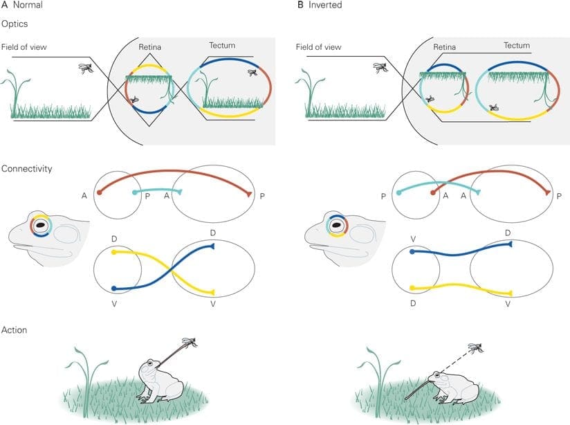
Roger Wolcott Sperry pioneered the inception of the Chemoaffinity Hypothesis after the 1960’s in a series of elegant experiments employing the retinotectal system of the African Clawed Frog, he sectioned the optic nerves and steered the eyes 180 degrees. He wished to know whether vision would be ordinary following regeneration or if the animal would eternally see the world ‘upside down’. If the latter held true, this could reveal that the nerves were somehow guided back to their original sites of termination; however, restoration of normal eyesight would mean that the nerves had resumed at new sites. Sperry showed that these creatures did indeed view the world ‘upside down’.
According to the experiment, initial�eye orientation gives that the top of the eye is Dorsal, and the underside is Ventral. Post-operation, the “top” of the eye is now Ventral, and the base is Dorsal. After a food source was supplied, the frog extended its tongue, meaning that the Dorsal-Ventral orientation of the eye still remained. In follow up experiments, the eye was dispersed and rotated 180� and the optic nerve was also cut to determine if this could affect the Dorsal-Ventral orientation. The results were identical. It was those studies which directed Sperry to suggest that complicated chemical codes, under genetic control, direct axons to their targets, his chemoaffinity hypothesis.
In his first theory, Sperry proposed that distinct cells bear different cell-surface proteins that serve as markers, a notion that demanded an unsatisfactorily high variety of proteins. He revised his model suggesting that double gradients of guidance cues in the afferent and target areas would enable proper axon targeting. There is now extensive experimental data to support the chemoaffinity hypothesis, as well as the requirement for gradients of receptor and/or ligand, such as ephrins and Eph receptors, in the projection and target regions is well established.
Roger Wolcott Sperry concluded that every individual optic nerve and tectal neuron used some kind of chemical markers that dictated their connectivity through development. He concluded that if the eye had been rotated, each optic fiber and every tectal neuron possessed cytochemical labels that uniquely denoted their neuronal kind and place and that optic fibers may use these labels to navigate to their own matching target cell, hence the visuomotor impairment.�Although certain aspects and details about Sperry’s model are unproven or incorrect, the basic notion of this chemoaffinity hypothesis has become dogma in developmental neurobiology.
Dr. Alex Jimenez’s Insight
Over the years, the principle to understand the establishment of neuronal connections has continued throughout the field of neurophysiology as well as prenatal development of the brain. Neuronal connections are believed to be established during the migration of growth cones guided by extracellular guidance cue gradients. Although this theory has been revisited countless of times, Roger Sperry was the first to explain how axons navigate to their correct targets in his chemoaffinity hypothesis. Countless experimental and clinical data now exists to support the chemoaffinity hypothesis.
The scope of our information is limited to chiropractic as well as to spinal injuries and conditions. To discuss the subject matter, please feel free to ask Dr. Jimenez or contact us at 915-850-0900 .
Curated by Dr. Alex Jimenez
Additional Topics: Sciatica
Sciatica is medically referred to as a collection of symptoms, rather than a single injury and/or condition. Symptoms of sciatic nerve pain, or sciatica, can vary in frequency and intensity, however, it is most commonly described as a sudden, sharp (knife-like) or electrical pain that radiates from the low back down the buttocks, hips, thighs and legs into the foot. Other symptoms of sciatica may include, tingling or burning sensations, numbness and weakness along the length of the sciatic nerve. Sciatica most frequently affects individuals between the ages of 30 and 50 years. It may often develop as a result of the degeneration of the spine due to age, however, the compression and irritation of the sciatic nerve caused by a bulging or herniated disc, among other spinal health issues, may also cause sciatic nerve pain.
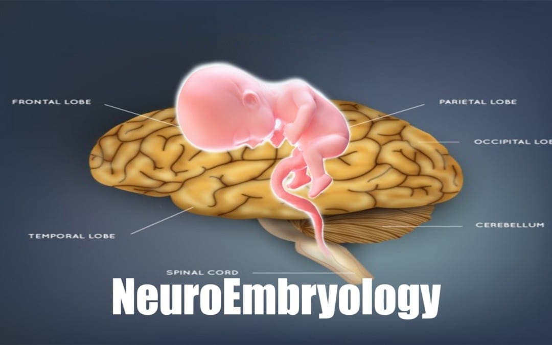
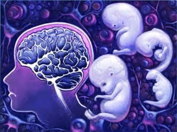
Neuroembryology: As an embryo all of the cells in the body are identical. But as time passes, some cells develop into neurons and others develop into skin, hair, or muscle cells.
Why do some cells become neurons? How do neurons become organized in the spinal cord and brain in order to allow us to walk, talk, see, recall life events, feel pain, keep balance, and think?
The answers will help us understand how we develop from an embryo into a full-grown person and how our body and brain constantly adapts, throughout our lives to the environments changes.
El Paso, TX. Chiropractor, Dr. Alexander Jimenez discusses neuroembryology, with the intent of educating patients, past and present, and the general public about the how the spine is connected to everything and where everything begins.
INTRODUCTION
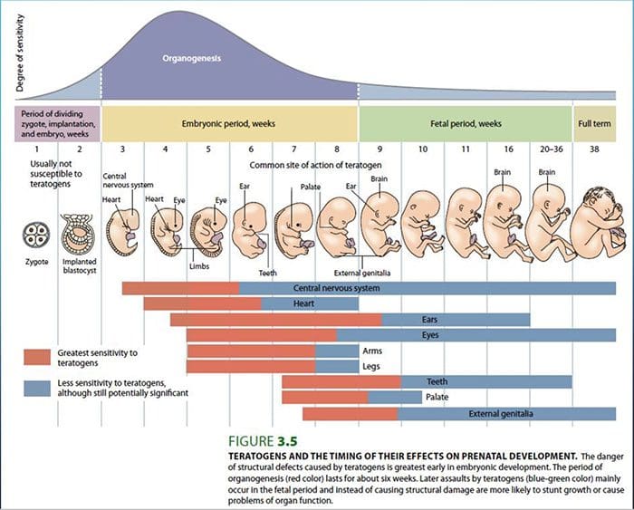
Focusing attention on the development of the nervous system can increase a clinician’s insight into brain functionality and treatment possibilities.
Dysfunctions that may be associated with developmental abnormalities of the brain may range from a mild reduction in cortical function to conditions such as autism and schizophrenia.
The development of the nervous system is influenced by both endogenous and exogenous mechanisms.
Endogenous referring to genetics, exogenous referring to the embryo�s environment.
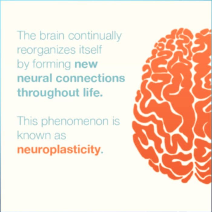
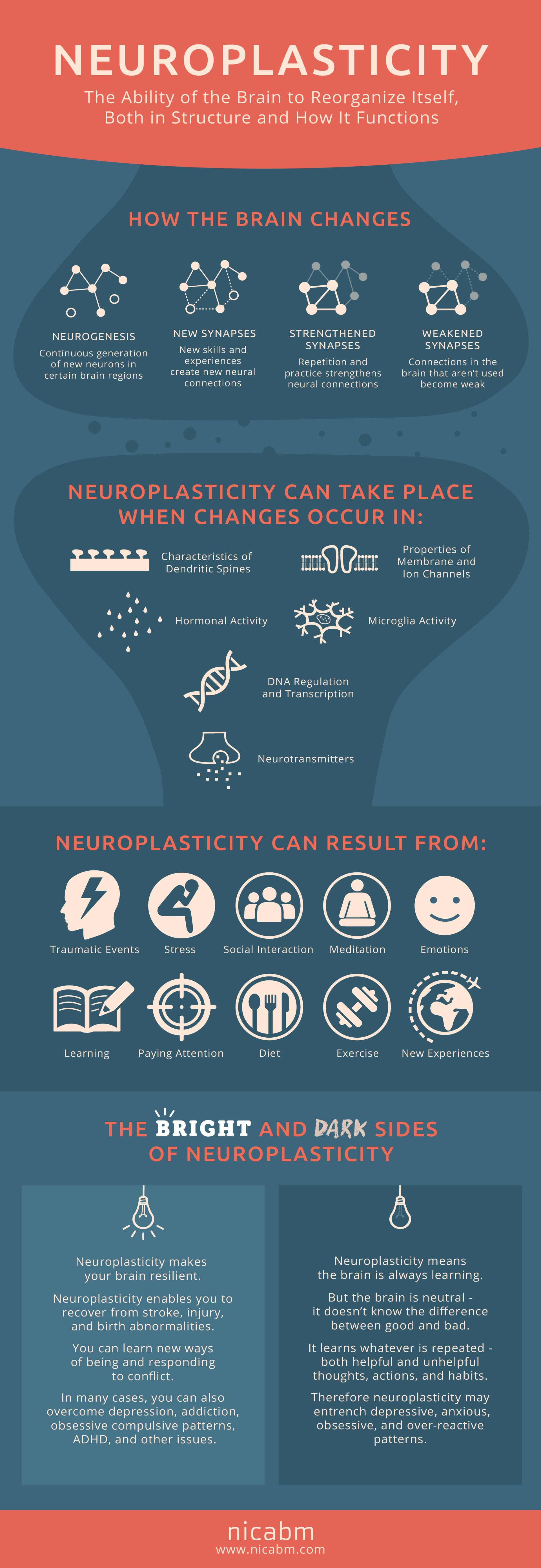
Development is an ongoing process….NEUROPLASTICITY
Neuroembryology: DEVELOPMENT
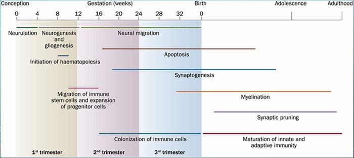
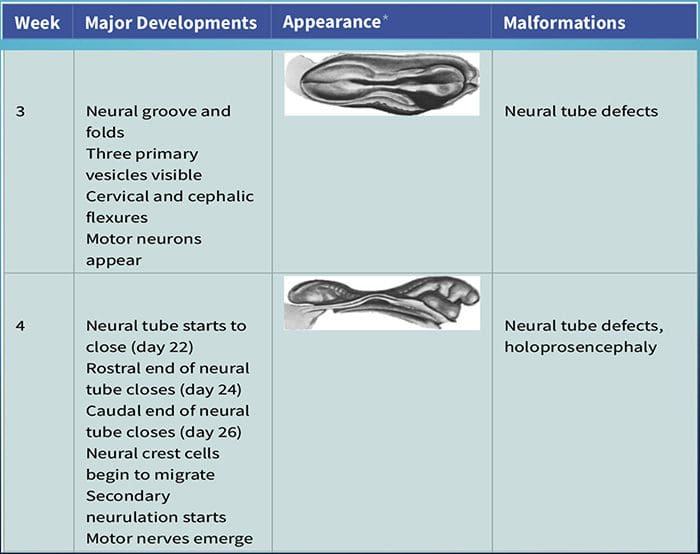
Development of the nervous system can first be identified at about 3 weeks after conception.
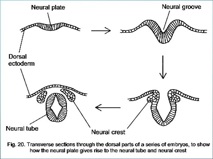
At 3 weeks, in response to underlying chemical signaling from the mesoderm, the neural plate forms, which quickly transitions into the neural groove.
At the beginning of the 4th week, the two folds forming the neural groove begin to fuse starting the formation of the neural tube
Fusion proceeds cranially and caudally and the entire neural tube is closed by the end of the 4th week.
This process is known as primary neurulation.
As the neural tube closes, it progressively separates from the ectodermal surface�and leaves behind neural crest cells.
Neural crest cells develop into the PNS.
The neural tube develops into virtually the entire CNS.
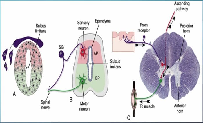
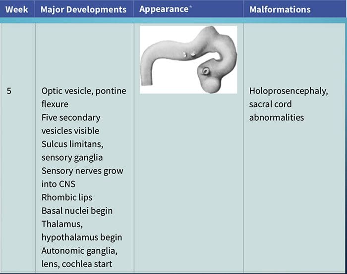
A longitudinal groove forms on the lateral wall of the neural tube�during the fourth week of development
This is known as the sulcus limitans, which separates the tube into dorsal and ventral halves.
The gray matter of the dorsal half forms an alar plate and the ventral half forms a basal plate.
This distinction is of great functional importance because the alar plate plays a role in sensory processing and the basal plate plays a role in motor output.
This similarity is seen in the adult spinal cord with the posterior gray matter receiving sensory input and the anterior gray matter producing motor output.
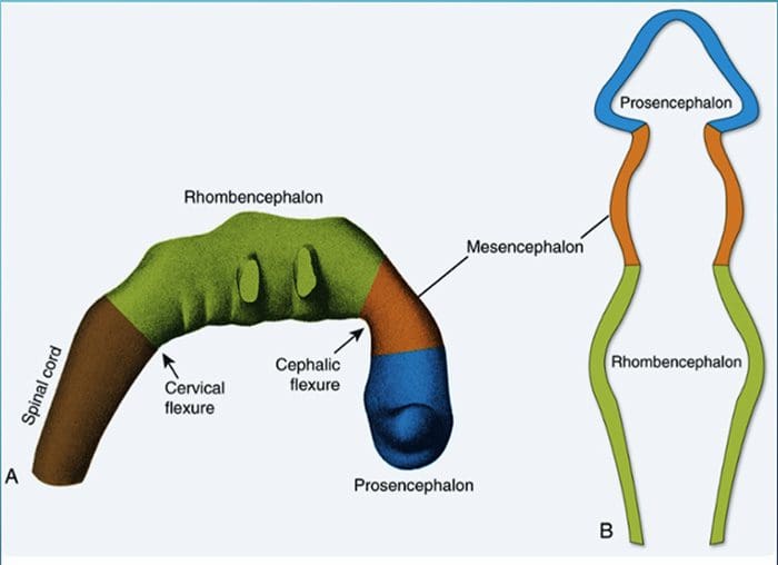
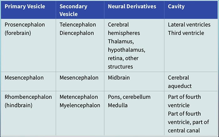
THREE PRIMARY VESICLES
Prosencephalon
Mesencephalon
Rhombencephalon
FIVE SECONDARY VESICLES
Telencephalon
Diencephalon
Mesencephalon (remains unchanged)
Metencephalon
Myelencephalon
ESTABLISHMENT OF NEURONAL CONNECTIONS
Neurons may have innate predetermined programs that lay out the basic patterns of connections to be formed initially in their development.
Theoretically, neurons gravitate to areas of attraction and move away from areas of repulsion � Chemoaffinity hypothesis
Several mechanisms allow axons to accurately find their way to their target destinations:
IFM's Find A Practitioner tool is the largest referral network in Functional Medicine, created to help patients locate Functional Medicine practitioners anywhere in the world. IFM Certified Practitioners are listed first in the search results, given their extensive education in Functional Medicine
 The cell needs three things to survive.
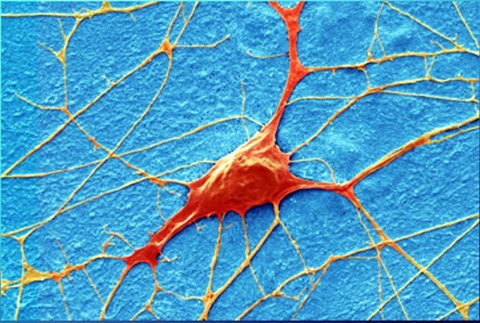
 The health of the neuron, however, plays a huge role in how neurons can produce receptor potentials, the endurance of the neuron and the ability to create plasticity.
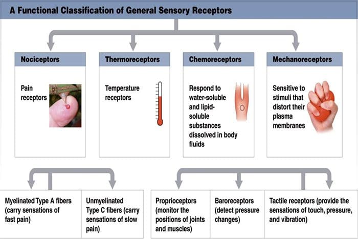 Chemoreceptors
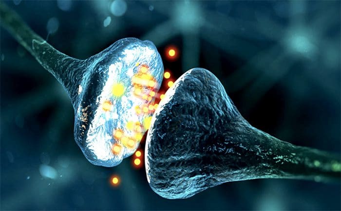 Although their morphologies vary widely, all receptors have three general parts:
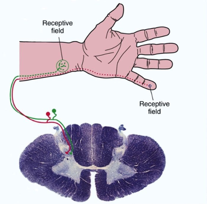
 Sensory receptors use ionotropic and metabotropic mechanisms to produce receptor potentials
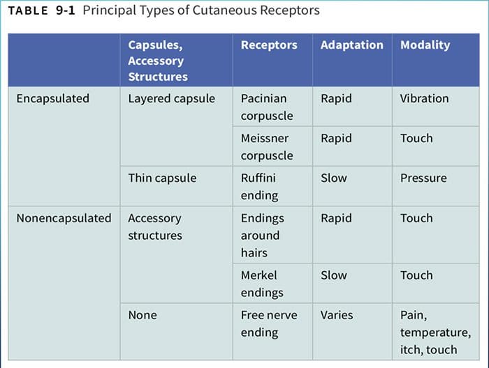
 Larger fibers conduct action potentials faster than do smaller fibers.
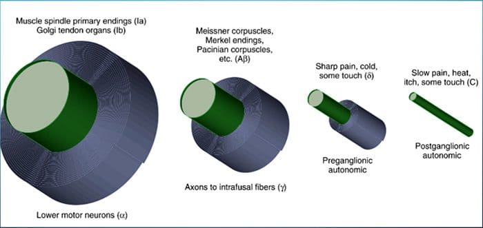

 junctions between muscles and tendons. They are similar to Ruffini endings in their basic organization, consisting of interwoven collagen bundles surrounded by a thin capsule (Fig. 9-16).
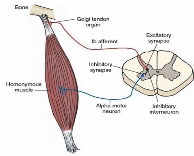 sensitive and can actually respond to the contraction of just a few muscle fibers.
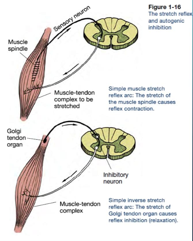


 CLINICAL SIGNIFICANCE OF NEURODEVELOPMENT