ClickCease
+1-915-850-0900 spinedoctors@gmail.com
Select Page

Clinical Case Series

Back Clinic Clinical Case Series. A clinical case series Is the most basic type of study design, in which researchers describe the experience of a group of people. Case series describe individuals who develop a particular new disease or condition. This type of study can provide compelling reading because they present a detailed account of the clinical experience of individual study subjects. Dr. Alex Jimenez conducts his own case series of studies.

A case study is a method of research that is commonly used in social sciences. It is a research strategy that investigates a phenomenon within a real context. They are based on an in-depth investigation of a single person, group, or event to explore the how of underlying problems/causes. It includes quantitative evidence and relies on multiple sources of evidence.

Case studies are an invaluable record of clinical practices of a profession. They do not provide specific guidance for the management of successive patients but they are a record of clinical interactions which help to frame questions for more rigorously designed clinical studies. They provide valuable teaching material, which demonstrates both classical and unusual information that can confront the practitioner. However, the majority of clinical interactions occur in the field and so it’s up to the practitioner to record and pass on the information. Guidelines are intended to assist the relative novice writer, practitioner, or student to efficiently navigate the study to publication.

A Case series is a descriptive study design and it’s just a series of cases of any particular disease or disease discrepancy that one might observe in clinical practice. These cases are described to suggest at best a hypothesis. However, there is no comparison group so there cannot be many conclusions about the disease or the disease process. Therefore, in terms of generating evidence regarding various aspects of a disease process, this is more of a starting point. For answers to any questions you may have please call Dr. Jimenez at 915-850-0900


Vertebral Artery Dissection Found During Chiropractic Examination

Vertebral Artery Dissection Found During Chiropractic Examination

Acknowledging the subsequent information below,�approximately more than 2 million people are injured in automobile accidents each year and among those incidents, the majority of the people involved are diagnosed with whiplash and/or neck injury by a healthcare professional. When the complex structure of the neck is subjected to trauma, tissue damage and other medical complications may occur. Vertebral artery dissection, or VAD, is characterized by a flap-like tear on the inner lining of the vertebral artery in charge of supplying blood to the brain. After the tear, blood can then enter the arterial wall and form a blood clot, thickening the artery wall and often impeding blood flow.


Through years of experience practicing chiropractic care,�VAD may often follow after trauma to the neck, such as that which occurs in an automobile accident, or whiplash injury. The symptoms of vertebral artery dissection include head and neck pain as well as intermittent or permanent stroke symptoms, such as difficulty speaking, impaired coordination and loss of vision. VAD, or vertebral artery dissection, is generally diagnosed with a contrast-enhanced CT or MRI scan.


Abstract


A 30-year-old woman presented to an emergency department with sudden onset of transient loss of left peripheral vision. Owing to a history of migraine headaches, she was released with a diagnosis of ocular migraine. Two days later, she sought chiropractic care for the chief symptom of severe neck pain. The chiropractor suspected the possibility of vertebral artery dissection (VAD). No manipulation was performed; instead, MR angiography (MRA) of the neck was obtained, which revealed an acute left VAD with early thrombus formation. The patient was placed on aspirin therapy. Repeat MRA of the neck 3?months later revealed resolution of the thrombus, without progression to stroke. This case illustrates the importance for all healthcare providers who see patients with neck pain and headache to be attentive to the symptomatic presentation of possible VAD in progress.


Background


Vertebral artery dissection (VAD) leading to stroke is an uncommon but potentially serious disorder. The incidence of stroke related to the vertebrobasilar system varies from 0.75 to 1.12/100?000 person-years. The pathological process in VAD typically involves dissection of the wall of the artery followed sometime later by thrombus formation, which may cause arterial occlusion or may lead to embolisation, causing occlusion of one or more of the distal branches off the vertebral artery, including the basilar artery, which can be catastrophic. VAD typically occurs in patients who have an inherent, transitory weakness in the arterial wall. In at least 80% of cases, the initial symptoms include neck pain with or without headache.


Many patients with VAD may in the early stages present to chiropractors seeking relief from neck pain and headache, without realising they are experiencing VAD. In many of these cases, the patient later develops a stroke. Until recently, it was assumed that the dissection (and subsequent stroke) was caused by cervical manipulative therapy (CMT). However, while early studies found an association between visits to a chiropractor and subsequent stroke related to VAD, recent data suggest that this relationship is not causal.


This case report is illustrative of the scenario in which a patient with an undiagnosed VAD in evolution consulted a chiropractor for neck pain and headache. After thorough history and examination, the chiropractor suspected VAD and did not perform CMT. Instead, the patient was referred for further evaluation, which detected a VAD in progress. Prompt diagnosis and anticoagulant treatment were thought to have averted progression to a stroke.


Case Presentation


A 30-year-old otherwise healthy woman consulted a chiropractor (DBF), reporting of right-sided neck pain in the suboccipital region. The patient reported that, 3?days previously, she had gone to the local hospital emergency department (ED) because of the sudden onset of loss of left peripheral vision. The visual symptoms interfered with her ability to see through her left eye; this was accompanied by �numbness� in her left eyelid. About 2?weeks prior to this ED visit, she had experienced an episode of acute left-sided neck pain with severe left-sided headache. She also related a history of migraine headache without prodrome. She was released from the ED with a tentative diagnosis of ocular migraine. She had never been previously diagnosed with ocular migraine, nor had she ever experienced any visual disturbances with her previous migraines.


Shortly after the left-sided ocular symptoms resolved, she suddenly developed right-sided neck pain without provocation, for which she sought chiropractic treatment. She also reported a transient episode of right-sided visual disturbance occurring that same day as well. This was described as sudden blurriness that was of short duration and resolved spontaneously earlier in the day of her presentation for chiropractic examination. When she presented for the initial chiropractic examination, she denied current visual disturbance. She said that she was not experiencing any numbness, paraesthesia or motor loss in the upper or lower extremities. She denied ataxia or difficulty with balance. Medical history was remarkable for childbirth 2� months prior to initial presentation. She stated that her migraine headaches were associated with her menstrual cycle. Family history was remarkable for a spontaneous ascending thoracic aortic aneurysm in her older sister, who was about 30?years of age when her aneurysm had occurred.


Investigations


Based on the history of sudden onset of severe upper cervical pain and headache with visual disturbance and ocular numbness, the DC was concerned about the possibility of early VAD. Urgent MR angiography (MRA) of the neck and head, along with MRI of the head, was ordered. No cervical spine examination or manipulation was performed because of the suspicion that the neck pain was related to VAD rather than to a �mechanical� cervical disorder.


MRA of the neck demonstrated that the left vertebral artery was small and irregular in calibre, extending from the C7 level cephalad to C2, consistent with dissection. There was a patent true lumen with a surrounding cuff of T1 hyper-intensity, consistent with dissection with subintimal thrombus within the false lumen (Figures 1 and ?2). MRI of the head with and without contrast, and MRA of the head without contrast, were both unremarkable. Specifically, there was no intracranial extension of dissection or evidence of infarction. MR perfusion of the brain revealed no focal perfusion abnormalities.


Figure 1 Axial Proton Density Image - Image 1

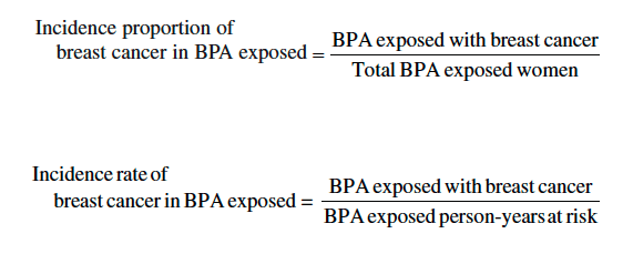
Figure 1: Axial proton density image demonstrates circumferential hyper-intensity surrounding the left cervical vertebral artery (representing the false lumen). Note decreased calibre of true lumen (black flow void) with respect to the right vertebral artery.


Figure 2 Axial Image from Three Dimensional Time of Flight MRA - Image 2

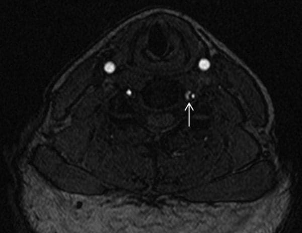
Figure 2: Axial image from three-dimensional time-of-flight MRA demonstrates T1 hypointense dissection flap separating the true lumen (lateral) from the false lumen (medial). MRA, MR angiography.


Differential Diagnosis


The ED released the patient with a tentative diagnosis of ocular migraine, due to her history of migraine headaches. However, the patient stated that the left-sided headache was atypical��like nothing I’ve ever experienced before.� Her previous migraines were associated with her menstrual cycle, but not with any vision changes. She had never been previously diagnosed with ocular migraine. MRA of the cervical region revealed that the patient actually had an acute dissection with thrombus formation in the left vertebral artery.


Treatment


Owing to the potential of impending stroke associated with an acute VAD with thrombus formation, the patient was admitted to the neurology stroke service for close neurological monitoring. During her admission, the patient did not experience any recurrence of neurological deficits and her headaches improved. She was discharged the following day with a diagnosis of left VAD and transient ischaemic attack. She was instructed to avoid vigorous exercise and trauma to the neck. Daily aspirin (325?mg) was prescribed, to be continued for 3�6?months after discharge.


Outcome and Follow-Up


After discharge from the stroke service, the patient had no recurrence of headache or visual disturbances, and her posterior neck pain symptoms resolved. Repeat imaging was performed 3?months after presentation, which demonstrated improved calibre of the cervical left vertebral artery with resolution of the thrombus within the false lumen (Figure 3). Imaging of the intracranial compartment remained normal, without evidence of interval infarction or perfusion asymmetry.


Figure 3 Maximum Intensity Projection MIP Images - Image 3

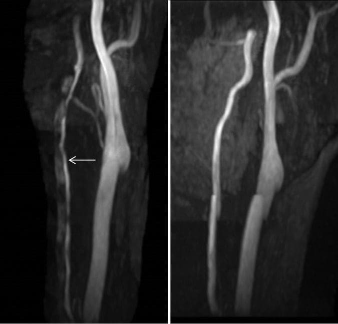
Figure 3: Maximum intensity projection (MIP) images from three-dimensional time-of-flight MRA (left image is at time of presentation and right image is at 3-month follow-up). The initial imaging demonstrates markedly diminutive calibre of the left vertebral artery


Discussion


The pathophysiological process of VAD is thought to start with degeneration of the tissues at the medial-adventitial border of the vertebral artery, leading to the development of microhaematomata within the wall of the artery and, eventually, arterial tear. This can lead to leakage of blood into the arterial wall, causing occlusion of the lumen with subsequent thrombus formation and embolisation, resulting in stroke related to one of the branches of the vertebral artery. This pathological process is similar to that of spontaneous carotid artery dissection, spontaneous thoracic aortic dissection and spontaneous coronary artery dissection. All these conditions tend to occur in younger adults and some have speculated that they may be part of a common inherited pathophysiological process. Notable in this case is the fact that the patient’s older sister had experienced a spontaneous thoracic aortic aneurysm (probably a dissection) at around the same age (30?years) as this patient was when she experienced her VAD.


While the dissection is often sudden, the luminal compromise and complications of VAD can develop gradually leading to variable symptoms and presentation, depending on the stage of the disease. The dissection itself, which develops some time before the onset of neural ischaemia, can cause stimulation of nociceptive receptors within the artery, producing pain that is most commonly felt in the upper cervical spine or head. Only after the pathophysiological process progresses to the point of complete arterial occlusion or thrombus formation with distal embolisation does the full manifestation of infarction occur. However, as illustrated in this case, neurological symptoms can develop early in the process, particularly in cases in which the true lumen demonstrates significant calibre decrease secondary to compression.


There are several interesting aspects to this case. First, it highlights the importance of spine clinicians being alert to the possibility that what may appear to be typical �mechanical� neck pain could be something potentially more sinister, such as VAD. The sudden onset of severe suboccipital pain, with or without headache, and accompanying brainstem related neurological symptoms, should alert the clinician to the possibility of VAD. As in the case reported here, patients with a history of migraine will typically describe the headache as different from their usual migraine. A careful neurological examination should be performed, looking for possible subtle neurological deficits, although the neurological examination will often be negative in the early stages of VAD.


Second, a triad of symptoms raised concern that the patient might be experiencing a VAD in progress. The symptom triad included: (1) spontaneous onset of severe upper cervical pain; (2) severe headache that was distinctly different from the patient’s usual migraine headaches; and (3) brainstem-related neurological symptoms (in the form of transient visual disturbance). Notably, careful neurological examination was negative. Nonetheless, the history was of sufficient concern to prompt immediate investigation.


When VAD is suspected but no frank signs of stroke are present, immediate vascular imaging is indicated. While the optimal imaging evaluation of VAD remains controversial, MRA or CTA are the diagnostic studies of choice given their excellent anatomic delineation and ability to evaluate for complications (including infarction and changes in brain perfusion). Some advocate the use of Doppler ultrasound; however, it has limited utility given the course of the vertebral artery in the neck and limited evaluation of the vertebral arteries cephalad to the origin. Additionally, ultrasound imaging is unlikely to allow visualisation of the dissection itself and thus can be negative in the absence of significant arterial occlusion.


Third, this case is interesting in light of the controversy about cervical manipulation as a potential �cause� of VAD. While case reports have presented patients who have experienced stroke related to VAD after cervical manipulation, and case�control studies have found a statistical association between visits to chiropractors and stroke related to VAD, further investigation has indicated that the association is not causal. Cassidy et al found that a patient who experiences stroke related to VAD is just as likely to have visited a primary care practitioner as to have visited a chiropractor prior to having the stroke. The authors suggested that the most likely explanation for the statistical association between visits to chiropractors and subsequent VAD is that a patient who experiences the initial symptoms of VAD (neck pain with or without headache) seeks medical attention for these symptoms (from a chiropractor, primary care practitioner, or another type of practitioner), then subsequently experiences the stroke, independent of any action taken by the practitioner.


It is important to note that, while there have been reported cases of carotid artery dissection after cervical manipulation, case�control studies have not found this association. The initial symptoms of carotid dissection (neurological symptoms, with neck and head pain less common than VAD), aortic dissection (sudden onset of severe, �tearing� pain) and coronary artery dissection (acute severe chest pain, ventricular fibrillation) are likely to cause the individual to immediately seek ED care, rather than seek chiropractic care. However, VAD has seemingly benign initial symptoms�neck pain and headache�which are symptoms that commonly cause patients to seek out chiropractic care. This may explain why only VAD is associated with visits to chiropractors, while these other types of dissections are not; patients with these other conditions, which have much more alarming symptoms, simply do not present to chiropractors.


This case is a good example of a patient with VAD in progress presenting to a chiropractor for the purpose of seeking relief from neck pain. Fortunately, the chiropractor was astute enough to ascertain that the patient’s symptoms were not suggestive of a �mechanical� cervical spine disorder, and appropriate diagnostic investigation was performed. However, if manipulation had been performed, the VAD that was already in progress from natural history may have been blamed on manipulation, after being detected on MRA imaging. Fortunately, in this case, the chiropractor was able to assist with early detection and treatment, and subsequently a stroke was likely averted.


Learning Points


  • A case is presented in which a patient saw a chiropractor, while seeking treatment for neck pain, and the history raised concern for possible vertebral artery dissection (VAD).
  • Rather than providing manipulative treatment, the chiropractor referred the patient for advanced imaging, which confirmed the diagnosis of VAD.
  • The case illustrates the importance of paying attention to subtle historical factors in patients with VAD.
  • It also serves as an example of a patient with a VAD in progress seeking the services of a chiropractor for the initial symptoms of the disorder.
  • In this case, early detection of the dissection occurred and the patient had a full recovery without any subsequent stroke.


Acknowledgments


The authors would like to acknowledge the assistance of Pierre Cote, DC, PhD, for his assistance with reviewing this manuscript.


Footnotes


Contributors: All the authors acknowledge that they have contributed the following to the submission of this manuscript: conception and design, drafting of the manuscript, critical revisions of the manuscript, literature review and references, and proof reading of the final manuscript.


Competing interests: None declared.


Patient consent: Obtained.


Provenance and peer review: Not commissioned; externally peer reviewed.


Information referenced from the National Center for Biotechnology Information (NCBI). The scope of our information is limited to chiropractic as well as to spinal injuries and conditions. To discuss the subject matter, please feel free to ask Dr. Jimenez or contact us at 915-850-0900 .


Cited by Dr. Alex Jimenez


Green-Call-Now-Button-24H-150x150-2-3.png


Additional Topics: Wellness


Overall health and wellness are essential towards maintaining the proper mental and physical balance in the body. From eating a balanced nutrition as well as exercising and participating in physical activities, to sleeping a healthy amount of time on a regular basis, following the best health and wellness tips can ultimately help maintain overall well-being. Eating plenty of fruits and vegetables can go a long way towards helping people become healthy.


blog picture of cartoon paperboy big news


IMPORTANT TOPIC: EXTRA EXTRA: Treating Sciatica Pain


Blank
References
1.�Debette S, Leys D.�Cervical-artery dissections: predisposing factors, diagnosis, and outcome.�Lancet Neurol�2009;8:668�78.�doi:10.1016/S1474-4422(09)70084-5[PubMed]
2.�Boyle E, Cote P, Grier AR et al.�Examining vertebrobasilar artery stroke in two Canadian provinces.�Spine�2008;33(4 Suppl):S170�5.�doi:10.1097/BRS.0b013e31816454e0[PubMed]
3.�Lee VH, Brown RD Jr, Mandrekar JN et al.�Incidence and outcome of cervical artery dissection: a population-based study.�Neurology�2006;67:1809�12.�doi:10.1212/01.wnl.0000244486.30455.71[PubMed]
4.�Schievink WI.�Spontaneous dissection of the cartoid and vertebral arteries.�N Engl J Med�2001;344:898�906.�doi:10.1056/NEJM200103223441206[PubMed]
5.�Volker W, Dittrich R, Grewe S et al.�The outer arterial wall layers are primarily affected in spontaneous cervical artery dissection.�Neurology�2011;76:1463�71.�doi:10.1212/WNL.0b013e318217e71c[PubMed]
6.�Gottesman RF, Sharma P, Robinson KA et al.�Clinical characteristics of symptomatic vertebral artery dissection: a systematic review.�Neurologist�2012;18:245�54.�doi:10.1097/NRL.0b013e31826754e1[PMC free article][PubMed]
7.�Cassidy JD, Boyle E, Cote P et al.�Risk of vertebrobasilar stroke and chiropractic care: results of a population-based case-control and case-crossover study.�Spine�2008;33(4�Suppl):S176�83.�doi:10.1097/BRS.0b013e3181644600[PubMed]
8.�Rothwell DM, Bondy SJ, Williams JI.�Chiropractic manipulation and stroke: a population-based case-control study.�Stroke�2001;32:1054�60.�doi:10.1161/01.STR.32.5.1054[PubMed]
9.�Smith WS, Johnston SC, Skalabrin EJ et al.�Spinal manipulative therapy is an independent risk factor for vertebral artery dissection.�Neurology�2003;60:1424�8.�doi:10.1212/01.WNL.0000063305.61050.E6[PubMed]
10.�Volker W, Besselmann M, Dittrich R et al.�Generalized arteriopathy in patients with cervical artery dissection.�Neurology�2005;64:1508�13.�doi:10.1212/01.WNL.0000159739.24607.98[PubMed]
11.�Evangelista A, Mukherjee D, Mehta RH et al.�Acute intramural hematoma of the aorta: a mystery in evolution.�Circulation�2005;111:1063�70.�doi:10.1161/01.CIR.0000156444.26393.80[PubMed]
12.�Tweet MS, Hayes SN, Pitta SR et al.�Clinical features, management, and prognosis of spontaneous coronary artery dissection.�Circulation�2012;126:579�88.�doi:10.1161/CIRCULATIONAHA.112.105718[PubMed]
13.�Choi S, Boyle E, Cote P et al.�A population-based case-series of Ontario patients who develop a vertebrobasilar artery stroke after seeing a chiropractor.�J Manipulative Physiol Ther�2011;34:15�22.�doi:10.1016/j.jmpt.2010.11.001[PubMed]
14.�Naggara O, Louillet F, Touze E et al.�Added value of high-resolution MR imaging in the diagnosis of vertebral artery dissection.�AJNR Am J Neuroradiol�2010;31:1707�12.�doi:10.3174/ajnr.A2165[PubMed]
15.�Haynes MJ, Vincent K, Fischhoff C et al.�Assessing the risk of stroke from neck manipulation: a systematic review.�Int J Clin Pract�2012;66:940�7.�doi:10.1111/j.1742-1241.2012.03004.x[PMC free article][PubMed]
16.�Nebelsieck J, Sengelhoff C, Nassenstein I et al.�Sensitivity of neurovascular ultrasound for the detection of spontaneous cervical artery dissection.�J Clin Neurosci�2009;16:79�82.�doi:10.1016/j.jocn.2008.04.005[PubMed]
17.�Bendick PJ, Jackson VP.�Evaluation of the vertebral arteries with duplex sonography.�J Vasc Surg1986;3:523�30.�doi:10.1016/0741-5214(86)90120-5[PubMed]
18.�Murphy DR.�Current understanding of the relationship between cervical manipulation and stroke: what does it mean for the chiropractic profession?Chiropr Osteopat�2010;18:22�doi:10.1186/1746-1340-18-22[PMC free article][PubMed]
19.�Engelter ST, Grond-Ginsbach C, Metso TM et al.�Cervical artery dissection: trauma and other potential mechanical trigger events.�Neurology�2013;80:1950�7.�doi:10.1212/WNL.0b013e318293e2eb[PubMed]
20.�Peters M, Bohl J, Th�mke F et al.�Dissection of the internal carotid artery after chiropractic manipulation of the neck.�Neurology�1995;45:2284�6.�doi:10.1212/WNL.45.12.2284[PubMed]
21.�Nadgir RN, Loevner LA, Ahmed T et al.�Simultaneous bilateral internal carotid and vertebral artery dissection following chiropractic manipulation: case report and review of the literature.�Neuroradiology2003;45:311�14.�doi:10.1007/s00234-003-0944-x[PubMed]
22.�Dittrich R, Rohsbach D, Heidbreder A et al.�Mild mechanical traumas are possible risk factors for cervical artery dissection.�Cerebrovasc Dis�2007;23:275�81.�doi:10.1159/000098327[PubMed]
23.�Chung CL, Cote P, Stern P et al.�The association between cervical spine manipulation and carotid artery dissection: a systematic review of the literature.�J Manipulative Physiol Ther�2014; doi:10.1016/j.jmpt.2013.09.005�doi:10.1016/j.jmpt.2013.09.005[PubMed]
24.�Thomas LC, Rivett DA, Attia JR et al.�Risk factors and clinical features of craniocervical arterial dissection.�Man Ther�2011;16:351�6.�doi:10.1016/j.math.2010.12.008[PubMed]
25.�Klineberg E, Mazanec D, Orr D et al.�Masquerade: medical causes of back pain.�Cleve Clin J Med2007;74:905�13.�doi:10.3949/ccjm.74.12.905[PubMed]
Close Accordion
What are Case Reports & Case Series?

What are Case Reports & Case Series?

The diagnosis of a variety of diseases has been effectively determined through clinical and experimental data. Research studies provide valuable information on the pathogenesis of many conditions and are often the primary source of information regarding new diseases or conditions. Case reports and case series are first level research studies, offering the most initial insights on a particular health issue through the personal experience of one or more people with a disease or condition. The following article describes the purpose of case reports and case series, and how they provide clinical and experimental data.


Learning Objectives


1. Case reports and case series describe the experience of one or more people with a disease.
2. Case reports and case series are often the first data alerting to a new disease or condition.
3. Case reports and case series have specific limitations:

  • a. Lack of a denominator to calculate rates of disease
  • b. Lack of a comparison group
  • c. Selecting study populations
  • d. Sampling variation


Case Reports and Case Series


Case reports and case series represent the most basic type of study design, in which researchers describe the experience of a single person (case report) or a group of people (case series). Typically, case reports and case series describe individuals who develop a particular new disease or condition. Case reports and case series can provide compelling reading because they present a detailed account of the clinical experience of individual study subjects. In contrast, studies that evaluate large numbers of individuals typically summarize the data using statistical measures, such as means and proportions.


Example 3.1. A case series describes 15 young women who develop breast cancer; 9 of these women report at least once weekly ingestion of foods packaged with the estrogenic chemical bisphenol A (BPA). Urine testing confirms the presence of BPA among all nine case women.


It is tempting to surmise from these data that BPA might be causally related to breast cancer. However, case reports/case series have important limitations that preclude inference of a causal relationship.

First, case reports/case series lack denominator data that are necessary to calculate the rate of disease. The denominator refers to the population from which the diseased subjects arose. For example, to calculate the incidence proportion or incidence rate of breast cancer among women exposed to BPA, the total number of women who were exposed to BPA or the total number of person-years at risk is needed.


Table 1 - Incidence Proportion & Incidence Rate


Disease rates are needed for comparison with historically reported disease rates, or with rates from a selected comparison group. Unfortunately, obtaining the necessary denominator data may not be easy. In this example, additional data sources are needed to determine the total number of BPA-exposed women from whom the breast cancer cases arose. The case series data alone cannot be used to calculate the rate of breast cancer because they do not include the total number of women who were exposed to BPA.


A second problem with case report/case series report data is the lack of a comparison group. The 60% prevalence of BPA exposure among women with breast cancer seems unusually high, but what is prevalence of BPA exposure among women without breast cancer? This comparison is critical for addressing the hypothesis that BPA might be a cause of breast cancer.


A third limitation of case reports/case series is that these studies often describe highly select individuals who may not represent the general population. For example, it is possible that the 15 breast cancer cases originated from a single hospital in a community with high levels of air pollution or other potential carcinogens. Under these conditions, a fair estimate of breast cancer incidence among non-BPA exposed women from the same community would be required to make an inference that BPA causes breast cancer.


A fourth limitation of case reports/case series is sampling variation. This concept will be explored in detail later in this book. The basic idea is that there is tremendous natural variation in disease development in humans. The fact that 9 of 15 women with breast cancer reported BPA exposure is interesting; however, this number may be very different in the next case series of 15 women with breast cancer simply due to chance. A precise estimate of the rate of a disease, independent from chance, can be obtained only by increasing the number of diseased subjects.


Recall the list of factors that are used to judge whether a factor may be a cause of disease:


1. Randomized evidence
2. Strength of association
3. Temporal relationship between exposure and outcome
4. Dose-response association
5. Biological plausibility


In general, case reports/case series rely almost exclusively on biological plausibility to make their case for causation. For the BPA and breast cancer case series, there is no randomized evidence, no measure of the strength of association between BPA and breast cancer, no reported dose�response association, and no evidence that BPA exposure preceded the development of breast cancer. The inference for causation derives completely from previous biological knowledge regarding the estrogenic effects of BPA.


Despite limitations of case series data, they may be highly suggestive of an important new association, disease process, or unintended side effect of a medication or treatment.


Example 3.2. In 2007, a case series described three cases of male prepubertal gynecomastia. The report included detailed information on each subjects� age, body size, serum levels of endogenous steroids, and known exposures to exogenous hormones. It was discovered that all three otherwise healthy boys had been exposed to some product containing lavender oil (lotion, shampoo, soap), and that in each case, the gynecomastia resolved upon discontinuation of the product. Subsequent in vitro studies demonstrated endocrine-disrupting activity of lavender oil. This novel case series data may lead to further investigations to determine whether lavender oil, a common ingredient in commercially available products, may be a cause of gynecomastia.


Example 3.3. A vaccine designed to prevent rotavirus infection was found to cause weakening of the intestinal muscle layers in animals. Following release of the vaccine, a number of cases of intussusception (when one portion of the bowel slides into the next) were reported in children who received the vaccine, with some fatal cases. The strong biological plausibility underlying this initial association, and knowledge that intussusception is otherwise rare in infants, was highly suggestive of a causal relationship and the vaccine was removed from the market.


Information referenced from B. Kestenbaum, Epidemiology and Biostatistics: An Introduction to Clinical Research, DOI 10.1007/978-0-387-88433-2_3, � Springer Science+Business Media, LLC 2009. The scope of our information is limited to chiropractic as well as to spinal injuries and conditions. To discuss the subject matter, please feel free to ask Dr. Jimenez or contact us at 915-850-0900 .


Referenced by Dr. Alex Jimenez


Green-Call-Now-Button-24H-150x150-2-3.png


Additional Topics: Wellness


Overall health and wellness are essential towards maintaining the proper mental and physical balance in the body. From eating a balanced nutrition as well as exercising and participating in physical activities, to sleeping a healthy amount of time on a regular basis, following the best health and wellness tips can ultimately help maintain overall well-being. Eating plenty of fruits and vegetables can go a long way towards helping people become healthy.


blog picture of cartoon paperboy big news


IMPORTANT TOPIC: EXTRA EXTRA: Treating Sciatica Pain


Management of Sciatica: Nonsurgical & Surgical Therapies

Management of Sciatica: Nonsurgical & Surgical Therapies

Consider the following, sciatica is a medical term used to describe a collective group of symptoms resulting from the irritation or compression of the sciatic nerve, generally due to an injury or aggravated condition. Sciatica is commonly characterized by radiating pain along the sciatic nerve, which runs down one or both legs from the lower back. The following case vignette discusses Mr. Winston’s medical condition, a 50-year-old bus driver who reported experiencing chronic, lower back and leg pain associated with sciatica during a 4-week time period. Ramya Ramaswami, M.B., B.S., M.P.H., Zoher Ghogawala, M.D., and James N. Weinstein, D.O., provide a comprehensive analysis of the various treatment options available to treat sciatica, including undergoing lumbar disk surgery and receiving nonsurgical therapy.


On a personal note, as a practicing doctor of chiropractic, choosing the correct treatment care for any type of injury or condition can be a personal and difficult decision. If the circumstances are favorable, the patient may determine what is the best form of treatment for their type of medical issue. While nonsurgical therapies, such as chiropractic care, can often be utilized to improve symptoms of sciatica, more severe cases of sciatica may require surgical interventions to treat the source of the issue. In most cases, nonsurgical therapies should be considered first, before turning to surgical therapies for sciatica.


Case Vignette


A Man with Sciatica Who is Considering Lumbar Disk Surgery


Ramya Ramaswami, M.B., B.S., M.P.H.


Mr. Winston, a 50-year-old bus driver, presented to your office with a 4-week history of pain in his left leg and lower back. He described a combination of severe sharp and dull pain that originated in his left buttock and radiated to the dorsolateral aspect of his left thigh, as well as vague aching over the lower lumbar spine. On examination, passive raising of his left leg off the table to 45 degrees caused severe pain that simulated his main symptom, and the pain was so severe that you could not lift his leg further. There was no leg or foot weakness. His body-mass index (the weight in kilograms divided by the square of the height in meters) was 35, and he had mild chronic obstructive pulmonary disease as a result of smoking one pack of cigarettes every day for 22 years. Mr. Winston had taken a leave of absence from his work because of his symptoms. You prescribed 150 mg of pregabalin per day, which was gradually increased to 600 mg daily because the symptoms had not abated.


Now, 10 weeks after the initial onset of his symptoms, he returns for an evaluation. The medication has provided minimal alleviation of his sciatic pain. He has to return to work and is concerned about his ability to complete his duties at his job. He undergoes magnetic resonance imaging, which shows a herniated disk on the left side at the L4�L5 root. You discuss options for the next steps in managing his sciatica. He is uncertain about invasive procedures such as lumbar disk surgery but feels limited by his symptoms of pain.


Treatment Options


Which of the following would you recommend for Mr. Winston?


  1. Undergo lumbar disk surgery.
  2. Receive nonsurgical therapy.


To aid in your decision making, each of these approaches is defended in a short essay by an expert in the field. Given your knowledge of the patient and the points made by the experts, which option would you choose?


Option 1: Undergo Lumbar Disk Surgery
Option 2: Receive Nonsurgical Therapy


1. Undergo Lumbar Disk Surgery


Zoher Ghogawala, M.D.


Mr. Winston�s case represents a common scenario in the management of symptomatic lumbar disk herniation. In this particular case, the patient�s symptoms and the physical examination are consistent with nerve-root compression and inflammation directly from an L4�L5 herniated disk on his left side. The patient does not have weakness but has ongoing pain and has been unable to work for the past 10 weeks despite receiving pregabalin. Two questions emerge: first, does lumbar disk surgery (microdiskectomy) provide outcomes that are superior to those with continued nonoperative therapy in patients with more than 6 weeks of symptoms; and second, does lumbar microdiskectomy improve the likelihood of return to work in patients with these symptoms?


The highest quality data on the topic come from the Spine Patient Outcomes Research Trial (SPORT). The results of the randomized, controlled trial are difficult to interpret because adherence to the assigned treatment strategy was suboptimal. Only half the patients who were randomly assigned to the surgery group actually underwent surgery within 3 months after enrollment, and 30% of the patients assigned to nonoperative treatment chose to cross over to the surgical group. In this study, the patients who underwent surgery had greater improvements in validated patient-reported outcomes. The treatment effect of microdiskectomy was superior to that of nonoperative treatment at 3 months, 1 year, and 2 years. Moreover, in an as-treated analysis, the outcomes among patients who underwent surgery were superior to those among patients who received nonoperative therapy. Overall, the results of SPORT support the use of microdiskectomy in this case.


Results of clinical trials are based on a comparison of treatment options in study populations and may or may not apply to individual patients. SPORT did not specify what type of nonoperative therapy was to be used. Physical therapy was used in 73% of the patients, epidural injections in 50%, and medical therapies (e.g., nonsteroidal antiinflammatory drugs) in more than 50%. In the case of Mr. Winston, pregabalin has been tried, but physical therapy and epidural glucocorticoid injections have not been attempted. Despite widespread use of physical therapy for the treatment of lumbar disk herniation, the evidence supporting its effectiveness is inconclusive, according to published guidelines of the North American Spine Society. On the other hand, there is evidence that transforaminal epidural glucocorticoid injection provides short-term relief (30 days) in patients with nerve-root symptoms directly related to a herniated disk. Overall, there is evidence, from SPORT and from a randomized trial from the Netherlands published in the Journal, that early surgery between 6 and 12 weeks after the onset of symptoms provides greater alleviation of leg pain and better overall pain relief than prolonged conservative therapy.


The ability to return to work has not been formally studied in comparisons of operative with nonoperative treatments for lumbar disk herniation. Registry data from the NeuroPoint-SD study showed that more than 80% of the patients who were working before disk herniation returned to work after surgery. The ability to return to work may be dependent on the type of vocation, since patients who are manual laborers may need more time to recover to reduce the risk of reherniation.


It is well recognized that many patients who have a symptomatic lumbar disk herniation will have improvement spontaneously over several months. Surgery can alleviate symptoms more quickly by immediately removing the offending disk herniation from the affected nerve root. The risk�benefit equation will vary among individual patients. In the case of Mr. Winston, obesity and mild pulmonary disease might increase the risk of complications from surgery, although in SPORT, 95% of surgical patients did not have any operative or postoperative complication. For Mr. Winston, a patient with pain that has persisted for more than 6 weeks, microdiskectomy is a rational option that is supported by high-quality evidence.


2. Receive Nonsurgical Therapy


James N. Weinstein, D.O.


This case involves a common presentation of low back pain radiating to the buttock and posterolateral thigh that might represent either referred mechanical pain or radiculopathy. Classic radiculopathy resulting from compression of a lower lumbar nerve root (L4, L5, or S1) results in pain that radiates distal to the knee and is often accompanied by weakness or numbness in the respective myotome or dermatome. In this case, the pain is proximal to the knee and is not associated with weakness or numbness. In SPORT, surgery resulted in faster recovery and a greater degree of improvement than nonoperative treatment in patients with pain that radiated distal to the knee and was accompanied by neurologic signs or symptoms. However, since Mr. Winston would not have met the inclusion criteria for SPORT, the results of diskectomy in this case would be somewhat unpredictable. He does not have radiculopathy that radiates below the knee, and he does not have weakness or numbness; nonoperative treatment should be exhausted before any consideration of a surgical procedure that in most cases has not been shown to be effective in patients with this type of presentation. In this issue of the Journal, Mathieson and colleagues report the results of a randomized, controlled trial that showed that pregabalin did not significantly alleviate pain related to sciatica. Mr. Winston has been treated only with pregabalin; therefore, other conservative options should be explored.


Saal and Saal reported that more than 80% of patients with radiculopathy associated with a lumbar disk herniation had improvement in a matter of months with exercise-based physical therapy. In the nonoperative SPORT cohort, patients had significant improvement from baseline, and approximately 60% of those with classic radiculopathy who initially received nonoperative treatment avoided surgery. Mr. Winston has had minimal treatment and has had symptoms for only 10 weeks. He should undergo a course of exercise-based physical therapy and a trial of a nonsteroidal antiinflammatory medication and may consider a lumbar epidural glucocorticoid injection. Although there is little evidence of the effectiveness of these nonoperative options alone, the combination of these treatments and the benign natural history of the patient�s condition could result in alleviation or resolution of symptoms. If these interventions � and time � do not resolve his symptoms, surgery could be considered as a final option, but it may not have long-term effectiveness and could in and of itself cause the possibility of more harm than good. Mr. Winston has risk factors, such as obesity and a history of smoking, that have been shown to contribute to poor surgical outcomes of certain spinal procedures.


Mr. Winston has symptoms of back pain that interfere with his quality of life. He would need to understand, through shared decision making, that a nonsurgical approach is likely to be more effective than surgery over time.


Information referenced from the National Center for Biotechnology Information (NCBI) and the New England Journal of Medicine (NEJM). The scope of our information is limited to chiropractic as well as to spinal injuries and conditions. To discuss the subject matter, please feel free to ask Dr. Jimenez or contact us at 915-850-0900 .


Cited by Dr. Alex Jimenez


Green-Call-Now-Button-24H-150x150-2-3.png


Additional Topics: Wellness


Overall health and wellness are essential towards maintaining the proper mental and physical balance in the body. From eating a balanced nutrition as well as exercising and participating in physical activities, to sleeping a healthy amount of time on a regular basis, following the best health and wellness tips can ultimately help maintain overall well-being. Eating plenty of fruits and vegetables can go a long way towards helping people become healthy.


blog picture of cartoon paperboy big news


IMPORTANT TOPIC: EXTRA EXTRA: Treating Sciatica Pain


Blank
References


  • 1. Weinstein JN, Tosteson TD, Lurie JD, et al. Surgical vs nonoperative treatment for lumbar disk herniation: the Spine Patient Outcomes Research Trial (SPORT): a randomized trial. JAMA 2006;296:24412450

  • 2. Weinstein JN, Lurie JD, Tosteson TD, et al. Surgical vs nonoperative treatment for lumbar disk herniation: the Spine Patient Outcomes Research Trial (SPORT) observational cohort. JAMA 2006;296:24512459

  • 3. Kreiner DS, Hwang SW, Easa JE, et al. An evidence-based clinical guideline for the diagnosis and treatment of lumbar disc herniation with radiculopathy. Spine J 2014;14:180191

  • 4. Ghahreman A, Ferch R, Bogduk N. The efficacy of transforaminal injection of steroids for the treatment of lumbar radicular pain. Pain Med 2010;11:11491168

  • 5. Peul WC, van Houwelingen HC, van den Hout WB, et al. Surgery versus prolonged conservative treatment for sciatica. N Engl J Med 2007;356:22452256

  • 6. Ghogawala Z, Shaffrey CI, Asher AL, et al. The efficacy of lumbar discectomy and single-level fusion for spondylolisthesis: results from the NeuroPoint-SD registry: clinical article. J Neurosurg Spine 2013;19:555563

  • 7. Deyo RA, Weinstein JN. Low back pain. N Engl J Med 2001;344:363370

  • 8. Lurie JD, Tosteson TD, Tosteson AN, et al. Surgical versus nonoperative treatment for lumbar disc herniation: eight-year results for the spine patient outcomes research trial. Spine (Phila Pa 1976) 2014;39:316

  • 9. Mathieson S, Maher CG, McLachlan AJ, et al. Trial of pregabalin for acute and chronic sciatica. N Engl J Med 2017;376:11111120

  • 10. Saal JA, Saal JS. Nonoperative treatment of herniated lumbar intervertebral disc with radiculopathy: an outcome study. Spine (Phila Pa 1976) 1989;14:431437

  • 11. Pinto RZ, Maher CG, Ferreira ML, et al. Drugs for relief of pain in patients with sciatica: systematic review and meta-analysis. BMJ 2012;344:e497e497

  • 12. Pearson A, Lurie J, Tosteson T, et al. Who should have surgery for an intervertebral disc herniation? Comparative effectiveness evidence from the Spine Patient Outcomes Research Trial. Spine 2012;37:140149

  • 13. Weeks WB, Weinstein JN. Patient-reported data can help people make better health care choices. Harvard Business Review. September 21, 2015


Close Accordion