ClickCease
+1-915-850-0900 [email protected]
Select Page

Injury Care

Back Clinic Injury Care Chiropractic and Physical Therapy Team. There are two approaches to injury care. They are active and passive treatment. While both can help get patients on the road toward recovery, only active treatment has a long-term impact and keeps patients moving. We focus on treating injuries sustained in auto accidents, personal injuries, work injuries, and sports injuries and provide complete interventional pain management services and therapeutic programs. Everything from bumps and bruises to torn ligaments and back pain. Passive Injury Care A doctor or a physical therapist usually gives passive injury care. It includes:
  • Acupuncture
  • Applying heat/ice to sore muscles
  • Pain medication
It’s a good starting point to help reduce pain, but passive injury care isn’t the most effective treatment. While it helps an injured person feel better in the moment, the relief doesn’t last. A patient won’t fully recover from injury unless they actively work to return to their normal life. Active Injury Care Active treatment also provided by a physician or physical therapist relies on the injured person’s commitment to work. When patients take ownership of their health, the active injury care process becomes more meaningful and productive. A modified activity plan will help an injured person transition to full function and improve their overall physical and emotional wellness.
  • Spine, neck, and back
  • Headaches
  • Knees, shoulders, and wrists
  • Torn ligaments
  • Soft tissue injuries (muscle strains and sprains)
What does active injury care involve? An active treatment plan keeps the body as strong and flexible as possible through a personalized work/transitional plan, which limits long-term impact and helps injured patients work toward a faster recovery. For example, in injury Medical & Chiropractic clinic’s injury care, a clinician will work with the patient to understand the cause of injury, then create a rehabilitation plan that keeps the patient active and brings them back to proper health in no time. For answers to any questions, you may have, please call Dr. Jimenez at 915-850-0900

Effectiveness of Exercise: Neck, Hip & Knee Injuries from Auto Accidents

Effectiveness of Exercise: Neck, Hip & Knee Injuries from Auto Accidents

Based on statistical findings, approximately more than three million people in the United States are injured in an automobile accident every year. In fact, auto accidents are considered to be one of the most common causes for trauma or injury. Neck injuries, such as whiplash, frequently occur due to the sudden back-and-forth movement of the head and neck from the force of the impact. The same mechanism of injury can also cause soft tissue injuries in other parts of the body, including the lower back as well as the lower extremities. Neck, hip, thigh and knee injuries are common types of injuries resulting from auto accidents.


Abstract


  • Objective: The purpose of this systematic review was to determine the effectiveness of exercise for the management of soft tissue injuries of the hip, thigh, and knee.
  • Methods: We conducted a systematic review and searched MEDLINE, EMBASE, PsycINFO, the Cochrane Central Register of Controlled Trials, and CINAHL Plus with Full Text from January 1, 1990, to April 8, 2015, for randomized controlled trials (RCTs), cohort studies, and case-control studies evaluating the effect of exercise on pain intensity, self-rated recovery, functional recovery, health-related quality of life, psychological outcomes, and adverse events. Random pairs of independent reviewers screened titles and abstracts and assessed risk of bias using the Scottish Intercollegiate Guidelines Network criteria. Best evidence synthesis methodology was used.
  • Results: We screened 9494 citations. Eight RCTs were critically appraised, and 3 had low risk of bias and were included in our synthesis. One RCT found statistically significant improvements in pain and function favoring clinicbased progressive combined exercises over a �wait and see� approach for patellofemoral pain syndrome. A second RCT suggests that supervised closed kinetic chain exercises may lead to greater symptom improvement than open chain exercises for patellofemoral pain syndrome. One RCT suggests that clinic-based group exercises may be more effective than multimodal physiotherapy in male athletes with persistent groin pain.
  • Conclusion: We found limited high-quality evidence to support the use of exercise for the management of soft tissue injuries of the lower extremity. The evidence suggests that clinic-based exercise programs may benefit patients with patellofemoral pain syndrome and persistent groin pain. Further high-quality research is needed. (J Manipulative Physiol Ther 2016;39:110-120.e1)
  • Key Indexing Terms: Knee; Knee Injuries; Hip; Hip Injuries; Thigh; Thigh Pain; Exercise


Soft tissue injuries of the lower limb are common. In the United States, 36% of all injuries presenting to emergency departments are sprains and/or strains of the lower extremity. Among Ontario workers, approximately 19% of all approved lost time compensation claims are related to lower extremity injuries. Moreover, 27.5% of Saskatchewan adults injured in a traffic collision report pain in the lower extremity. Soft tissue injuries of the hip, thigh, and knee are costly and place a significant economic and disability burden on workplaces and compensation systems. According to the US Department of Labor Bureau of Statistics, the median time off work for lower extremity injuries was 12 days in 2013. Knee injuries were associated with the longest work absenteeism (median, 16 days).


Most soft tissue injuries of the lower limb are managed conservatively, and exercise is commonly used to treat these injuries. Exercise aims to promote good physical health and restore normal function of the joints and surrounding soft tissues through concepts which include range of motion, stretching, strengthening, endurance, agility, and proprioceptive exercises. However, the evidence about the effectiveness of exercise for managing soft tissue injuries of the lower limb is unclear.


Previous systematic reviews have investigated the effectiveness of exercise for the management of soft tissue injuries of the lower extremity. Reviews suggest that exercise is effective for the management of patellofemoral pain syndrome and groin injuries but not for patellar tendinopathy. To our knowledge, the only review reporting on the effectiveness of exercise for acute hamstring injuries found little evidence to support stretching, agility, and trunk stability exercises.
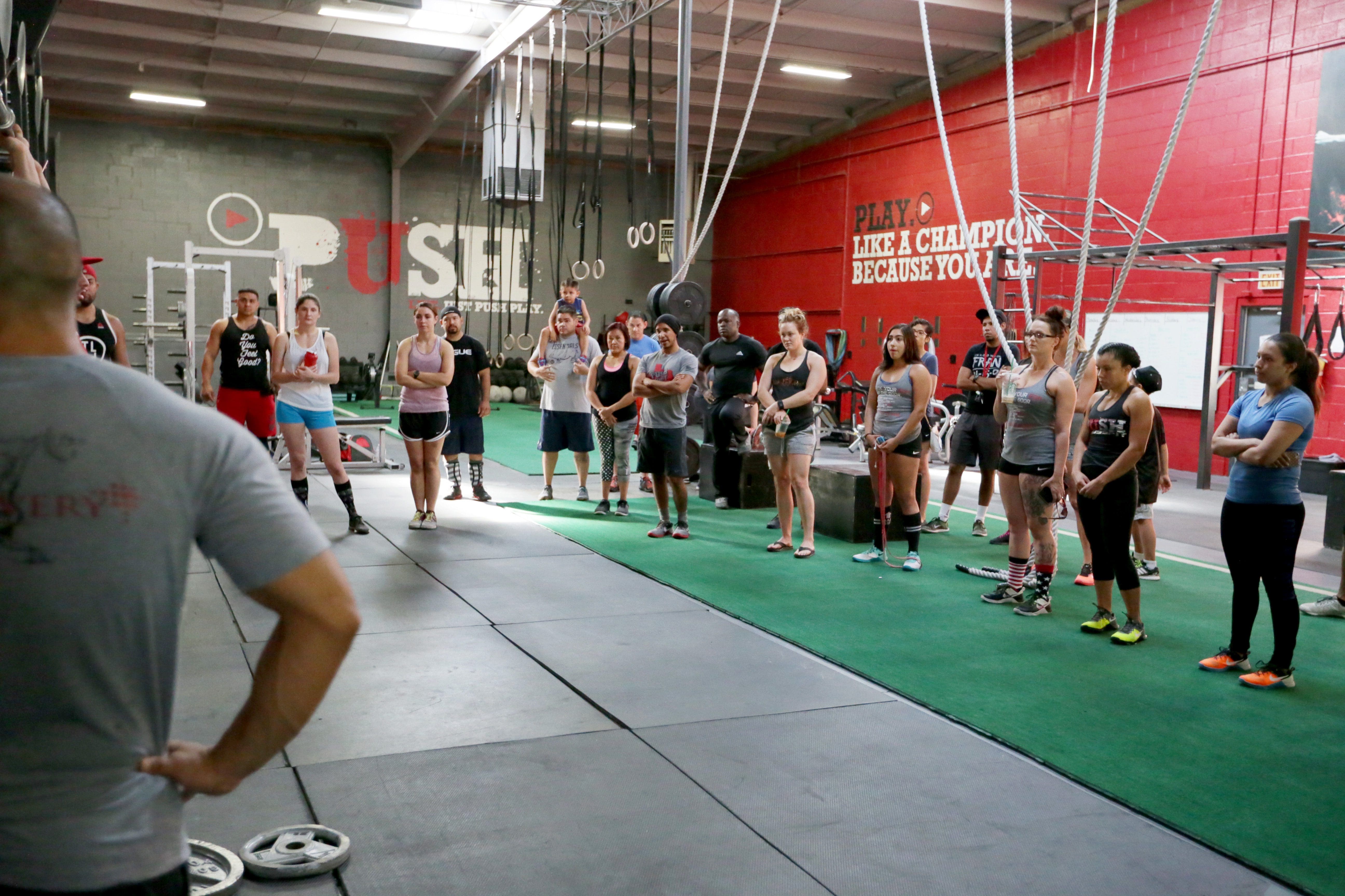
 

Image of trainer demonstrating rehabilitation exercises.


The purpose of our systematic review was to investigate the effectiveness of exercise compared to other interventions, placebo/sham interventions, or no intervention in improving self-rated recovery, functional recovery (eg, return to activities, work, or school), or clinical outcomes (eg, pain, health-related quality of life, depression) of patients with soft tissue injuries of the hip, thigh, and knee.


Methods


Registration


This systematic review protocol was registered with the International Prospective Register of Systematic Reviews on March 28, 2014 (CRD42014009140).


Eligibility Criteria


Population. Our review targeted studies of adults (?18 years) and/or children with soft tissue injuries of the hip, thigh, or knee. Soft tissue injuries include but are not limited to grade I to II sprains/strains; tendonitis; tendinopathy; tendinosis; patellofemoral pain (syndrome); iliotibial band syndrome; nonspecific hip, thigh, or knee pain (excluding major pathology); and other soft tissue injuries as informed by available evidence. We defined the grades of sprains and strains according to the classification proposed by the American Academy of Orthopaedic Surgeons (Tables 1 and 2). Affected soft tissues in the hip include the supporting ligaments and muscles crossing the hip joint into the thigh (including the hamstrings, quadriceps, and adductor muscle groups). Soft tissues of the knee include the supporting intra-articular and extra-articular ligaments and muscles crossing the knee joint from the thigh including the patellar tendon. We excluded studies of grade III sprains or strains, acetabular labral tears, meniscal tears, osteoarthritis, fractures, dislocations, and systemic diseases (eg, infection, neoplasm, inflammatory disorders).
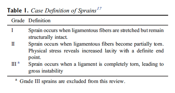
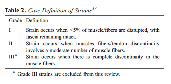
 

Table 1 Case Definition of Sprains


Table 2 Case Definition of Strains


Interventions. We restricted our review to studies that tested the isolated effect of exercise (ie, not part of a multimodal program of care). We defined exercise as any series of movements aimed at training or developing the body by routine practice or as physical training to promote good physical health.


Comparison Groups. We included studies that compared 1 or more exercise interventions to one another or one exercise intervention to other interventions, wait list, placebo/sham interventions, or no intervention.


Outcomes. To be eligible, studies had to include one of the following outcomes: (1) self-rated recovery; (2) functional recovery (eg, disability, return to activities, work, school, or sport); (3) pain intensity; (4) health-related quality of life; (5) psychological outcomes such as depression or fear; and (6) adverse events.


Study Characteristics. Eligible studies met the following criteria: (1) English language; (2) studies published between January 1, 1990, and April 8, 2015; (3) randomized controlled trials (RCTs), cohort studies, or case-control studies which are designed to assess the effectiveness and safety of interventions; and (4) included an inception cohort of a minimum of 30 participants per treatment arm with the specified condition for RCTs or 100 participants per group with the specified condition in cohort studies or case-control studies. Studies including other grades of sprains or strains in the hip, thigh, or knee had to provide separate results for participants with grades I or II sprains/strains to be included.


We excluded studies with the following characteristics: (1) letters, editorials, commentaries, unpublished manuscripts, dissertations, government reports, books and book chapters, conference proceedings, meeting abstracts, lectures and addresses, consensus development statements, or guideline statements; (2) study designs including pilot studies, cross-sectional studies, case reports, case series, qualitative studies, narrative reviews, systematic reviews (with or without meta-analyses), clinical practice guidelines, biomechanical studies, laboratory studies, and studies not reporting on methodology; (3) cadaveric or animal studies; and (4) studies on patients with severe injuries (eg, grade III sprains/strains, fractures, dislocations, full ruptures, infections, malignancy, osteoarthritis, and systemic disease).


Information Sources


We developed our search strategy with a health sciences librarian (Appendix 1). The Peer Review of Electronic Search Strategies (PRESS) Checklist was used by a second librarian to review the search strategy for completeness and accuracy. We searched MEDLINE and EMBASE, considered to be the major biomedical databases, and PsycINFO, for psychological literature through Ovid Technologies, Inc; CINAHL Plus with Full Text for nursing and allied health literature through EBSCOhost; and the Cochrane Central Register of Controlled Trials through Ovid Technologies, Inc, for any studies not captured by the other databases. The search strategy was first developed in MEDLINE and subsequently adapted to the other bibliographic databases. Our search strategies combined controlled vocabulary relevant to each database (eg, MeSH for MEDLINE) and text words relevant to exercise and soft tissue injuries of the hip, thigh, or knee including grade I to II sprain or strain injuries (Appendix 1). We also hand searched the reference lists of previous systematic reviews for any additional relevant studies.


Study Selection


A 2-phase screening process was used to select eligible studies. Random pairs of independent reviewers screened citation titles and abstracts to determine the eligibility of studies in phase 1. Screening resulted in studies being classified as relevant, possibly relevant, or irrelevant. In phase 2, the same pairs of reviewers independently screened the possibly relevant studies to determine eligibility. Reviewers met to reach consensus on the eligibility of studies and resolve disagreements. A third reviewer was used if consensus could not be reached.
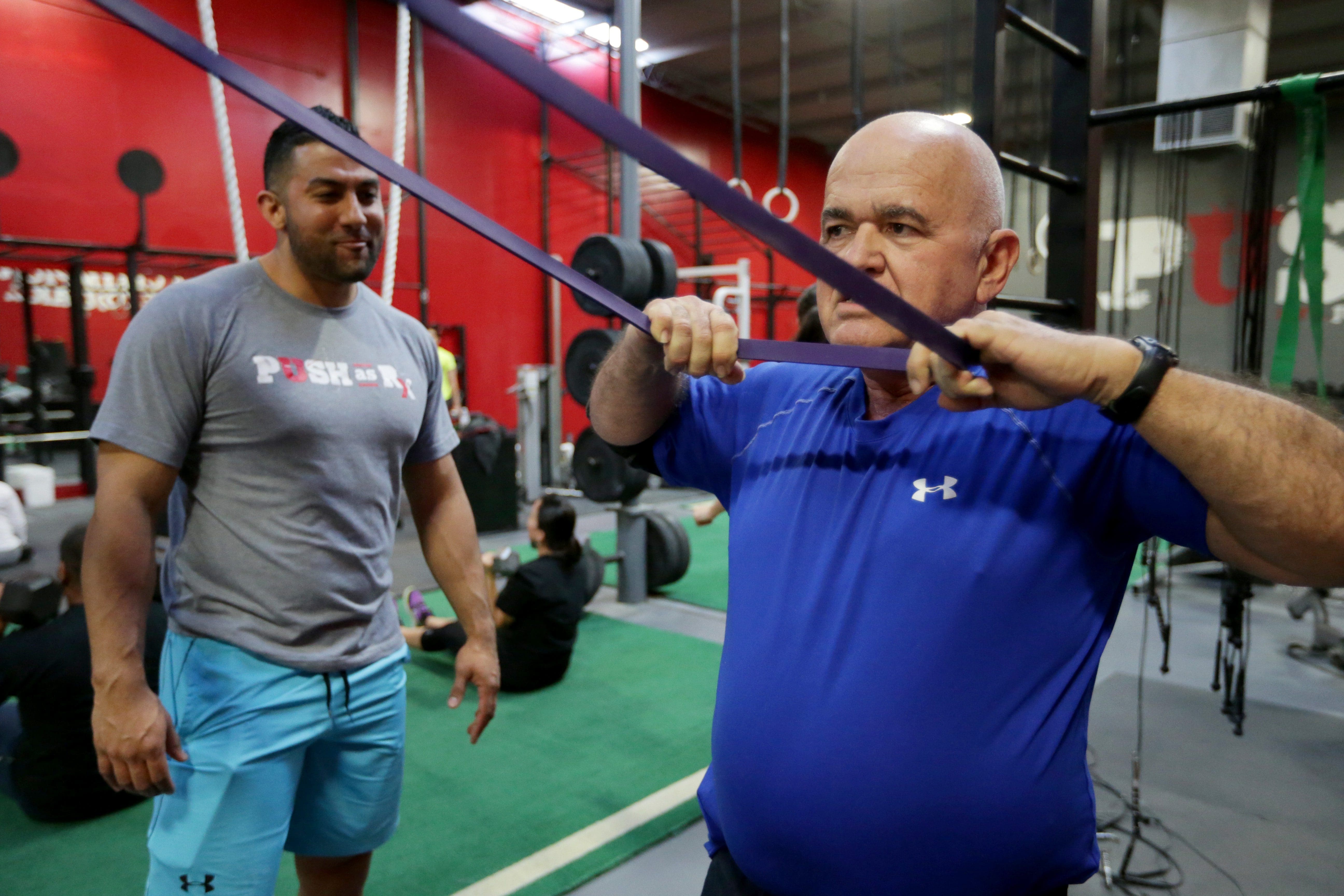
 

Image of older patient engaging in upper rehabilitation exercises with a personal trainer.


Assessment of Risk of Bias


Independent reviewers were randomly paired to critically appraise the internal validity of eligible studies using the Scottish Intercollegiate Guidelines Network (SIGN) criteria. The impact of selection bias, information bias, and confounding on the results of a study was qualitatively evaluated using the SIGN criteria. These criteria were used to guide reviewers in making an informed overall judgment on the internal validity of studies. This methodology has been previously described. A quantitative score or a cutoff point to determine the internal validity of studies was not used for this review.


The SIGN criteria for RCTs were used to critically appraise the following methodological aspects: (1) clarity of the research question, (2) randomization method, (3) concealment of treatment allocation, (4) blinding of treatment and outcomes, (5) similarity of baseline�characteristics between/among treatment arms, (6) cointervention contamination, (7) validity and reliability of outcome measures, (8) follow-up rates, (9) analysis according to intention-to-treat principles, and (10) comparability of results across study sites (where applicable). Consensus was reached through reviewer discussion. Disagreements were resolved by an independent third reviewer when consensus could not be reached. The risk of bias of each appraised study was also reviewed by a senior epidemiologist (PC). Authors were contacted when additional information was needed to complete the critical appraisal. Only studies with low risk of bias were included in our evidence synthesis.


Data Extraction and Synthesis of Results


Data were extracted from studies (DS) with low risk of bias to create evidence tables. A second reviewer independently checked the extracted data. We stratified results based on the duration of the condition (recent onset [0-3 months], persistent [N3 months], or variable duration [recent onset and persistent combined]).


We used standardized measures to determine the clinical importance of changes reported in each trial for common outcome measures. These include a between-group difference of 2/10 points on the Numeric Rating Scale (NRS), 2/10 cm difference on the Visual Analog Scale (VAS), and 10/100 point difference on the Kujala Patellofemoral scale, otherwise known as the Anterior Knee Pain Scale.


Statistical Analyses


Agreement between reviewers for the screening of articles was computed and reported using the ? statistic and 95% confidence interval (CI). Where available, we used data provided in the studies with a low risk of bias to measure the association between the tested interventions and the outcomes by computing the relative risk (RR) and its 95% CI. Similarly, we computed differences in mean changes between groups and 95% CI to quantify the effectiveness of interventions. The calculation of 95% CIs was based on the assumption that baseline and follow-up outcomes were highly correlated (r = 0.80).


Reporting


This systematic review was organized and reported based on the Preferred Reporting Items for Systematic Reviews and Meta-Analyses statement.

 

Dr. Alex Jimenez's Insight

As a doctor of chiropractic, automobile accident injuries are one of the most common reasons people seek chiropractic care. From neck injuries, such as whiplash, to headaches and back pain, chiropractic can be utilized to safely and effectively restore the integrity of the spine after a car crash. A chiropractor like myself will often use a combination of spinal adjustments and manual manipulations, as well as a variety of other non-invasive treatment methods,�to gently correct any spinal misalignments resulting from an auto accident injury. Whiplash and other types of neck injuries occur when the complex structures along the cervical spine are stretched beyond their natural range of movement due to the sudden back-and-forth movement of the head and neck from the force of the impact. Back injury, particularly in the lower spine, are also common as a result of an automobile accident. When the complex structures along the lumbar spine are damaged or injured, symptoms of sciatica may radiate down the lower back, into the buttocks, hips, thighs, legs and down into the feet. Knee injuries may also occur upon impact during an auto accident. Exercise is frequently used with chiropractic care to help promote recovery as well as improve strength, flexibility and mobility. Rehabilitation exercises are offered to patients to further restore the integrity of their body. The following research studies demonstrate that exercise, compared to non-invasive treatment options, is a safe and effective treatment method for individuals suffering with neck and lower extremity injury from a car crash.


Results


Study Selection


We screened 9494 citations based on the title and abstract (Figure 1). Of these, 60 full-text publications were screened, and 9 articles were critically appraised. The primary reasons for ineligibility during full text screening were (1) ineligible study design, (2) small sample size (n b 30 per treatment arm), (3) multimodal interventions not allowing isolation of the effectiveness of exercise, (4) ineligible study population, and (5) interventions not meeting our definition of exercise (Figure 1). Of those critically appraised, 3 studies (reported in 4 articles) had low risk of bias and were included in our synthesis. The interrater agreement for the screening of the articles was ? = 0.82 (95% CI, 0.69-0.95). The percentage agreement for the critical appraisal of studies was 75% (6/8 studies). Disagreement was resolved through discussion for 2 studies. We contacted authors from 5 studies during critical appraisal to request additional information and 3 responded.
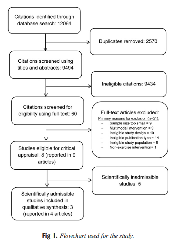
 

Figure 1 Flowchart Used for the Study


Study Characteristics


The studies with low risk of bias were RCTs. One study, conducted in the Netherlands, examined the effectiveness of a standardized exercise program compared to a �wait and see� approach in participants with patellofemoral pain syndrome of variable duration. A second study, with outcomes reported in 2 articles, compared the benefit of closed vs open kinetic chain exercises in individuals with�variable duration patellofemoral pain syndrome in Belgium. The final study, conducted in Denmark, investigated active training compared to a multimodal physiotherapy intervention for the management of persistent adductor-related groin pain.


Two RCTs used exercise programs that combined strengthening exercises with balance or agility training for the lower extremity. Specifically, the strengthening exercises consisted of both isometric and concentric contractions of the quadriceps, hip adductor, and gluteal muscles for the management of patellofemoral pain46 and hip adductors and muscles of the trunk and pelvis for adductor-related groin pain. The exercise programs ranged from 646 to 1243 weeks in duration and were supervised and clinic based with additional daily home exercises. The exercise programs were compared to a �wait and see� approach or to multimodal physiotherapy. The third RCT compared 2 different 5-week protocols which combined either closed or open kinetic chain strengthening and stretching exercises for the lower extremity musculature.


Meta-analysis was not performed due to heterogeneity of accepted studies with respect to patient populations, interventions, comparators, and outcomes. Principles of best evidence synthesis were used to develop evidence statements and perform a qualitative synthesis of findings from studies with low risk of bias.


Risk of Bias Within Studies


The studies with low risk of bias had a clearly defined research question, used appropriate blinding methods where possible, reported adequate similarity of baseline characteristics between treatment arms, and performed an intention-to-treat analyses where applicable (Table 3). The RCTs had follow-up rates greater than 85%. However, these studies also had methodological limitations: insufficient detail describing methods for allocation concealment (1/3), insufficient detail describing methods of randomization (1/3), the use of outcome measures that have not been demonstrated to be valid or reliable (ie, muscle length and successful treatment) (2/3), and clinically important differences in baseline characteristics (1/3).
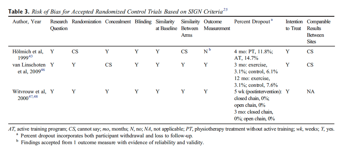
 

Table 3 Risk of Bias for Accepted Randomized Control Trials Based on SIGN Criteria


Of 9 relevant articles, 5 were deemed to have high risk of bias. These studies had the following limitations: (1) poor or unknown randomization methods (3/5); (2) poor or unknown allocation concealment methods (5/ 5); (3) outcome assessor not blinded (4/ 5); (4) clinically important differences in baseline characteristics (3/5); (5) dropouts not reported, insufficient information regarding dropouts per group or large differences in dropout rates between treatment arms (N15%) (3/5); and (6) a lack of information about or no intention-to-treat analysis (5/5).


Summary of Evidence


Patellofemoral Pain Syndrome of Variable Duration. Evidence from 1 RCT suggests that a clinic-based progressive exercise program may provide short- and long-term benefit over usual care for the management of patellofemoral pain syndrome of variable duration. van Linschoten et al randomized participants with a clinical diagnosis of patellofemoral pain syndrome of 2 months to 2 years duration to (1) a clinic-based exercise program (9 visits over 6 weeks) consisting of progressive, static, and dynamic strengthening exercises for the quadriceps, adductor, and gluteal muscles and balance and flexibility exercises, or (2) a usual care �wait and see� approach. Both groups received standardized information, advice, and home-based isometric exercises for the quadriceps based on recommendations from Dutch General Practitioner guidelines (Table 4). There�were statistically significant differences favoring the exercise group for (1) pain (NRS) at rest at 3 months (mean change difference 1.1/10 [95% CI, 0.2-1.9]) and 6 months (mean change difference 1.3/10 [95% CI, 0.4-2.2]); (2) pain (NRS) with activity at 3 months (mean change difference 1.0/10 [95% CI, 0.1-1.9]) and 6 months (mean change difference 1.2/10 [95% CI, 0.2-2.2]); and (3) function (Kujala Patellofemoral Scale [KPS]) at 3 months (mean change difference 4.9/100 [95% CI, 0.1-9.7]). However, none of these differences were clinically important. Furthermore, there were no significant differences in the proportion of participants reporting recovery (fully recovered, strongly recovered), but the exercise group was more likely to report improvement at 3-month follow-up (odds ratio [OR], 4.1 [95% CI, 1.9-8.9]).
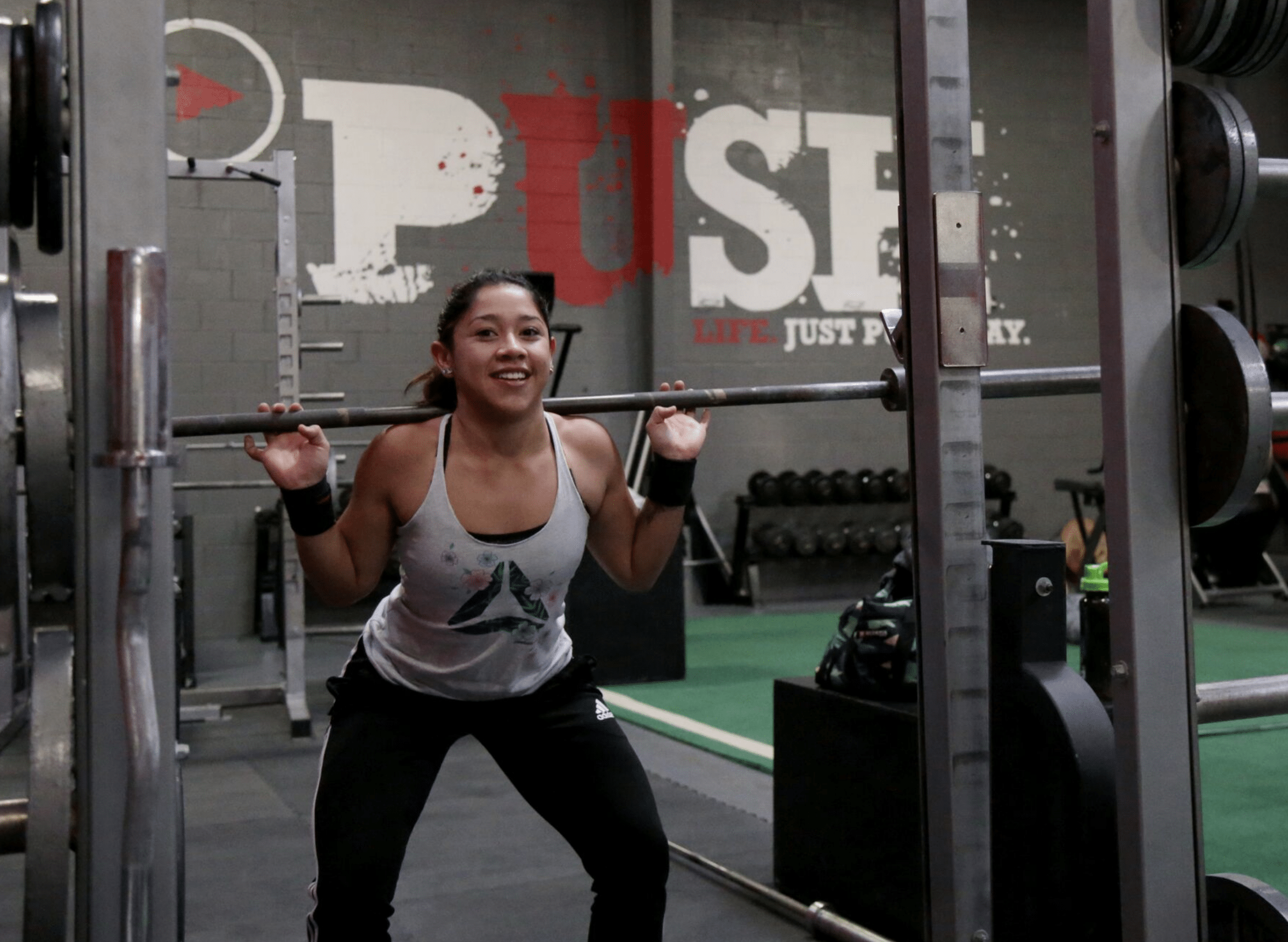
 

Image of patient engaging in rehabilitation exercises.


Evidence from a second RCT suggests that physiotherapist- supervised closed kinetic chain leg exercises (where the foot remains in constant contact with a surface) may provide short-term benefit compared to supervised open kinetic chain exercises (where the limb moves freely) for some patellofemoral pain syndrome symptoms (Table 4). All participants trained for 30 to 45 minutes, 3 times per week for 5 weeks. Both groups were instructed to perform static lower limb stretching after each training session. Those randomized to closed chain exercises performed supervised (1) leg presses, (2) knee bends, (3) stationary biking, (4) rowing, (5) step-up and step-down exercises, and (6) progressive jumping exercises. Open chain exercise participants performed (1) maximal quad muscle contraction, (2) straight-leg raises, (3) short arc movements from 10� to full knee extension, and (4) leg adduction. Effect sizes were not reported, but the authors reported statistically significant differences favoring closed kinetic chain exercise at 3 months for (1) frequency of locking (P = .03), (2) clicking sensation (P = .04), (3) pain with isokinetic testing (P = .03), and (4) pain during night (P = .02). The clinical significance of these results is unknown. There were no statistically significant differences between groups for any other pain or functional measures at any follow-up period.
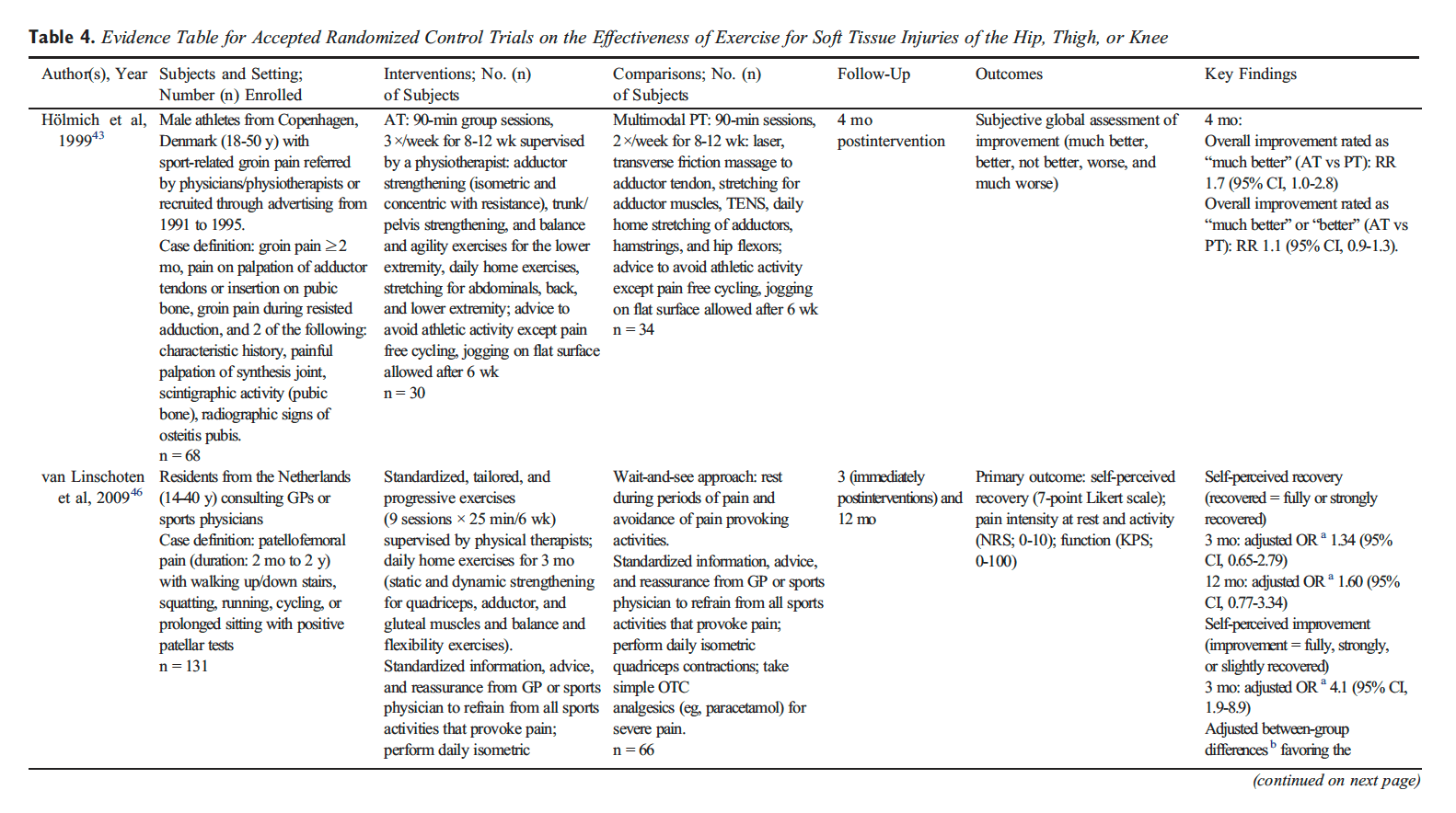
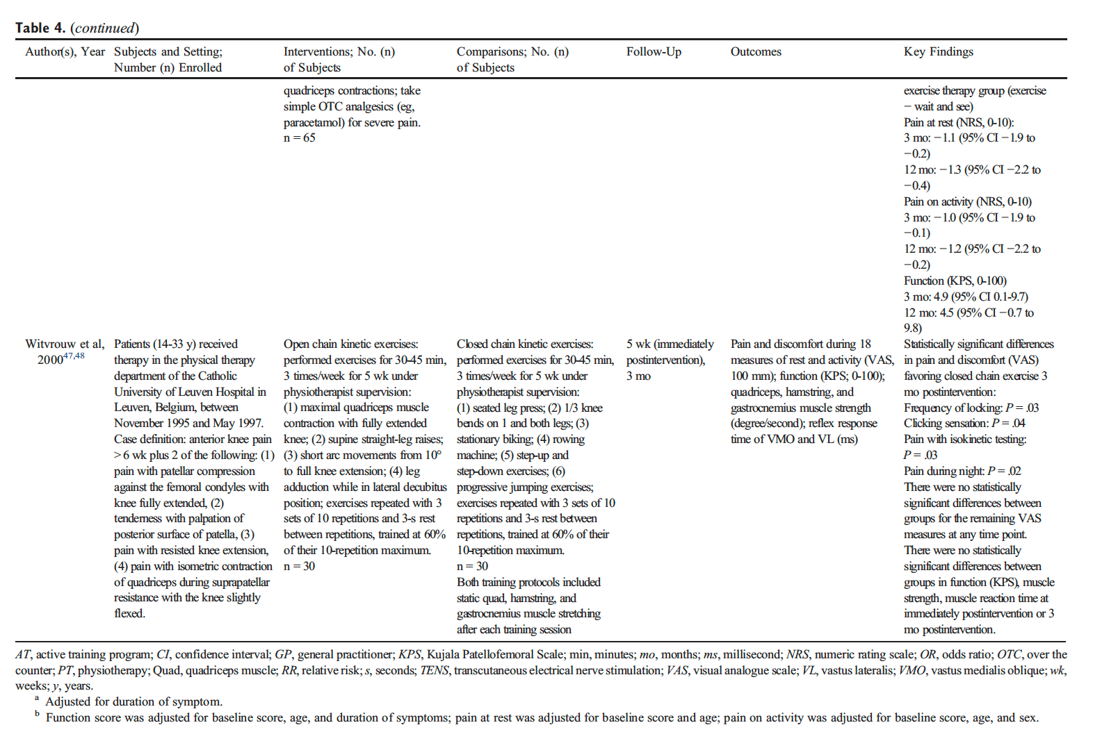
 

Table 4 Evidence Table for Accepted Randomized Control Trials on the Effectiveness of Exercise for Soft Tissue Injuries of the Hip, Thigh, or Knee


Table 4 Evidence Table for Accepted Randomized Control Trials on the Effectiveness of Exercise for Soft Tissue Injuries of the Hip, Thigh, or Knee


Persistent Adductor-Related Groin Pain


Evidence from 1 RCT suggests that a clinic-based group exercise program is more effective than a multimodal program of care for persistent adductor-related groin pain. H�lmich et al studied a group of male athletes with a clinical diagnosis of adductor-related groin pain of greater than 2 months duration (median duration, 38-41 weeks; range, 14-572 weeks) with or without osteitis pubis. Participants were randomized to (1) a clinic-based group exercise program (3 sessions per week for 8-12 weeks) consisting of isometric and concentric resistance strengthening exercises for the adductors, trunk, and pelvis; balance and agility exercises for the lower extremity; and stretching for the abdominals, back, and lower extremity (with the exception of the adductor muscles) or (2) a multimodal physiotherapy program (2 visits per week for 8-12 weeks) consisting of laser; transverse friction massage; transcutaneous electrical nerve stimulation (TENS); and stretching for the adductors, hamstrings, and hip flexors (Table 4). Four months after the intervention, the exercise group was more likely to report that their condition was �much better� (RR, 1.7 [95% CI, 1.0-2.8]).


Adverse Events


None of the included studies commented on the frequency or nature of adverse events.


Discussion


Summary of Evidence


Our systematic review examined the effectiveness of exercise for the management of soft tissue injuries of the hip, thigh, or knee. Evidence from 1 RCT suggests that a clinic-based progressive combined exercise program may offer additional short- or long-term benefit compared to providing information and advice for the management of patellofemoral pain syndrome of variable duration. There is also evidence that supervised closed kinetic chain exercises may be beneficial for some patellofemoral pain syndrome symptoms compared to open kinetic chain exercises. For persistent adductor-related groin pain, evidence from 1 RCT suggests that a clinic-based group exercise program is more effective than a multimodal program of care. Despite the common and frequent use of exercise prescription, there is limited high-quality evidence to inform the use of exercise for the management of soft tissue injuries of the lower extremity. Specifically, we did not find high-quality studies on exercise for the management of some of the more commonly diagnosed conditions including patellar tendinopathy, hamstring sprain and strain injuries, hamstring tendinopathy, trochanteric bursitis, or capsular injuries of the hip.
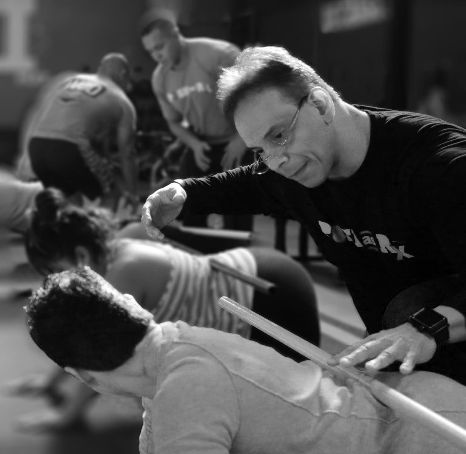
 

Image of Dr. Jimenez demonstrating rehabilitation exercises to patient.


Previous Systematic Reviews


Our results are consistent with findings from previous systematic reviews, concluding that exercise is effective for the management of patellofemoral pain syndrome and groin pain. However, the results from previous systematic reviews examining the use of exercise for the management of patellar tendinopathy and acute hamstring injuries are inconclusive. One review noted strong evidence for use of eccentric training, whereas others reported uncertainty of whether isolated eccentric exercises were beneficial for tendinopathy compared to other forms of exercise. Furthermore, there is limited evidence of a positive effect from stretching, agility and trunk stability exercises, or slump stretching for the management of acute�hamstring injuries. Differing conclusions between systematic reviews and the limited number of studies deemed admissible in our work may be attributed to differences in methodology. We screened reference lists of previous systematic reviews, and most studies included in the reviews did not meet our inclusion criteria. Many studies accepted in other reviews had small sample sizes (b30 per treatment arm). This increases the risk of residual confounding while also reducing the effect size precision. Furthermore, a number of systematic reviews included case series and case studies. These types of studies are not designed to assess the effectiveness of interventions. Finally, previous reviews included studies where exercise was part of a multimodal intervention, and as a consequence, the isolated effect of exercise could not be ascertained. Of the studies that satisfied our selection criteria, all were critically appraised in our review, and only 3 had low risk of bias and were included in our synthesis.


Strengths


Our review has many strengths. First, we developed a rigorous search strategy that was independently reviewed by a second librarian. Second, we defined clear inclusion and exclusion criteria for the selection of possibly relevant studies and only considered studies with adequate sample sizes. Third, pairs of trained reviewers screened and critically appraised eligible studies. Fourth, we used a valid set of criteria (SIGN) to critically appraise studies. Finally, we restricted our synthesis to studies with low risk of bias.


Limitations and Recommendations for Future Research


Our review also has limitations. First, our search was limited to studies published in the English language. However, previous reviews have found that the restriction of systematic reviews to English language studies has not led to a bias in reported results. Second, despite our broad definition of soft tissue injuries of the hip, thigh, or knee, our search strategy may not have captured all potentially relevant studies. Third, our review may have missed potentially relevant studies published before 1990. We aimed to minimize this by hand searching the reference lists of previous systematic reviews. Finally, critical appraisal requires scientific judgment that may differ between reviewers. We minimized this potential bias by training reviewers in the use of the SIGN tool and using a consensus process to determine study admissibility. Overall, our systematic review highlights a deficit of strong research in this area.


High-quality studies on the effectiveness of exercise for the management of soft tissue injuries of the lower extremity are needed. Most studies included in our review (63%) had a high risk of bias and could not be included in our synthesis. Our review identified important gaps in the literature. Specifically, studies are needed to inform the specific effects of exercises, their long-term effects, and the optimal doses of intervention. Furthermore, studies are needed to determine the relative effectiveness of different types of exercise programs and if the effectiveness varies for soft tissue injuries of the hip, thigh, and knee.


Conclusion


There is limited high-quality evidence to inform the use of exercise for the management of soft tissue injuries of the hip, thigh, and knee. The current evidence suggests that a clinic-based progressive combined exercise program may lead to improved recovery when added to information and advice on resting and avoiding pain provoking activities for the management of patellofemoral pain syndrome. For persistent adductor-related groin pain, a supervised clinic- based group exercise program is more effective than multimodal care in promoting recovery.


Funding Sources and Potential Conflicts of Interest


This study was funded by the Ontario Ministry of Finance and the Financial Services Commission of Ontario (RFP no. OSS_00267175). The funding agency was not involved in the collection of data, data analysis, interpretation of data, or drafting of the manuscript. The research was undertaken, in part, thanks to funding from the Canada Research Chairs program. Pierre C�t� has previously received funding from a Grant from the Ontario Ministry of Finance; consulting for the Canadian Chiropractic Protective Association; speaking and/or teaching arrangements for the National Judicial Institute and Soci�t� des M�decins Experts du Quebec; trips/travel, European Spine Society; board of directors, European Spine Society; grants: Aviva Canada; fellowship support, Canada Research Chair Program�Canadian Institutes of Health Research. No other conflicts of interest were reported for this study.


Contributorship Information


  • Concept development (provided idea for the research): D.S., C.B., P.C., J.W., H.Y., S.V.
  • Design (planned the methods to generate the results): D.S., C.B., P.C., H.S., J.W., H.Y., S.V.
  • Supervision (provided oversight, responsible for organization and implementation, writing of the manuscript): D.S., P.C.
  • Data collection/processing (responsible for experiments, patient management, organization, or reporting data): D.S., C.B., H.S., J.W., D.e.S., R.G., H.Y., K.R., J.C., K.D., P.C., P.S., R.M., S.D., S.V.
  • Analysis/interpretation (responsible for statistical analysis, evaluation, and presentation of the results): D.S., C.B., P.C., H.S., M.S., K.R., L.C.
  • Literature search (performed the literature search): A.T.V.
  • Writing (responsible for writing a substantive part of the manuscript): D.S., C.B., P.C., H.S.
  • Critical review (revised manuscript for intellectual content, this does not relate to spelling and grammar checking): D.S., P.C., H.S., J.W., D.e.S., R.G., M.S., A.T.V., H.Y., K.R., J.C., K.D., L.C., P.S., S.D., R.M., S.V.


Practical Applications


  • There is evidence to suggest that clinic-based exercises may benefit patients with patellofemoral pain syndrome or adductor-related groin pain.
  • Supervised progressive exercises may be beneficial for patellofemoral pain syndrome of variable duration compared to information/advice.
  • Supervised closed kinetic chain exercises may provide more benefit compared to open kinetic chain exercises for some patellofemoral pain syndrome symptoms.
  • Self-rated improvement in persistent groin pain is higher after a clinic-based group exercise program compared to multimodal physiotherapy.


Are Non-Invasive Interventions Effective for the Management of Headaches Associated with Neck Pain?


Furthermore,�other non-invasive interventions, as well as non-pharmacological interventions, are also commonly utilized to help treat symptoms of neck pain and headaches associated with neck injuries, such as whiplash, caused by automobile accidents. As mentioned before, whiplash is one of the most common types of neck injuries resulting from auto accidents. Chiropractic care, physical therapy and exercise, can be used to improve the symptoms of neck pain, according to the following research studies.


Abstract


Purpose


To update findings of the 2000�2010 Bone and Joint Decade Task Force on Neck Pain and its Associated Disorders and evaluate the effectiveness of non-invasive and non-pharmacological interventions for the management of patients with headaches associated with neck pain (i.e., tension-type, cervicogenic, or whiplash-related headaches).


Methods


We searched five databases from 1990 to 2015 for randomized controlled trials (RCTs), cohort studies, and case�control studies comparing non-invasive interventions with other interventions, placebo/sham, or no interventions. Random pairs of independent reviewers critically appraised eligible studies using the Scottish Intercollegiate Guidelines Network criteria to determine scientific admissibility. Studies with a low risk of bias were synthesized following best evidence synthesis principles.


Results


We screened 17,236 citations, 15 studies were relevant, and 10 had a low risk of bias. The evidence suggests that episodic tension-type headaches should be managed with low load endurance craniocervical and cervicoscapular exercises. Patients with chronic tension-type headaches may also benefit from low load endurance craniocervical and cervicoscapular exercises; relaxation training with stress coping therapy; or multimodal care that includes spinal mobilization, craniocervical exercises, and postural correction. For cervicogenic headaches, low load endurance craniocervical and cervicoscapular exercises; or manual therapy (manipulation with or without mobilization) to the cervical and thoracic spine may also be helpful.
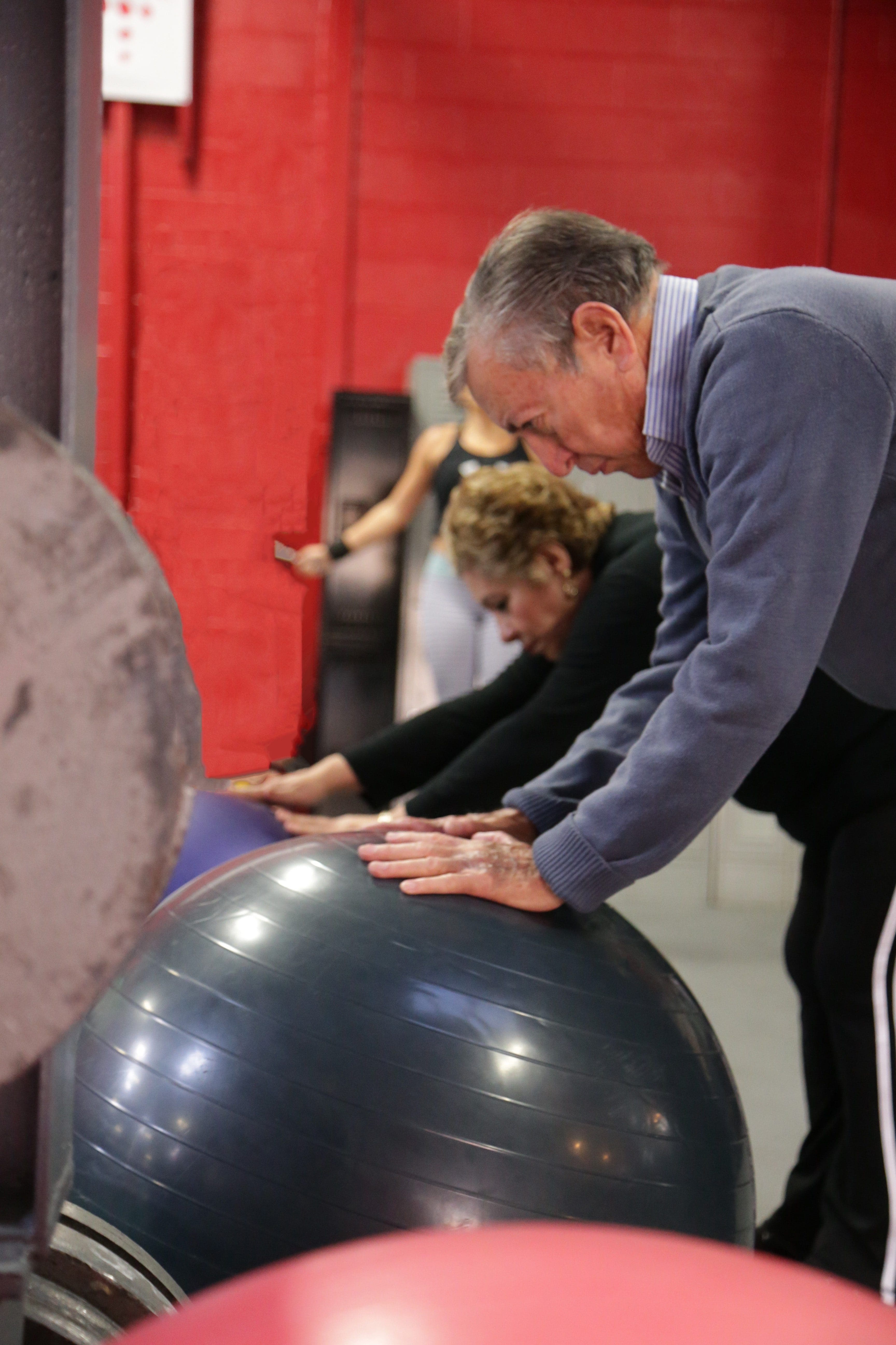
 

Image of elderly couple participating in low-impact rehabilitation exercises.


Conclusions


The management of headaches associated with neck pain should include exercise. Patients who suffer from chronic tension-type headaches may also benefit from relaxation training with stress coping therapy or multimodal care. Patients with cervicogenic headache may also benefit from a course of manual therapy.


Keywords


Non-invasive interventions, Tension-type headache, Cervicogenic headache, Headache attributed to whiplash injury, Systematic review


Notes


Acknowledgments


We would like to acknowledge and thank all of the individuals who have made important contributions to this review: Robert Brison, Poonam Cardoso, J. David Cassidy, Laura Chang, Douglas Gross, Murray Krahn, Michel Lacerte, Gail Lindsay, Patrick Loisel, Mike Paulden, Roger Salhany, John Stapleton, Angela Verven, and Leslie Verville. We would also like to thank Trish Johns-Wilson at the University of Ontario Institute of Technology for her review of the search strategy.


Compliance with Ethical Standards


Conflict of Interest


Dr. Pierre C�t� has received a grant from the Ontario government, Ministry of Finance, funding from the Canada Research Chairs program, personal fees from National Judicial Institute for lecturing, and personal fees from European Spine Society for teaching. Drs. Silvano Mior and Margareta Nordin have received reimbursement for travel expenses to attend meetings for the study. The remaining authors report no declarations of interest.


Funding


This work was supported by the Ontario Ministry of Finance and the Financial Services Commission of Ontario [RFP# OSS_00267175]. The funding agency had no involvement in the study design, collection, analysis, interpretation of data, writing of the manuscript or decision to submit the manuscript for publication. The research was undertaken, in part, thanks to funding from the Canada Research Chairs program to Dr. Pierre C�t�, Canada Research Chair in Disability Prevention and Rehabilitation at the University of Ontario Institute of Technology.


In conclusion,�exercise included in chiropractic care and other non-invasive interventions should be utilized as an essential part of treatment to further help improve the symptoms of neck injury as well as that of hip, thigh and knee injury. According to the above research studies, exercise, or physical activity, is beneficial towards speeding up recovery time for patients with automobile accident injuries and for restoring strength, flexibility and mobility to the affected structures of the spine. Information referenced from the National Center for Biotechnology Information (NCBI). The scope of our information is limited to chiropractic as well as to spinal injuries and conditions. To discuss the subject matter, please feel free to ask Dr. Jimenez or contact us at 915-850-0900 .


Curated by Dr. Alex Jimenez

 

Green-Call-Now-Button-24H-150x150-2-3.png


Additional Topics: Sciatica


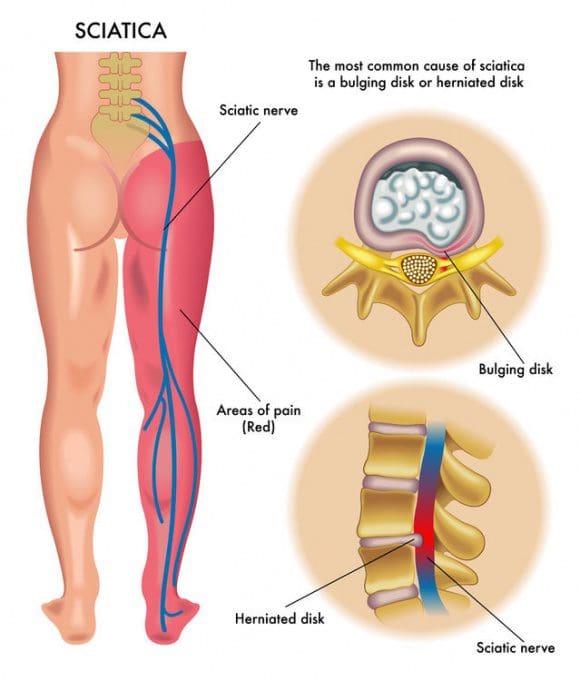
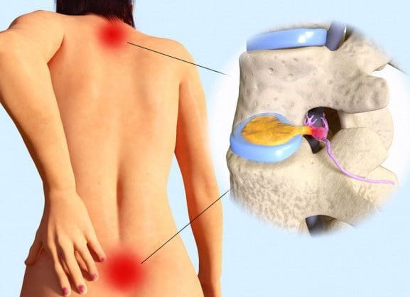
Sciatica is referred to as a collection of symptoms rather than a single type of injury or condition. The symptoms are characterized as radiating pain, numbness and tingling sensations from the sciatic nerve in the lower back, down the buttocks and thighs and through one or both legs and into the feet. Sciatica is commonly the result of irritation, inflammation or compression of the largest nerve in the human body, generally due to a herniated disc or bone spur.


blog picture of cartoon paperboy big news


IMPORTANT TOPIC: EXTRA EXTRA: Treating Sciatica Pain


Blank
References

1. Lambers K, Ootes D, Ring D. Incidence of patients with lower
extremity injuries presenting to US emergency departments by
anatomic region, disease category, and age. Clin Orthop Relat
Res 2012;470(1):284-90.
2. Workplace Safety and Insurance Board. By the numbers: 2014
WSIB statistical report. Injury profile�schedule 1; historical
and supplementary data on leading part of body injuries.
[cited June 22, 2015]; Available from: http://www.
wsibstatistics.ca/en/s1injury/s1part-of-body/ 2014.
3. Hincapie CA, Cassidy JD, C�t� P, Carroll LJ, Guzman J.
Whiplash injury is more than neck pain: a population-based
study of pain localization after traffic injury. J Occup Environ
Med 2010;52(4):434-40.
4. Bureau of Labor Statistics US Department of Labor. Nonfatal
occupational injuries and illnesses requiring days away from
work. Table 5. Washington, DC 2014 [June 22, 2015];
Available from: http://www.bls.gov/news.release/archives/
osh2_12162014.pdf 2013.
5. New ZealandGuidelinesDevelopmentGroup. The diagnosis and
management of soft tissue knee injuries: internal derangements.
Best practice evidence-based guideline. Wellington: Accident
Compensation Corporation; 2003 [[June 22, 2015]; Available
from: http://www.acc.co.nz/PRD_EXT_CSMP/groups/
external_communications/documents/guide/wcmz002488.pdf].
6. Bizzini M, Childs JD, Piva SR, Delitto A. Systematic review of
the quality of randomized controlled trials for patellofemoral pain
syndrome. J Orthop Sports Phys Ther 2003;33(1):4-20.
7. Crossley K, Bennell K, Green S, McConnell J. A systematic
review of physical interventions for patellofemoral pain
syndrome. Clin J Sport Med 2001;11(2):103-10.
8. Harvie D, O�Leary T, Kumar S. A systematic review of
randomized controlled trials on exercise parameters in the
treatment of patellofemoral pain: what works? J Multidiscip
Healthc 2011;4:383-92.
9. Lepley AS, Gribble PA, Pietrosimone BG. Effects of electromyographic
biofeedback on quadriceps strength: a systematic
review. J Strength Cond Res 2012;26(3):873-82.
10. Peters JS, Tyson NL. Proximal exercises are effective in treating
patellofemoral pain syndrome: a systematic review. Int J Sports
Phys Ther 2013;8(5):689-700.
11. Wasielewski NJ, Parker TM, Kotsko KM. Evaluation of
electromyographic biofeedback for the quadriceps femoris: a
systematic review. J Athl Train 2011;46(5):543-54.
12. Kristensen J, Franklyn-Miller A. Resistance training in musculoskeletal
rehabilitation: a systematic review. Br J Sports Med
2012;46(10):719-26.
13. Larsson ME, Kall I, Nilsson-Helander K. Treatment of patellar
tendinopathy�a systematic review of randomized controlled
trials. Knee Surg Sports Traumatol Arthrosc 2012;20(8):1632-46.
14. Malliaras P, Barton CJ, Reeves ND, Langberg H. Achilles and
patellar tendinopathy loading programmes: a systematic review
comparing clinical outcomes and identifying potential mechanisms
for effectiveness. Sports Med 2013;43(4):267-86.
15. Wasielewski NJ, KotskoKM. Does eccentric exercise reduce pain
and improve strength in physically active adults with symptomatic
lower extremity tendinosis? A systematic review. J Athl Train
2007;42(3):409-21.
16. Reurink G, Goudswaard GJ, Tol JL, Verhaar JA, Weir A, Moen
MH. Therapeutic interventions for acute hamstring injuries: a
systematic review. Br J Sports Med 2012;46(2):103-9.
17. American Academy of Orthopaedic Surgeons. Sprains, strains,
and other soft-tissue injuries. [updated July 2007 March 11,
2013]; Available from: http://orthoinfo.aaos.org/topic.cfm?topic=
A00304 2007.
18. Abenhaim L, Rossignol M, Valat JP, et al. The role of activity in
the therapeutic management of back pain. Report of
the International Paris Task Force on Back Pain. Spine 2000;
25(4 Suppl):1S-33S.
19. McGowan J, Sampson M, Lefebvre C. An evidence
based checklist for the Peer Review of Electronic Search Strategies
(PRESS EBC). Evid Based Library Inf Pract 2010;5(1):149-54.
20. Sampson M, McGowan J, Cogo E, Grimshaw J, Moher D,
Lefebvre C. An evidence-based practice guideline for the peer
review of electronic search strategies. J Clin Epidemiol 2009;
62(9):944-52.
21. Almeida MO, Silva BN, Andriolo RB, Atallah AN, Peccin MS.
Conservative interventions for treating exercise-related musculotendinous,
ligamentous and osseous groin pain. Cochrane
Database Syst Rev 2013;6:CD009565.
22. Ellis R, Hing W, Reid D. Iliotibial band friction syndrome�a
systematic review. Man Ther 2007;12(3):200-8.
23. Machotka Z, Kumar S, Perraton LG. A systematic review of the
literature on the effectiveness of exercise therapy for groin pain in
athletes. SportsMed Arthrosc Rehabil Ther Technol 2009;1(1):5.
24. Moksnes H, Engebretsen L, Risberg MA. The current evidence
for treatment of ACL injuries in children is low: a systematic
review. J Bone Joint Surg Am 2012;94(12):1112-9.
25. Harbour R, Miller J. A new system for grading recommendations
in evidence based guidelines. BMJ 2001;323(7308):
334-6.
26. Carroll LJ, Cassidy JD, Peloso PM, Garritty C, Giles-Smith L.
Systematic search and review procedures: results of the WHO
Collaborating Centre Task Force on Mild Traumatic Brain
Injury. J Rehabil Med 2004(43 Suppl):11-4.
27. Carroll LJ, Cassidy JD, Peloso PM, et al. Methods for the best
evidence synthesis on neck pain and its associated disorders: the
Bone and Joint Decade 2000-2010 Task Force on Neck Pain
and Its Associated Disorders. JManipulative Physiol Ther 2009;
32(2 Suppl):S39-45.
28. C�t� P, Cassidy JD, Carroll L, Frank JW, Bombardier C. A
systematic review of the prognosis of acute whiplash and a new
conceptual framework to synthesize the literature. Spine (Phila
Pa 1976) 2001;26(19):E445-58.
29. Hayden JA, Cote P, Bombardier C. Evaluation of the quality of
prognosis studies in systematic reviews. Ann Intern Med 2006;
144(6):427-37.
30. Hayden JA, van der Windt DA, Cartwright JL, Cote P,
Bombardier C. Assessing bias in studies of prognostic factors.
Ann Intern Med 2013;158(4):280-6.
31. Spitzer WO, Skovron ML, Salmi LR, et al. Scientific
monograph of the Quebec Task Force on Whiplash-Associated
Disorders: redefining �whiplash� and its management. Spine
1995;20(8 Suppl):1S-73S.
32. van der Velde G, van Tulder M, Cote P, et al. The sensitivity of
review results to methods used to appraise and incorporate trial
quality into data synthesis. Spine (Phila Pa 1976) 2007;32(7):
796-806.
33. Slavin RE. Best evidence synthesis: an intelligent alternative to
meta-analysis. J Clin Epidemiol 1995;48(1):9-18.
34. Hinman RS, McCrory P, Pirotta M, et al. Efficacy of
acupuncture for chronic knee pain: protocol for a randomised
controlled trial using a Zelen design. BMCComplement Altern
Med 2012;12:161.
35. Crossley KM, Bennell KL, Cowan SM, Green S. Analysis of
outcome measures for persons with patellofemoral pain: which
are reliable and valid? Arch Phys Med Rehabil 2004;85(5):
815-22.
36. Cohen J. A coefficient of agreement for nominal scales. Educ
Psychol Meas 1960;20(1):37-46.
37. Abrams KR, Gillies CL, Lambert PC. Meta-analysis of
heterogeneously reported trials assessing change from baseline.
Stat Med 2005;24(24):3823-44.
38. Follmann D, Elliott P, Suh I, Cutler J. Variance imputation for
overviews of clinical trials with continuous response. J Clin
Epidemiol 1992;45(7):769-73.
39. Moher D, Liberati A, Tetzlaff J, Altman DG. Preferred
reporting items for systematic reviews and meta-analyses: the
PRISMA statement. BMJ 2009;339:b2535.
40. Askling CM, Tengvar M, Thorstensson A. Acute hamstring
injuries in Swedish elite football: a prospective randomised
controlled clinical trial comparing two rehabilitation protocols.
Br J Sports Med 2013;47(15):953-9.
41. Dursun N, Dursun E, Kilic Z. Electromyographic biofeedbackcontrolled
exercise versus conservative care for patellofemoral
pain syndrome. Arch Phys Med Rehabil 2001;82(12):1692-5.
42. Harrison EL, Sheppard MS, McQuarry AM. A randomized
controlled trial of physical therapy treatment programs in
patellofemoral pain syndrome. Physiother Can 1999;1999:93-100.
43. Holmich P, Uhrskou P, Ulnits L, et al. Effectiveness of active
physical training as treatment for long-standing adductor-related
groin pain in athletes: randomised trial. Lancet 1999;353(9151):
439-43.
44. Lun VM, Wiley JP, Meeuwisse WH, Yanagawa TL. Effectiveness
of patellar bracing for treatment of patellofemoral pain
syndrome. Clin J Sport Med 2005;15(4):235-40.
45. Malliaropoulos N, Papalexandris S, Papalada A, Papacostas E.
The role of stretching in rehabilitation of hamstring injuries: 80
athletes follow-up. Med Sci Sports Exerc 2004;36(5):756-9.
46. van Linschoten R, van Middelkoop M, Berger MY, et al.
Supervised exercise therapy versus usual care for patellofemoral
pain syndrome: an open label randomised controlled trial. BMJ
2009;339:b4074.
47. Witvrouw E, Cambier D, Danneels L, et al. The effect of exercise
regimens on reflex response time of the vasti muscles in patients
with anterior knee pain: a prospective randomized intervention
study. Scand J Med Sci Sports 2003;13(4):251-8.
48. Witvrouw E, Lysens R, Bellemans J, Peers K, Vanderstraeten G.
Open versus closed kinetic chain exercises for patellofemoral
pain. A prospective, randomized study. Am J Sports Med 2000;
28(5):687-94.
49. Johnson AP, Sikich NJ, Evans G, et al. Health technology
assessment: a comprehensive framework for evidence-based
recommendations in Ontario. Int J Technol Assess Health Care
2009;25(2):141-50.
Close Accordion
Car Crash Victims: 6 Chiropractic Tips

Car Crash Victims: 6 Chiropractic Tips

Crash: Few instances shatter our normal world into pieces more quickly than an automobile accident. Never expected, a wreck causes bodily injury, stress, and, in some cases, ongoing financial litigation issues.

Unfortunately, the vast number of vehicles on the road today, as well as drivers’ penchant for distracted driving, dramatically increases an individual’s chances of being involved in a crash. If you already suffer from an injury or medical condition, you must do your part to ensure it is not aggravated or exacerbated.

If a car crash happens to you, it’s essential to recognize and follow these six tips to keep you safe and your injuries to a minimum.

Car Crash: Immediately Take Stock Of The Situation

The way you react seconds after a crash impacts the situation tremendously. Determine what area you are injured, and if you are in imminent danger in the vehicle.

For example, if the automobile is on fire, or you are sinking into a lake, rescue yourself as quickly as possible. Otherwise, stay inside your vehicle.

Analyze Your Injured Areas

How injured do you appear to be? Keep in mind you are not a doctor. So, even if you feel fine, your neck or back could still have been impacted. Identify which areas of your body hurts, and the intensity of the pain.

Wait For The Authorities

Stay calm inside your vehicle and wait for the police and ambulance to arrive. This is imperative if your vehicle has flipped and you are hanging from your seatbelt.

Many head and neck injuries result from automobile occupants releasing their seat belts after a crash that has left them upside down.

crash

Inform The Emergency Technicians

Once help arrives, it’s vital to explain to them, if you can, the areas of injury. If you have previously suffered from injury or medical condition to your neck, back, or spine, let them know that, too.

This information helps them formulate the form of extraction and emergency treatment that minimizes the chance of creating further harm. Be calm and specific when you relay the information, using simple language and the 1-10 pain scale to describe your level of discomfort.

Visit Your Chiropractor

If your injuries are deemed minimal and you are released, be happy and grateful that you were not hurt worse! Then, make an appointment with your chiropractor, and explain the nature of the wreck.

Certain injuries take a few days to show up, and the crash could have impacted bones, joints, and ligaments that went undiscovered during the initial after-crash exam. Ask for a complete examination, and talk with your chiropractor about any treatment deemed necessary.

Minimize The Chances Of Another Automobile Accident

While you cannot control being in a wreck, you can take measures to guard against the occurrence, and give yourself a greater chance to avoid injury. Always wear your seatbelt, avoid distracted driving (this means your cell phone), maintain your vehicle’s brakes and tires, and understand the current traffic laws. Commit to driving at a safe speed depending on the weather conditions, and never, ever drive after imbibing alcohol.

Being in an automobile accident is scary business, and we hope it never happens to you. There is increased risk to individuals who already deal with medical conditions or bodily injuries from sports, work, or falls.

However, by maintaining a clear head and following these six tips, you can minimize the chance of being seriously injured in many car wreck situations and return to your normal life quickly, putting this awful incident behind you.

Basketball Hall Of Famer Nancy Lieberman Rear Ended

This article is copyrighted by Blogging Chiros LLC for its Doctor of Chiropractic members and may not be copied or duplicated in any manner including printed or electronic media, regardless of whether for a fee or gratis without the prior written permission of Blogging Chiros, LLC.

Soccer Injuries: Avoid/Treat With Chiropractic Care

Soccer Injuries: Avoid/Treat With Chiropractic Care

Soccer is one of the most popular team sports in the United States, and offers an excellent form of exercise to children and adults alike. Unfortunately, the nature of the sport, the repeated movement and the chance of collision, add up to quite a few opportunities for injury.

Lower and upper extremity injuries, overuse injuries, and head, neck, and face injuries are commonplace. According to Stanford Children’s Health, “88,000 children 8-14 were treated in an emergency room for soccer-related injuries.”

Soccer players who take certain precautionary measures decrease their chances of injury. Let�s look at three ways you can avoid injury as a soccer player:

#1: Soccer: Use Proper Equipment

Donning proper fitting cleats, uniforms, and shin guards decrease the risk of being hurt in the first place. Make adjustments often, especially if the player is growing rapidly or fluctuates in weight.

#2: Get Checked Out By A Chiropractor Pre-Season

Soccer players who allow their fitness less to lapse increase the chance of injury. Visit a Doctor of Chiropractic to ensure there are no underlying issues with participating in strenuous activity. A chiropractor is also able to make sure the spine is aligned and muscles and joints are strong and functioning properly.

#3: Pay Attention To The Surroundings

A field that is not kept up well offers a greater chance of turning an ankle or falling. It’s vital to check out the playing area beforehand and note any uneven areas that could cause a player to trip.

In addition, consider the weather. Muddy, slick fields create extra issues, and particularly hot temperatures make players run the risk of dehydration or heat stroke. Prepare for weather issues in advance of the game.

If, even though you take all of these precautions, you still end up injured, there are several options for treatment. The injury is hopefully mild and heals on its own after a few days of rest. More serious injuries require a doctor visit, and one of these three treatments.

First, ice and elevate it: Keep weight off the injured area as much as possible, and elevate it with pillows. Use an ice bag wrapped in a towel to keep down swelling and inflammation. If the injury is painful, over the counter medication helps reduce discomfort.

Then, take a break: The last decision you want to make is to begin playing too soon and re-injure yourself. With more serious injuries, sitting out of a few games, or even an entire season, is a choice that promotes healing and health. Talk to your chiropractor about the timeframe the injury needs to be able to recover correctly, and follow his or her advice.

Finally, keep your chiropractic adjustments: Chiropractors are trained in treating the neuromusculoskeletal system as a whole. Many of the injuries suffered from soccer show an improvement after a few chiropractic visits.

soccer

Spinal and joint alignment, muscle healing, and tendon relaxation are all techniques chiropractors employ to promote and hasten healing. Additionally, chiropractors give insight on valuable ways to use nutrition and exercise to keep the body functioning at optimum capacity, to avoid re-injury.

Enjoying physical activity is essential to maintain a routine that provides a healthy, active lifestyle, and joining a soccer team is a great choice for children as well as adults. Knowing the advance precautions to put in place to avoid injury will help keep you strong and safe.

If, however, you or your child end up hurt, these forms of treatment will lessen healing time and get you back in the game at full speed. So give us a call to schedule your next appointment before you get back out on the pitch.

Chiropractic Treatment For Concussions

This article is copyrighted by Blogging Chiros LLC for its Doctor of Chiropractic members and may not be copied or duplicated in any manner including printed or electronic media, regardless of whether for a fee or gratis without the prior written permission of Blogging Chiros, LLC.

Baseball Injuries: Chiropractic Works Wonders

Baseball Injuries: Chiropractic Works Wonders

Baseball Injuries: The crack of a ball against your bat, good! The crack of a back or shoulder, bad!

Baseball, the nationwide pass time, heats up in summer. From little league on up, individuals enjoy swinging the bat and running the bases. Unfortunately, the movements baseball requires can wreak havoc on a person’s body, leaving them with strained backs, hurt shoulders, and pulled muscles. According to Livestrong, there are over 600,000 injuries from playing baseball per year, and 5-14 year olds suffer from 117,000 of them.

The aspects of the game � running, sliding, twisting, and jumping � cause the body to maneuver into awkward positions. If you or a loved one has slid into first and felt a pop, or twisted to catch a fly ball and felt a snap, chiropractic care offers several ways to help put you on the road to a complete recovery.

Baseball Injuries: Manage Pain

Baseball injuries frequently involve large muscles, resulting in a high degree of pain. Chiropractic treatment offers relief from severe pain of many injuries involving the spine, muscles, and joints.

Using spinal adjustments, a chiropractor is able to help the body align itself properly, and sooth the injured area. Once the body functions normally, pain is diminished. Sometimes this can be accomplished in one visit, while other injuries require a few sessions before pain diminishes.

Increase Mobility

A strained neck, pulled back, or overextended knee may cause the individual serious issues in being able to move. Limping around slowly is nobody’s idea of fun!

Chiropractic adjustments are proven to help reduce inflammation and improve mobility in many injury cases. If that last baseball game has you unable to put weight on your knee, your neck won’t twist without pain, or your shoulder won’t lift your arm, it may be time to visit a chiropractor for evaluation.

baseball injuriesPromote Healing

The premise of chiropractic care is to treat the entire body as a whole, not just the injured part. As the body becomes better aligned, it functions at a higher capacity, and begins to heal itself.

Most injuries resulting from playing baseball, such as tendonitis, strained muscles, torn rotator cuffs, and the like, heal faster with chiropractic care. Increased blood flow to the injured area, and less pressure on the body part (usually due to misalignment of the spine) allow the injured area to regenerate, and a greater chance to begin healing faster than without chiropractic care.

Avoid Medication

Even over-the-counter pain and anti-inflammatory drugs cause side effects in certain individuals. Allergies, stomach issues, and other reasons result in many injured people steering clear of medication to relieve pain and inflammation.

Chiropractic care provides these individuals a drug-free, less invasive way to help manage the pain and promote healing. More people every year are choosing to visit a chiropractor for pain relief instead of popping pain medicine.

Before jumping into a baseball game, or even if you or your children play regularly, remember that prevention is worth its weight in gold. Take care of your body by stretching beforehand, being aware of your physical limits, maintaining proper posture, and staying hydrated during the game. With a little extra effort, the chance of a baseball injury can be dramatically decreased.

Summer fun often includes activities such as baseball, whether in a league or in your back yard with your family. Avoid suffering from an injury as best you can, and, if you or your child end up with an achy knee, twisted elbow, or smarting shoulder, call you chiropractor to schedule a thorough evaluation as quickly as possible.

Prevention, Recognition & Management Of Youth Sports Injuries

This article is copyrighted by Blogging Chiros LLC for its Doctor of Chiropractic members and may not be copied or duplicated in any manner including printed or electronic media, regardless of whether for a fee or gratis without the prior written permission of Blogging Chiros, LLC.

Safe Physiotherapy Interventions in Cervical Disc Herniations

Safe Physiotherapy Interventions in Cervical Disc Herniations

Recognizing clinical and experimental evidence, physiotherapy is a healthcare profession that helps restore and maintain function to individuals affected by injury, disease or disability by using mechanical force and movements, manual therapy, exercise and electrotherapy, as well as through patient education and advice. The terms physiotherapy and physical therapy are used interchangeably to describe the same healthcare profession. Physiotherapy is recommended for a variety of injuries and conditions, and it can help support overall health and wellness for people of all ages.


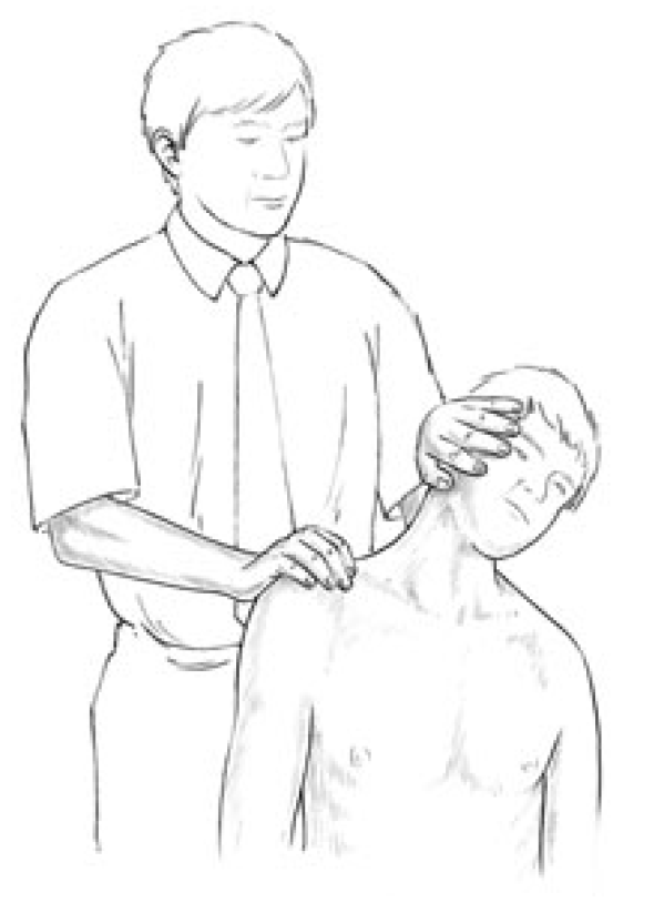
For further notice,�physiotherapy services may be offered alongside chiropractic care, to provide a cautious and gentle manipulation and/or mobilization of the cervical and thoracic spine in the instance of a large cervical disc herniation. Cervical disc herniations can cause pain and discomfort, numbness and weakness in the neck, shoulders, chest, arms and hands.

Abstract


A 34-year-old woman was seen in a physiotherapy department with signs and symptoms of cervical radiculopathy. Loss of cervical lordosis and a large paracentral to intraforaminal disc prolapse (8?mm) at C5�C6 level was reported on MRI. She was taking diclofenac sodium, tramadol HCl, diazepam and pregabalin for the preceding 2?months and no significant improvement, except temporary relief, was reported. She was referred to physiotherapy while awaiting a surgical opinion from a neurosurgeon. In physiotherapy she was treated with mobilisation of the upper thoracic spine from C7 to T6 level. A cervical extension exercise was performed with prior voluntary extension of the thoracic spine and elevated shoulders. She was advised to continue the same at home. General posture advice was given. Signs and symptoms resolved within the following four sessions of treatment over 3?weeks. Surgical intervention was subsequently deemed unnecessary.


Background


Surgical interventions are commonly recommended in large cervical prolapsed discs and the importance of non-aggressive physiotherapy interventions is less recognised and poorly understood. We present interventions that were associated with resolution of symptoms of radiculopathy resulting from a larger cervical herniated disc. These interventions, if applied correctly, may help to reduce the number of surgeries required for cervical prolapsed discs.


Case Presentation


The patient was a 34-year-old woman. She was seen in the physiotherapy department with a complaint of left-sided neck and shoulder pain. The pain was radiating to her left arm and there was associated numbness. The duration of symptoms was more than 2?months with no history of trauma. The pain was present on waking in the morning and gradually increased during the day. She was otherwise a healthy woman. Neck movements were aggravating the symptoms. She was seen in the acute hospital accident and emergency department (A&E) twice since onset and had been taking diclofenac sodium, tramadol HCl, diazepam and pregabalin. An MRI was planned and a request was sent for physiotherapy during the MRI waiting period. A neurosurgical review was requested by the A&E consultant upon receipt of the MRI report 7?weeks later.


Patient examination in the physiotherapy department revealed a normal gait pattern, her left arm held in front of her chest with the left shoulder slightly elevated. Her active range of neck motion was restricted and was painful on the left side. Flexion and rotation to the left were aggravating her arm and shoulder pain. Strength deficits were noted in the left elbow flexors and wrist extensors (4/5) when compared with the right side. There was paraesthesia along the radial border of the forearm and thumb regions. The brachioradialis reflex was diminished and biceps reflex was sluggish. Triceps and plantar reflexes were normal. Passive intervertebral movements were tender at C5�C6 level and were reproducing the pain. Sustained pressure at C7 and below was easing the pain and also improving the neck range of motion. The patient was deemed to have C6 radiculopathy. The MRI report, available 2?weeks after the commencement of physiotherapy, confirmed the diagnosis.


Investigations


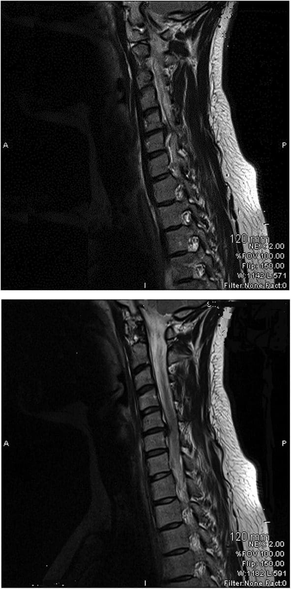
The findings from the plain cervical x-ray were unremarkable. MRI showed (Figure 1) loss of cervical spine lordosis, a left paracentral to intraforaminal lesion with 8?mm hernia, which indented the cord and obstructed the left paracentral recess and neural foramen.


Figure 1 Loss of Cervical Spine Lordosis and Large Disc Herniation at C5 and C6 on MRI

Figure 1: Loss of cervical spine lordosis and large disc herniation at C5 and C6 on MRI.


Differential Diagnosis


  • Cervical myelopathy.


Treatment


The patient received pharmacological treatment for the initial two symptomatic months, which included diclofenic sodium, tramadol, diazepam and pregabalin (lyrica) tablet. Physiotherapy was started after 2?months. Physiotherapy intervention consisted of mobilisation of the thoracic spine, resisted cervical extension exercises, a home programme of exercises and advice regarding the posture.


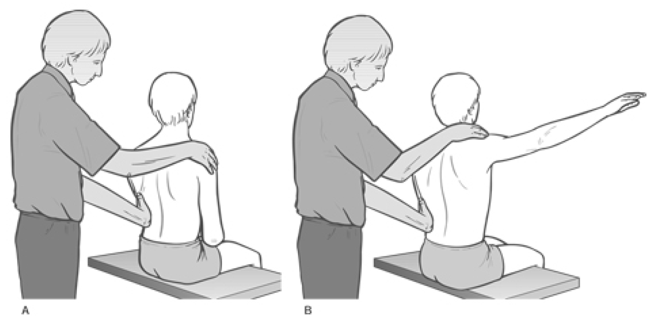
Mobilisation of the thoracic spine was administered in the prone lying position from C7 toT6 level. Mild intensity oscillations (15?reps) in an anterosuperior direction were directly applied to each of the spinal segments, through the thumb over the spinous processes, during the first visit. The applied force was enough to appreciate intervertebral movement in each segment and without significant pain. High-intensity oscillations (10�20) were applied during the subsequent treatment sessions. The patient was asked for symptom feedback during treatment.


Cervical spine extension exercises were carried out in a sitting position. The patient was asked to extend her thoracic spine with lungs fully inflated and shoulders elevated followed by extension of her cervical spine. Head extension was moderately resisted by the therapist near the end range of extension for 5�10?s and brought back to neutral after each resisted movement. The resisted movement was repeated at least three times with intervals of 30?s. The patient was asked to perform the same exercise at home every hour during the day.


The patient was educated regarding the rationale of extension exercises, sitting and lying posture and their effects on the spine. The duration of each session was approximately 20�25?min.


Dr. Alex Jimenez's Insight

Surgical interventions are generally recommended and widely considered for large cervical disc herniations. Although less recognized and often misunderstood, however, physiotherapy can be just as effective towards improving herniated discs in the cervical spine, excluding the need for surgery, according to the research study. Pharmacological treatments are also commonly used to help temporarily reduce symptoms alongside physiotherapy interventions. Cautious and gentle, spinal manipulation and mobilization of the cervical spine should be performed in the case of large cervical disc herniations to avoid aggravating the injury and/or condition. As recommended by a physiotherapist, or other healthcare professional experienced in physiotherapy, proper exercise can restore the function of the cervical spine and prevent regression of large prolapsed discs along the spine. Through appropriate physiotherapy intervention as well as through patient safety and compliance, the retraction of the cervical herniated discs is possible.


Outcome and Follow-Up


Pharmacological interventions were helpful to reduce the patient's pain on a temporary basis. Symptoms were recurring and resolution was not sustainable. The symptoms started improving after the first physiotherapy session and continued to improve during the subsequent sessions. It fully resolved in four sessions extended over 3?weeks. The patient was reviewed 4?months after the resolution of symptoms and there was no recurrence of symptoms. She was reviewed by a neurosurgeon and the surgical option was withdrawn.


Discussion


Stiffness of the thoracic spine has been linked to the painful pathologies of the cervical spine, and manipulation of the thoracic spine has been shown to improve painful symptoms and mobility of the cervical spine. However, cervical disc herniations of greater than 4?mm are considered inappropriate for physiotherapy interventions such as traction and manipulation. Spinal manipulation refers to a passive movement thrust of high velocity and low amplitude, usually applied at the end range of movement and is beyond the patient's control. Manipulation of the cervical spine is an aggressive procedure, which carries various risks and is often associated with worsening of symptoms. Manipulation was not considered in the treatment options for this patient because of the risks associated with it, and also because of patient's anxiety and lack of MRI-confirmed diagnosis.


Active extension of the thoracic spine increases the range of motion of the cervical spine and, in these authors� clinical experience, relieves minor neck symptoms. Conversely, thoracic spine kyphosis, such as slouch sitting, restricts the mobility of the cervical spine and aggravates the painful symptoms. A good sitting posture is constituted by a slightly extended thoracic spine. Therefore, active extension of the thoracic spine prior to cervical extension may improve cervical movements and restore cervical curvature.


It is believed that excessive pressure during flexion on the anterior aspect of the intervertebral discs pushes the nucleus pulposus posteriorly and causes herniations. Conversely, cervical lordosis might have the reverse effect�that is, decreases pressure on the anterior aspect of the discs and may create a suction effect which retracts the herniated contents. Therefore, a combination of short duration and repeated movements at the end of extension may serve as a suction pump and possibly retract the extruded content of the disc. Active cervical extension exercises, with an extended thoracic spine posture, may have been the key element in a home exercise programme to restore lordosis of the cervical spine and relieve radiculopathy symptoms in the current case. This may possibly have been due to the retraction of the herniated discs.


Spinal mobilisation refers to a gentle, oscillatory, passive movement of a spinal segment. These are applied to a spinal segment to gently increase the passive range of motion. It allows the patient to report aggravation of pain and to resist any unwanted movements. No mobilisation treatment was administered at C5�C6 level as palpation at this level was aggravating the symptoms. Segments below this level were mobilised with emphasis at C7�T1 level. Any treatment at the affected segment was likely to irritate the nerve root and thereby increase the inflammatory process.


Various interventions are reported for the treatment of prolapsed discs. Saal et al reported the use of traction, specific physical therapy exercise, oral anti-inflammatory medication and patient education in the treatment of 26 patients with herniated cervical discs (<4?mm) and reported significant improvement in outcomes for 24 patients. They observed that surgery for disc herniations occurs when a patient has significant myotomal weakness, severe pain or pain that persists beyond an arbitrary conservative treatment period of 2�8?weeks.


Spontaneous regressions of cervical disc protrusions are reported in the literature. However, spontaneous regressions of herniated cervical discs are speculated to be rare. Various factors related to regression are hypothesised and theorised. Pan et al summarised the factors related to the resorption of herniated disc as: the age of the patients; dehydration of the expanded nucleus pulposus; resorption of haematoma; revascularisation; penetration of herniated cervical disc fragments through the posterior longitudinal ligament; size of disc herniations; and existence of cartilage and annulus fibrosus tissue in the herniated material. Some studies on spontaneous regressions of discs reported that the patients were receiving physiotherapy. Physiotherapy interventions are not defined in any of these studies, however. Therefore, it is possible that disc regressions in these studies may be due to similar physiotherapy interventions as described here, or the patients were practising techniques and adopting postures as reported in the current case.


Learning Points


  • Thoracic spine mobilisation improves cervical spine biomechanics and can be considered in conjunction with other interventions in all painful conditions of the cervical spine.
  • Active extension of the thoracic spine facilitates movements of the cervical spine and may help regression of large prolapsed discs.
  • There is a possibility of retraction of herniated cervical discs through appropriate physiotherapy intervention.
  • Patient education ensures safety and compliance to therapist advice.
  • Meticulous assessment and patient feedback guides the therapist in selection of intensity of mobilisation.


Footnotes


Competing interests: None.


Patient consent: Obtained.


In conclusion,�physiotherapy, or physical therapy, is used to treat various injuries, diseases and disabilities, through the use of mechanical force and movements, manual therapy, exercise, electrotherapy, and through patient education and advice to restore and maintain function. As in the case above, physiotherapy can be recommended and considered as treatment before referring to surgical interventions of large cervical disc herniations. Information referenced from the National Center for Biotechnology Information (NCBI). The scope of our information is limited to chiropractic as well as to spinal injuries and conditions. To discuss the subject matter, please feel free to ask Dr. Jimenez or contact us at 915-850-0900 .


Curated by Dr. Alex Jimenez


Green-Call-Now-Button-24H-150x150-2-3.png


Additional Topics: Sciatica


Sciatica is referred to as a collection of symptoms rather than a single type of injury or condition. The symptoms are characterized as radiating pain, numbness and tingling sensations from the sciatic nerve in the lower back, down the buttocks and thighs and through one or both legs and into the feet. Sciatica is commonly the result of irritation, inflammation or compression of the largest nerve in the human body, generally due to a herniated disc or bone spur.


blog picture of cartoon paperboy big news


IMPORTANT TOPIC: EXTRA EXTRA: Treating Sciatica Pain


Blank
References
1.�Norlander S, Gustavsson BA, Lindell J, et al.�Reduced mobility in the cervico-thoracic motion segment�a risk factor for musculoskeletal neck-shoulder pain: a two-year prospective follow-up study.�Scand J Rehabil Med�1997;29:167�74.�[PubMed]
2.�Walser RF, Meserve BB, Boucher TR.�The effectiveness of thoracic spine manipulation for the management of musculoskeletal conditions: a systematic review and meta-analysis of randomized clinical trials.�J Man Manipulative Ther�2009;17:237�46.�[PMC free article]�[PubMed]
3.�Krauss J, Creighton D, Ely JD, et al.�The immediate effects of upper thoracic translatoric spinal manipulation on cervical pain and range of motion: a randomized clinical trial.�J Man Manipulative Ther2008;16:93�9.�[PMC free article]�[PubMed]
4.�Saal JS, Saal JA, Yurth EF.�Nonoperative management of herniated cervical intervertebral disc with radiculopathy.�Spine (Phila Pa 1976)�1996;21:1877�83.�[PubMed]
5.�Murphy DR, Beres JL.�Cervical myelopathy: a case report of a �near-miss� complication to cervical manipulation.�J Manipulative Physiol Ther�2008;31:553�7.�[PubMed]
6.�Leon-Sanchez A, Cuetter A, Ferrer G.�Cervical spine manipulation: an alternative medical procedure with potentially fatal complications.�South Med J�2007;100:201�3.�[PubMed]
7.�Scannell JP, McGill SM.�Disc prolapse: evidence of reversal with repeated extension.�Spine (Phila Pa 1976)�2009;34:344�50.�[PubMed]
8.�Gurkanlar D, Yucel E, Er U, et al.�Spontaneous regression of cervical disc herniations.�Minim Invasive Neurosurg�2006;49:179�83.�[PubMed]
9.�Mochida K, Komori H, Okawa A, et al.�Regression of cervical disc herniation observed on magnetic resonance images.�Spine (Phila Pa 1976)�1998;23:990�5; discussion 6�7.�[PubMed]
10.�Song JH, Park HK, Shin KM.�Spontaneous regression of a herniated cervical disc in a patient with myelopathy. Case report.�J Neurosurg�1999;90(1 Suppl):138�40.�[PubMed]
11.�Westmark RM, Westmark KD, Sonntag VK.�Disappearing cervical disc. Case report.�J Neurosurg1997;86:289�90.�[PubMed]
12.�Pan H, Xiao LW, Hu QF.�Spontaneous regression of herniated cervical disc fragments and its clinical significance.�Orthop Surg�2010;2:77�9.�[PubMed]
13.�Teplick JG, Haskin ME.�Spontaneous regression of herniated nucleus pulposus.�AJR Am J Roentgenol1985;145:371�5.�[PubMed]
Close Accordion
Cause Of Sciatica: Six Sources

Cause Of Sciatica: Six Sources

Cause Of Sciatica: Several lumbar spine (lower back) disorders can cause sciatica. Sciatica is often described as mild to intense pain in the left or right leg. Sciatica is caused by compression of one or more of the 5 sets of nerve roots in the lower back. Sometimes doctors call sciatica a radiculopathy. Radiculopathy is a medical term used to describe pain, numbness, tingling, and weakness in the arms or legs caused by a nerve root problem. If the nerve problem is in the neck, it is called a cervical radiculopathy. However, since sciatica affects the low back, it is called a lumbar radiculopathy.

Pathways To Sciatic Nerve Pain

Five sets of paired nerve roots in the lumbar spine combine to create the sciatic nerve. Starting at the back of the pelvis (sacrum), the sciatic nerve runs from the back, under the buttock, and downward through the hip area into each leg. Nerve roots are not “solitary” structures but are part of the body’s entire nervous system capable of transmitting pain and sensation to other parts of the body. Radiculopathy occurs when compression of a nerve root from a disc rupture (herniated disc) or bone spur (osteophyte) occurs in the lumbar spine prior to it joining the sciatic nerve.

sciatica causeWhat Causes Sciatic Nerve Compression?
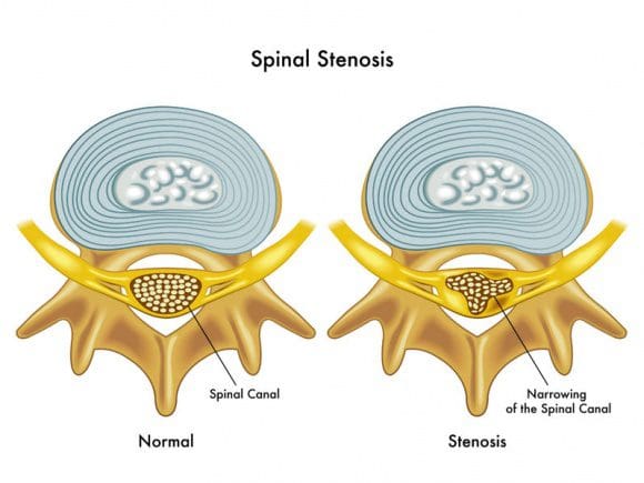
Several spinal disorders can cause spinal nerve compression and sciatica or lumbar radiculopathy. The 6 most common are:

  • a bulging or herniated disc
  • lumbar spinal stenosis
  • spondylolisthesis
  • trauma
  • piriformis syndrome
  • spinal tumors

Sciatica�Cause:�6 Leading Sources

Several lumbar spine (lower back) disorders can cause sciatica. Sciatica is often described as mild to intense pain in the left or right leg. Sciatica is caused by compression of one or more of the 5 sets of nerve roots in the lower back. Sometimes doctors call sciatica a radiculopathy. Radiculopathy is a medical term used to describe pain, numbness, tingling, and weakness in the arms or legs caused by a nerve root problem. If the nerve problem is in the neck, it is called a cervical radiculopathy. However, since sciatica affects the low back, it is called a lumbar radiculopathy.

Sciatica Cause #1: Lumbar Bulging Disc Or Herniated Disc

sciatica cause

A bulging disc is also known as a contained disc disorder. This means the gel-like center (nucleus pulposus) remains “contained” within the tire-like outer wall (annulus fibrosus) of the disc.

A herniated disc occurs when the nucleus breaks through the annulus fibrosus. It is called a “non-contained” disc disorder. Whether a disc bulges or herniates, disc material can press against an adjacent nerve root and compress delicate nerve tissue and cause sciatica.

The consequences of a herniated disc are worse. Not only does the herniated disc cause direct compression of the nerve root against the interior of the bony spinal canal, but the disc material itself also contains an acidic, chemical irritant (hyaluronic acid) that causes nerve inflammation. In both cases, nerve compression and irritation cause inflammation and pain, often leading to extremity numbness, tingling, and muscle weakness.

Herniated disc is a relatively common condition that can occur anywhere along the spine, but most often affects the lower back or neck region. Also known as a slipped disc or ruptured disc, a herniated disc develops when one of the cushion-like pads between the vertebrae moves out of position and presses on adjacent nerves.

Herniated discs are typically caused by overuse injuries or trauma to the spine; however, disc conditions can also develop as a result of the normal aging process. It is also known that there is a genetic factor that contributes to the development of disc degeneration and herniated disc. In most cases, a herniated disc in the lower back will heal within six months, as the size of herniation shrinks with time via resorption. Surgery may be needed if medication, physical therapy and other treatments fail.

What Is A Disc?

Spinal discs are cushion-like pads located between the vertebrae. Without these �shock absorbers,� the bones in the spine would grind against one another. In addition to giving the spine flexibility and making movements such as twisting and bending possible, discs protect the spine by absorbing the impact of trauma and body weight. Each disc has a strong outer layer called annulus fibrosus and a soft, gel-like center, called nucleus pulposus. There are fibers on the outside of each disc that attach to adjacent vertebrae and hold the disc in place. A herniated disc occurs when the outer layer tears or ruptures and the gel-like center leaks into the spinal canal.

The spinal canal has just enough space to house the spinal cord and spinal fluid. When a disc herniates and spills into the spinal canal, it can cause compression of the nerves or spinal cord. Intense, debilitating pain and alterations in sensation often occur. In addition, the gel-like substance inside the disc releases chemical irritants that contribute to nerve inflammation and pain.

What Causes A Herniated Disc?

As we age, the spinal discs gradually lose fluid volume. This process starts at about age 30 and progresses slowly, over time. As the discs dry out, microscopic cracks or tears can form on the outer surface, causing it to become brittle, weak and more susceptible to injury. The most common causes of herniated disc are:

  • Wear and tear: Discs dry out and aren�t as flexible as they once were.
  • Repetitive movements: Work, lifestyle, and certain sports activities that put stress on the spine, especially the lower back, further weaken an already vulnerable area.
  • Lifting the wrong way: Never lift while bent at the waist. Proper lifting entails lifting with your legs and a straight back.
  • Injury: High-impact trauma can cause the disc to bulge, tear or rupture.
  • Obesity: Carrying excess weight puts an undue amount of strain on the spine.
  • Genetics: There are some genes that are more commonly present in individuals with disc degeneration. More research is needed to investigate the role of these genes�they could be targets of biological treatment in the future.

What Are The Symptoms Of A Herniated Disc?

Pain from a herniated disc can vary, depending on the location and severity of the injury. It is typically felt on one side of the body.

If the injury is minimal, little or no pain may be felt. If the disc ruptures, pain can be severe and unrelenting. Pain may radiate to an extremity in a specific nerve root distribution if significant nerve impingement has occurred. For example, sciatica is frequently caused by a herniated disc in the lower back. Herniated disc can manifest itself with a range of symptoms, including:

  • Dull ache to severe pain
  • Numbness, tingling, burning
  • Muscle weakness; spasm; altered reflexes
  • Loss of bowel or bladder control (Note: These symptoms constitute a medical emergency. If they occur, seek medical attention immediately).

How Is A Herniated Disc Diagnosed?

History and physical examination point to a diagnosis of herniated disc. A herniated disc is likely if low back pain is accompanied by radiating leg pain in a nerve root distribution with positive straight leg raising test (ie, elevating the leg while lying down causes radiating pain down the leg), and other neurologic deficits such as numbness, weakness, and altered reflexes.

Imaging studies are usually ordered to confirm a diagnosis of herniated disc. X-rays are not the imaging medium of choice because soft tissues (eg, discs, nerves) are hard to capture with this technology. However, they may be used as an initial tool to rule out other disorders such as a growth or fracture. Confirmation of the suspicion of herniated disc is generally accomplished with:

  • Magnetic Resonance Imaging (MRI): This technology reveals the spinal cord, surrounding soft tissue and nerves. It is the best imaging study to support the diagnosis of a herniated disc.
  • Nerve Conduction Studies (NCS) and Electromyogram (EMG): These studies use electrical impulses to measure the degree of damage to the nerve/s caused by compression from a herniated disc and other conditions that cause nerve impingement can be ruled out. NCS and EMG are not routine tests to diagnose herniated disc.

Herniated discs sometimes heal on their own through a process called resorption. This means that the disc fragments are absorbed by the body. Most people suffering from herniated disc respond well to conservative treatment and do not require surgery.

Sciatica Cause #2: Lumbar Spinal Stenosis

sciatica causeSpinal stenosis is a nerve compression disorder most often affecting older adults. Leg pain similar to sciatica may occur as a result of lumbar spinal stenosis. The pain is usually positional, often brought on by activities such as standing or walking and relieved by sitting down.
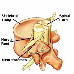
Spinal nerve roots branch outward from the spinal cord through passageways called neural foramina comprised of bone and ligaments. Between each set of vertebral bodies, located on the left and right sides, is a foramen. Nerve roots pass through these openings and extend outward beyond the spinal column to innervate other parts of the body. When these passageways become narrow or clogged causing nerve compression, the term foraminal stenosis is used.

What Is Spinal Stenosis?

A clue to answering this question is found in the meaning of each word. Spinal refers to the spine. Stenosis is a medical term used to describe a condition where a normal-size opening has become narrow. Spinal stenosis may affect the cervical (neck), thoracic (chest), or lumbar (lower back) spines.

The most commonly area affected is the lumbar spine followed by the cervical spine.

Visualize Spinal Stenosis?

Consider a water pipe. Over time, rust and debris builds up on the walls of the pipe, thereby narrowing the passageway that normally allows water to freely flow. In the spine, the passageways are the spinal canal and the neuroforamen. The spinal canal is a hollow vertical hole that contains the spinal cord. The neuroforamen are the passageways that are naturally created between the vertebrae through which spinal nerve roots exit the spinal canal.
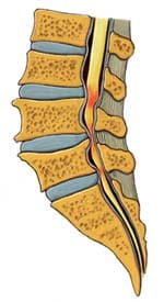
sciatica causeThe neuroforamen are the passageways that are naturally created between the vertebrae through which spinal nerve roots exit the spinal canal.

Illustration above: The spine’s bony structures encase and protect the spinal cord. Small nerve roots shoot off from the spinal cord and exit the spinal canal through passageways called neuroforamen.

Lumbar (low back) spinal stenosis is illustrated below. Notice the narrowed areas in the spinal canal (reddish-colored areas). As the canal space narrows, the spinal cord and nearby nerve roots are squeezed causing different types of symptoms. The medical term is nerve compression.

sciatica causeAnatomy Overview Can Help You Understand Spinal Stenosis
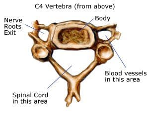
The spine is a column of connected bones called vertebrae. There are 24 vertebrae in the spine, plus the sacrum and tailbone (coccyx). Most adults have 7 vertebrae in the neck (the cervical vertebrae), 12 from the shoulders to the waist (the thoracic vertebrae), and 5 in the lower back (the lumbar vertebrae). The sacrum is made up of 5 vertebrae between the hipbones that are fused into one bone. The coccyx is made up of small fused bones at the tail end of the spine.

Lamina and Spinous Processes: At the back (posterior) of each vertebra, you have the lamina, a bony plate that protects your spinal canal and spinal cord. Your vertebrae also have several bony tabs that are called spinous processes; those processes are attachment points for muscles and ligaments.

Ligaments, Especially the Ligamentum Flavum: Vertebrae are connected by ligaments, which keep the vertebrae in their proper place. The ligamentum flavum is a particularly important ligament. Not only does it help stabilize your spine, it also protects your spinal cord and nerve roots. Plus, the ligamentum flavum is the strongest ligament in your spine.

The ligamentum flavum is a dynamic structure, which means that it adapts its shape as you move your body. When you’re sitting down and leaning forward, the ligamentum flavum is stretched out; that gives your spinal canal more room for the spinal nerves. When you stand up and lean back, though, the ligamentum flavum becomes shorter and thicker; that means there’s less room for the spinal nerves. (This dynamic capability helps explain why people with spinal stenosis find that sitting down feels better than standing or walking.)

Discs: In between each vertebra are tough fibrous shock-absorbing pads called the intervertebral discs. Each disc is made up of a tire-like outer band (annulus fibrosus) and a gel-like inner substance (nucleus pulposus).

Spinal Nerves and the Spinal Cord: Nerves are also an important part of your spinal anatomy�after all, they’re what sends messages from your brain to the rest of your body. The spinal cord, the thick bundle of nerves that extends downward from the brain, passes through a ring in each vertebra. Those rings line up into a channel called the spinal canal.

Between each vertebra, two nerves branch out of the spinal cord (one to the right and one to the left). Those nerves exit the spine through openings called the foramen and travel to all parts of your body.

sciatica causeNormally, the spinal channel is wide enough for the spinal cord, and the foramen are wide enough for the nerve roots. But either or both can become narrowed, and cause spinal stenosis.
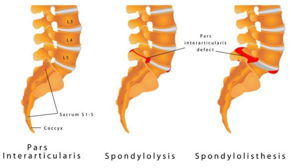
Sciatica Cause #3: Spondylolisthesis

Spondylolisthesis is a disorder that most often affects the lumbar spine. It is characterized by one vertebra slipping forward over an adjacent vertebra. When a vertebra slips and is displaced, spinal nerve root compression occurs and often causes sciatic leg pain. Spondylolisthesis is categorized as developmental (found at birth, develops during childhood) or acquired from spinal degeneration, trauma or physical stress (eg, lifting weights).

Spondylolisthesis occurs when one vertebra slips forward over the vertebra below it. The term is pronounced spondy-low-lis-thesis and is derived from the Greek language: spondylo means vertebra and listhesis means to slip. There are several types or causes of spondylolisthesis; a few are listed below.

sciatica cause


  • Congenital spondylolisthesis means the disorder is present at birth.
  • Isthmic spondylolisthesis occurs when a defect, such as a fracture occurs in a bony supporting vertebral structure at the back of the spine.
  • Degenerative spondylolisthesis is more common and is often associated with degenerative disc disease, wherein the discs (eg, due to the effects of growing older) lose hydration and resilency.

How Spondylolisthesis May Develop

The lumbar spine is exposed to directional pressures while it carries, absorbs, and distributes most of your body’s weight at rest and during activity. In other words, while your lumbar spine is carrying and absorbing body weight, it also moves in different directions (eg, rotate, bend forward). Sometimes, this combination causes excessive stress to the vertebra and/or its supporting structures, and may lead to a vertebral body slipping forward over the vertebrae beneath.

Who May Be At Risk

If a family member (eg, mother, father) has spondylolisthesis, your risk for developing the disorder may be greater. Some activities make you more susceptible to spondylolisthesis. Gymnasts, linemen in football, and weight lifters all put significant pressure and weight on their low backs. Think about gymnasts and the positions they put their body in: They practically bend in half backwards�that’s an extreme arched back. They also twist through the air quickly when doing flips and then land, absorbing the impact through their legs and low back. Those movements put substantial stress on the spine, and spondylolisthesis can develop as a result of repeated excessive strains and stress.

X-Ray View Of Spondylolisthesis

The x-ray below shows you a good example of a lumbar spondylolisthesis. Look at the area the arrow is pointing to: You can see that the vertebra above the arrow isn’t in line with the vertebra below it. It’s slipped forward; it’s spondylolisthesis.

sciatica causeArrow Points To A Lumbar Spondylolisthesis
Grading Spondylolisthesis

Doctors “grade” the severity of a spondylolisthesis using five descriptive categories. Although there are several factors your doctor considers when evaluating your spondylolisthesis, the grading scale (below) is based on the far forward a vertebral body has slid forward over the vertera beneath. Often, the doctor uses a lateral (side view) x-ray to examine and grade a spondyloisthesis. Grade I is a smaller slip than Grade IV or V.

  • Grade I: Less than 25% slip
  • Grade II: 25% to 49% slip.
  • Grade III: 50% to 74% slip.
  • Grade IV: 75% to 99% slip.
  • Grade V: The vertebra that has fallen forward off the vertebra below it. This is the most severe type of spondylolisthesis and is termed spondyloptosis.

Sciatica Cause #4: Trauma

Sciatica can result from direct nerve compression caused by external forces to the lumbar or sacral spinal nerve roots. Examples include motor vehicle accidents, falling down, football and other sports. The impact may injure the nerves or, occasionally, fragments of broken bone may compress the nerves.

Sciatica Cause #5: Piriformis Syndrome

Piriformis syndrome is named for the piriformis muscle and the pain caused when the muscle irritates the sciatic nerve. The piriformis muscle is located in the lower part of the spine, connects to the thighbone, and assists in hip rotation. The sciatic nerve runs beneath the piriformis muscle. Piriformis syndrome develops when muscle spasms develop in the piriformis muscle thereby compressing the sciatic nerve. It may be difficult to diagnose and treat due to the lack of x-ray or magnetic resonance imaging (MRI) findings.

If you�ve ever felt pain in the hip, pain in the center of the butt, or pain down the back of the leg, you are likely suffering, at least partially, with piriformis syndrome. The piriformis is a muscle which runs from your sacrum (mid-line base of spine) to the outer hip bone (trochanter). This muscle truly works overtime on anyone who runs at all.

sciatica causeThe muscles in and around the gluteal region help with three areas
  • rotation of the hip and leg;
  • balance while one foot is off the ground; and
  • stability for the pelvic region.

Needless to say, all of these characteristics are highly needed by runners (and everyone else, when you come to think of it).

Injuries To The Piriformis

This muscle is a prime candidate for repetitive motion injury (RMI). RMI occurs when a muscle is asked to perform beyond it�s level of capability, not given enough time to recover, and asked to perform again. The typical response from a muscle in this situation is to tighten, which is a defensive response of the muscle. This tightness, however, manifests itself in several ways to a runner.

The first symptom suggesting piriformis syndrome would be pain in and around the outer hip bone. The tightness of the muscle produces increased tension between the tendon and the bone which produces either direct discomfort and pain or an increased tension in the joint producing a bursitis. Again, a bursitis is an inflammation of the fluid filled sac in a joint caused by an elevation of stress and tension within that joint.

The second symptom suggesting piriformis syndrome would be pain directly in the center of the buttocks. Although this is not as common as the other two symtpoms, this pain can be elicited with direct compression over the belly of the buttocks area. A tight muscle is a sore muscle upon compression due to a reduced blood flow to that muscle.

The third symptom suggesting piriformis syndrome would be a sciatic neuralgia, or pain from the buttocks down the back of the leg and sometimes into different portions of the lower leg. We have an article that can teach you more about how piriformis syndrome and sciatica are related.

The sciatic nerve runs right through the belly of the piriformis muscle and if the piriformis muscle contracts from being overused, the sciatic nerve now becomes strangled, producing pain, tingling and numbness.

Simple Physiology

Any muscle repetitively used needs to have an opportunity to recover. This recovery can either be on Nature�s clock, or can be facilitated and sped up with proper knowledge and treatment. Since the muscle is tightening due to overuse, continued use will only make it worse. This injured muscle needs to relax and have increased blood flow encouraged to it for more rapid healing. This tightness that exists also reduces the normal blood flow going to the muscle reducing the speed with which the muscle can recover. To encourage fresh, oxygen-rich blood to the muscle is the most powerful means of getting the muscle to begin to relax and function normally. Multiple massages per day to this area is greatly encouraged.

The next step in this “recovery” process is to use a tennis ball under the butt and hip area. While sitting down on the floor, roll away from the side of involvement and place a tennis ball just inside the outer hip bone under the butt area. As you begin to allow your weight onto the tennis ball, note areas of increased pain and soreness. Trigger points will tend to accumulate in a repetitively used muscle, and until these toxins are manually broken up and eliminated, the muscle will have an artificial ceiling with regard to flexibility potential and recovery potential. So, if it�s sore and hurts while your sitting on it, you�re doing a good job. Let the ball work under each spot for 15-20 seconds before moving it to another area. Once you�ve been on the ball for 4-5 minutes, now put the ankle of the involved leg over the knee of the non-involved leg (crossing your legs). Now place the tennis ball just inside the outer hip bone again and work the tendon of the piriformis muscle. While this pain is typically excruciating and takes some time to effectively reduce, the benefits here are huge. Be patient, be consistent and good things will happen.

Additional Treatments

Due to the fact that the sciatic neuralgia and the hip bursitis or tendonitis are both inflammatory in nature, ice, or cryotherapy, over the involved area 15-20 minutes at a time will be beneficial. This should be done multiple times per day.

Stretching of the hip muscles should not be done until the acute pain is gone. At that point in time, begin with gentle stretching, such as the cross-legged stretch while pulling up on the knee. The muscle should have increased flexibility before an active return to running.

Finally, I�m always discouraging the use of pharmaceutical anti-inflammatories. Not only do they greatly aggravate the intestines, but they also suggest an artificial wellness that can lead to bigger problems. Proteolytic enzymes, such as bromelain, are both natural and extremely beneficial with no side effects.

Conclusion: The piriformis muscle is pretty important for all of us.

Sciatica Cause #6: Spinal Tumors

Spinal tumors are abnormal growths that are either benign or cancerous (malignant). Fortunately, spinal tumors are rare. However, when a spinal tumor develops in the lumbar region, there is a risk for sciatica to develop as a result of nerve compression.

If you think you have sciatica, call your doctor or chiropractor. The first step toward relieving pain is a proper diagnosis.

Written by Jean-Jacques Abitbol, MD; Reviewed by Brian R. Subach, MD

Howard S. An, MD, Stewart G. Eidelson, MD; Reviewed by Howard S. An, MD, Jason M. Highsmith, MD

Timothy J. Maggs, D.C.; Reviewed by Edward C. Benzel, MD

Check out our sister page at SPINEUNIVERSE

Chiropractic Care Benefits Juvenile Arthritis Patients

Assessment and Treatment of Upper Trapezius

Assessment and Treatment of Upper Trapezius

These assessment and treatment recommendations represent a synthesis of information derived from personal clinical experience and from the numerous sources which are cited, or are based on the work of researchers, clinicians and therapists who are named (Basmajian 1974, Cailliet 1962, Dvorak & Dvorak 1984, Fryette 1954, Greenman 1989, 1996, Janda 1983, Lewit 1992, 1999, Mennell 1964, Rolf 1977, Williams 1965).


Clinical Application of Neuromuscular Techniques: Upper Trapezius


Lewit (1999) simplifies the need to assess for shortness by stating, �The upper trapezius should be treated if tender and taut.� Since this is an almost universal state in modern life, it seems that everyone requires MET application to this muscle. Lewit also notes that a characteristic mounding of the muscle can often be observed when it is very short, producing the effect of �Gothic shoulders�, similar to the architectural supports of a Gothic church tower (see Fig. 2.13).


Assessment for Shortness of Upper Trapezius (13) (Fig. 4.30)


Figure 4 30 Assessment of the Relative Shortness of the Right Side Upper Trapezius Image 1


Figure 4.30 Assessment of the relative shortness of the right side upper trapezius. One side is compared with the other (for both the range of unforced motion and the nature of the end-feel of motion) to ascertain the side most in need of MET attention.


Test for upper trapezius for shortness (a) See scapulohumeral rhythm test (Ch. 5) which helps identify excessive activity or inappropriate tone in levator scapula and upper trapezius, which, because they are postural muscles, indicates shortness (Fig 5.13A, B). Greenman (1996) describes a functional �firing sequence� assessment which identifies general imbalance and dysfunction involving the upper and lower fixators of the shoulder (Fig. 4.31).


Figure 4 31 Palpation Assessment for Upper and Lower Fixators of the Shoulder Image 2


Figure 4.31 Palpation assessment for upper and lower fixators of the shoulder, including upper trapezius (Greenman 1996).


The patient is seated and the practitioner stands behind. The practitioner rests his right hand over the right shoulder area to assess firing sequence of muscles. The other hand can be placed either on the mid-thoracic region, mainly on the side being assessed, or spanning the lower back to palpate quadratus firing. The assessment should be performed at least twice so that various hand positions are used for different muscles (as in Fig. 4.31).


Greenman bases his description on Janda (1983), who notes the �correct� sequence for shoulder abduction, when seated, as involving: supraspinatus, deltoid, infraspinatus, middle and lower trapezius and finally contralateral quadratus. In dysfunctional states the most common substitutions are said to involve: shoulder elevation by levator scapulae and upper trapezius, as well as early firing by quadratus lumborum, ipsilateral and contralateral.


Inappropriate activity of the upper fixators results in shortness, and of the lower fixators in weakness and possible lengthening (see Ch. 2 for discussion of postural/phasic, etc. muscle characteristics).


Test for upper trapezius for shortness (b) The patient is seated and the practitioner stands behind with one hand resting on the shoulder of the side to be tested and stabilising it. The other hand is placed on the ipsilateral side of the head and the head/neck is taken into contralateral sidebending without force while the shoulder is stabilised (see Fig. 4.30).


The same procedure is performed on the other side with the opposite shoulder stabilised. A comparison is made as to which sidebending manoeuvre produced the greater range and whether the neck can easily reach 45� of side-flexion in each direction, which it should. If neither side can achieve this degree of sidebend, then both trapezius muscles may be short. The relative shortness of one, compared with the other, is evaluated.


Test for upper trapezius for shortness (c) The patient is seated and the practitioner stands behind with a hand resting over the muscle on the side to be assessed. The patient is asked to extend the arm at the shoulder joint, bringing the flexed arm/elbow backwards. If the upper trapezius is stressed on that side it will inappropriately activate during this movement. Since it is a postural muscle, shortness in it can then be assumed (see discussion of postural muscle characteristics in Ch. 3).


Test of upper trapezius for shortness (d) The patient is supine with the neck fully (but not forcefully) sidebent contralaterally (away from the side being assessed). The practitioner is standing at the head of the table and uses a cupped hand contact on the ipsilateral shoulder (i.e. on the side being tested) to assess the ease with which it can be depressed (moved caudally) (Fig. 4.32).


Figure 4 32 MET Treatment of Right Side Upper Trapezius Muscle Image 3


Figure 4.32 MET treatment of right side upper trapezius muscle. A Posterior fibres, B middle fibres, C anterior fibres. Note that stretching in this (or any of the alternative positions which access the middle and posterior fibres) is achieved following the isometric contraction by means of an easing of the shoulder away from the stabilised head, with no force being applied to the neck and head itself.


There should be an easy �springing� sensation as the practitioner pushes the shoulder towards the feet, with a soft end-feel to the movement. If depression of the shoulder is difficult or if there is a harsh, sudden end-point, upper trapezius shortness is confirmed.


This same assessment (always with full lateral flexion) should be performed with the head fully rotated away from the side being treated, half turned away from the side being treated, and slightly turned towards the side being treated, in order to respectively assess the relative shortness and functional efficiency of posterior, middle and anterior subdivisions of the upper portion of trapezius.


MET Treatment of Chronically Shortened Upper Trapezius


MET treatment of upper trapezius, method (a) (Fig. 4.32) In order to treat all the fibres of upper trapezius, MET needs to be applied sequentially. The upper trapezius is subdivided here into anterior, middle and posterior fibres. The neck should be placed into different positions of rotation, coupled with the sidebending as described in the assessment description above, for precise treatment of the various fibres.


The patient lies supine, arm on the side to be treated lying alongside the trunk, head/neck sidebent away from the side being treated to just short of the restriction barrier, while the practitioner stabilises the shoulder with one hand and cups the ear/mastoid area of the same side of the head with the other:


  • With the neck fully sidebent and fully rotated contralaterally, the posterior fibres of upper trapezius are involved in the contraction (see below). This will facilitate subsequent stretching of this aspect of the muscle.
  • With the neck fully sidebent and half rotated, the middle fibres are involved in the contraction.
  • With the neck fully sidebent and slightly rotated towards the side being treated the anterior fibres of upper trapezius are being treated.


The various contractions and subsequent stretches can be performed with practitioner�s arms crossed, hands stabilising the mastoid area and shoulder.


The patient introduces a light resisted effort (20% of available strength) to take the stabilised shoulder towards the ear (a shrug movement) and the ear towards the shoulder. The double movement (or effort towards movement) is important in order to introduce a contraction of the muscle from both ends simultaneously. The degree of effort should be mild and no pain should be felt. The contraction is sustained for 10 seconds (or so) and, upon complete relaxation of effort, the practitioner gently eases the head/neck into an increased degree of sidebending and rotation, where it is stabilised, as the shoulder is stretched caudally.


When stretching is introduced the patient can usefully assist in this phase of the treatment by initiating, on instruction, the stretch of the muscle (�as you breathe out please slide your hand towards your feet�). This reduces the chances of a stretch reflex being initiated. Once the muscle is being stretched, the patient relaxes and the stretch is held for 10�30 seconds.


CAUTION: No stretch should be introduced from the cranial end of the muscle as this could stress the neck. The head is stabilised at its side-flexion and rotation barrier.


Disagreement


There is some disagreement as to the head/neck rotation position as described in the treatment method above, which calls (for posterior and middle fibres) for sidebending and rotation away from the affected side.


Liebenson (1996), suggests that the patient �lies supine with the head supported in anteflexion and laterally flexed away and rotated towards the side of involvement�.


Lewit (1985b) suggests: �The patient is supine � the therapist fixes the shoulder from above with one hand, sidebending the head and neck with the other hand so as to take up the slack. He then asks the patient to look towards the side away from which the head is bent, resisting the patient�s automatic tendency to move towards the side of the lesion.� (This method is described below.)


The author has used the methods described above with good effect and urges readers to try these approaches as well as those of Liebenson and Lewit, and to evaluate results for themselves.


MET treatment of acutely shortened upper trapezius, method (b) Lewit suggests the use of eye movements to facilitate initiation of PIR before stretching, an ideal method for acute problems in this region.


The patient is supine, while the practitioner fixes the shoulder and the sidebent (away from the treated side) head and neck at the restriction barrier and asks the patient to look, with the eyes only (i.e. not to turn the head), towards the side away from which the neck is bent.


This eye movement is maintained, as is a held breath, while the practitioner resists the slight isometric contraction that these two factors (eye movement and breath) will have created.


On exhalation and complete relaxation, the head/neck is taken to a new barrier and the process repeated. If the shoulder is brought into the equation, this is firmly held as it attempts to lightly push into a shrug. After this 10 second contraction the muscle will have released somewhat and slack can again be taken out as the head is repositioned before a repetition of the procedure commences.


Dr. Alex Jimenez offers an additional assessment and treatment of the hip flexors as a part of a referenced clinical application of neuromuscular techniques by Leon Chaitow and Judith Walker DeLany. The scope of our information is limited to chiropractic and spinal injuries and conditions. To discuss the subject matter, please feel free to ask Dr. Jimenez or contact us at 915-850-0900 .


By Dr. Alex Jimenez


Green-Call-Now-Button-24H-150x150-2-3.png


Additional Topics: Wellness


Overall health and wellness are essential towards maintaining the proper mental and physical balance in the body. From eating a balanced nutrition as well as exercising and participating in physical activities, to sleeping a healthy amount of time on a regular basis, following the best health and wellness tips can ultimately help maintain overall well-being. Eating plenty of fruits and vegetables can go a long way towards helping people become healthy.


blog picture of cartoon paperboy big news


WELLNESS TOPIC: EXTRA EXTRA: Managing Workplace Stress


Blank
References
1.�Ballou SK, Keefer L. Multicultural considerations in the diagnosis and management of irritable bowel syndrome: a selective summary.�Eur J Gastroenterol Hepatol.�2013;25:1127�1133.�[PubMed]
2.�Gonzales Gamarra RG, Ruiz S�nchez JG, Le�n Jim�nez F, Cubas Benavides F, D�az V�lez C. [Prevalence of irritable bowel syndrome in the adult population of the city of Chiclayo in 2011]�Rev Gastroenterol Peru.�2012;32:381�386.�[PubMed]
3.�Gwee KA, Lu CL, Ghoshal UC. Epidemiology of irritable bowel syndrome in Asia: something old, something new, something borrowed.�J Gastroenterol Hepatol.�2009;24:1601�1607.�[PubMed]
4.�Krogsgaard LR, Engsbro AL, Bytzer P. The epidemiology of irritable bowel syndrome in Denmark. A population-based survey in adults ? 50 years of age.�Scand J Gastroenterol.�2013;48:523�529.�[PubMed]
5.�Ibrahim NK, Battarjee WF, Almehmadi SA. Prevalence and predictors of irritable bowel syndrome among medical students and interns in King Abdulaziz University, Jeddah.�Libyan J Med.�2013;8:21287.[PMC free article]�[PubMed]
6.�Lee YY, Waid A, Tan HJ, Chua AS, Whitehead WE. Rome III survey of irritable bowel syndrome among ethnic Malays.�World J Gastroenterol.�2012;18:6475�6480; discussion p. 6479.�[PMC free article][PubMed]
7.�Ghoshal UC, Abraham P, Bhatt C, Choudhuri G, Bhatia SJ, Shenoy KT, Banka NH, Bose K, Bohidar NP, Chakravartty K, et al. Epidemiological and clinical profile of irritable bowel syndrome in India: report of the Indian Society of Gastroenterology Task Force.�Indian J Gastroenterol.�2008;27:22�28.�[PubMed]
8.�Bures J, Cyrany J, Kohoutova D, F�rstl M, Rejchrt S, Kvetina J, Vorisek V, Kopacova M. Small intestinal bacterial overgrowth syndrome.�World J Gastroenterol.�2010;16:2978�2990.�[PMC free article][PubMed]
9.�Dibaise JK, Young RJ, Vanderhoof JA. Enteric microbial flora, bacterial overgrowth, and short-bowel syndrome.�Clin Gastroenterol Hepatol.�2006;4:11�20.�[PubMed]
10.�Bouhnik Y, Alain S, Attar A, Flouri� B, Raskine L, Sanson-Le Pors MJ, Rambaud JC. Bacterial populations contaminating the upper gut in patients with small intestinal bacterial overgrowth syndrome.�Am J Gastroenterol.�1999;94:1327�1331.�[PubMed]
11.�Dukowicz AC, Lacy BE, Levine GM. Small intestinal bacterial overgrowth: a comprehensive review.�Gastroenterol Hepatol (N Y)�2007;3:112�122.�[PMC free article]�[PubMed]
12.�Posserud I, Stotzer PO, Bj�rnsson ES, Abrahamsson H, Simr�n M. Small intestinal bacterial overgrowth in patients with irritable bowel syndrome.�Gut.�2007;56:802�808.�[PMC free article]�[PubMed]
13.�Ghoshal U, Ghoshal UC, Ranjan P, Naik SR, Ayyagari A. Spectrum and antibiotic sensitivity of bacteria contaminating the upper gut in patients with malabsorption syndrome from the tropics.�BMC Gastroenterol.�2003;3:9.�[PMC free article]�[PubMed]
14.�Carrara M, Desideri S, Azzurro M, Bulighin GM, Di Piramo D, Lomonaco L, Adamo S. Small intestine bacterial overgrowth in patients with irritable bowel syndrome.�Eur Rev Med Pharmacol Sci.�2008;12:197�202.�[PubMed]
15.�Ghoshal UC, Ghoshal U, Das K, Misra A. Utility of hydrogen breath tests in diagnosis of small intestinal bacterial overgrowth in malabsorption syndrome and its relationship with oro-cecal transit time.�Indian J Gastroenterol.�2006;25:6�10.�[PubMed]
16.�Ghoshal UC, Kumar S, Mehrotra M, Lakshmi C, Misra A. Frequency of small intestinal bacterial overgrowth in patients with irritable bowel syndrome and chronic non-specific diarrhea.�J Neurogastroenterol Motil.�2010;16:40�46.�[PMC free article]�[PubMed]
17.�Pimentel M, Chow EJ, Lin HC. Eradication of small intestinal bacterial overgrowth reduces symptoms of irritable bowel syndrome.�Am J Gastroenterol.�2000;95:3503�3506.�[PubMed]
18.�Sachdeva S, Rawat AK, Reddy RS, Puri AS. Small intestinal bacterial overgrowth (SIBO) in irritable bowel syndrome: frequency and predictors.�J Gastroenterol Hepatol.�2011;26 Suppl 3:135�138.�[PubMed]
19.�Park H. The role of small intestinal bacterial overgrowth in the pathophysiology of irritable bowel syndrome.�J Neurogastroenterol Motil.�2010;16:3�4.�[PMC free article]�[PubMed]
20.�Ghoshal UC, Shukla R, Ghoshal U, Gwee KA, Ng SC, Quigley EM. The gut microbiota and irritable bowel syndrome: friend or foe?�Int J Inflam.�2012;2012:151085.�[PMC free article]�[PubMed]
21.�Brown AC. Ulcerative colitis, Crohn�s disease and irritable bowel syndrome patients need fecal transplant research and treatment.�J Crohns Colitis.�2014;8:179.�[PubMed]
22.�Sampath K, Levy LC, Gardner TB. Fecal transplantation: beyond the aesthetic.�Gastroenterology.�2013;145:1151�1153.�[PubMed]
23.�Grace E, Shaw C, Whelan K, Andreyev HJ. Review article: small intestinal bacterial overgrowth--prevalence, clinical features, current and developing diagnostic tests, and treatment.�Aliment Pharmacol Ther.�2013;38:674�688.�[PubMed]
24.�Yakoob J, Abbas Z, Khan R, Hamid S, Awan S, Jafri W. Small intestinal bacterial overgrowth and lactose intolerance contribute to irritable bowel syndrome symptomatology in Pakistan.�Saudi J Gastroenterol.�2011;17:371�375.�[PMC free article]�[PubMed]
25.�Gwee KA, Bak YT, Ghoshal UC, Gonlachanvit S, Lee OY, Fock KM, Chua AS, Lu CL, Goh KL, Kositchaiwat C, et al. Asian consensus on irritable bowel syndrome.�J Gastroenterol Hepatol.�2010;25:1189�1205.�[PubMed]
26.�Lacy BE, Gabbard SL, Crowell MD. Pathophysiology, evaluation, and treatment of bloating: hope, hype, or hot air?�Gastroenterol Hepatol (N Y)�2011;7:729�739.�[PMC free article]�[PubMed]
27.�Harder H, Serra J, Azpiroz F, Passos MC, Aguad� S, Malagelada JR. Intestinal gas distribution determines abdominal symptoms.�Gut.�2003;52:1708�1713.�[PMC free article]�[PubMed]
28.�Kumar S, Misra A, Ghoshal UC. Patients with irritable bowel syndrome exhale more hydrogen than healthy subjects in fasting state.�J Neurogastroenterol Motil.�2010;16:299�305.�[PMC free article][PubMed]
29.�Hungin AP, Mulligan C, Pot B, Whorwell P, Agr�us L, Fracasso P, Lionis C, Mendive J, Philippart de Foy JM, Rubin G, Winchester C, de Wit N. Systematic review: probiotics in the management of lower gastrointestinal symptoms in clinical practice -- an evidence-based international guide.�Aliment Pharmacol Ther.�2013;38:864�886.�[PMC free article]�[PubMed]
30.�Attar A, Flouri� B, Rambaud JC, Franchisseur C, Ruszniewski P, Bouhnik Y. Antibiotic efficacy in small intestinal bacterial overgrowth-related chronic diarrhea: a crossover, randomized trial.�Gastroenterology.�1999;117:794�797.�[PubMed]
31.�Marcelino RT, Fagundes-Neto U. [Hydrogen test (H2) in the air expired for the diagnosis of small bowel bacterial overgrowth]�Arq Gastroenterol.�1995;32:191�198.�[PubMed]
32.�Santavirta J. Lactulose hydrogen and [14C]xylose breath tests in patients with ileoanal anastomosis.�Int J Colorectal Dis.�1991;6:208�211.�[PubMed]
33.�Ghoshal UC. How to interpret hydrogen breath tests.�J Neurogastroenterol Motil.�2011;17:312�317.[PMC free article]�[PubMed]
34.�Yang CY, Chang CS, Chen GH. Small-intestinal bacterial overgrowth in patients with liver cirrhosis, diagnosed with glucose H2 or CH4 breath tests.�Scand J Gastroenterol.�1998;33:867�871.�[PubMed]
35.�Ghoshal UC, Ghoshal U, Ayyagari A, Ranjan P, Krishnani N, Misra A, Aggarwal R, Naik S, Naik SR. Tropical sprue is associated with contamination of small bowel with aerobic bacteria and reversible prolongation of orocecal transit time.�J Gastroenterol Hepatol.�2003;18:540�547.�[PubMed]
36.�Lu CL, Chen CY, Chang FY, Lee SD. Characteristics of small bowel motility in patients with irritable bowel syndrome and normal humans: an Oriental study.�Clin Sci (Lond)�1998;95:165�169.�[PubMed]
37.�Sciarretta G, Furno A, Mazzoni M, Garagnani B, Malaguti P. Lactulose hydrogen breath test in orocecal transit assessment. Critical evaluation by means of scintigraphic method.�Dig Dis Sci.�1994;39:1505�1510.�[PubMed]
38.�Rao SS, Camilleri M, Hasler WL, Maurer AH, Parkman HP, Saad R, Scott MS, Simren M, Soffer E, Szarka L. Evaluation of gastrointestinal transit in clinical practice: position paper of the American and European Neurogastroenterology and Motility Societies.�Neurogastroenterol Motil.�2011;23:8�23.[PubMed]
39.�Yu D, Cheeseman F, Vanner S. Combined oro-caecal scintigraphy and lactulose hydrogen breath testing demonstrate that breath testing detects oro-caecal transit, not small intestinal bacterial overgrowth in patients with IBS.�Gut.�2011;60:334�340.�[PubMed]
40.�Rana SV, Sharma S, Sinha SK, Kaur H, Sikander A, Singh K. Incidence of predominant methanogenic flora in irritable bowel syndrome patients and apparently healthy controls from North India.�Dig Dis Sci.�2009;54:132�135.�[PubMed]
41.�Dridi B, Henry M, El Kh�chine A, Raoult D, Drancourt M. High prevalence of Methanobrevibacter smithii and Methanosphaera stadtmanae detected in the human gut using an improved DNA detection protocol.�PLoS One.�2009;4:e7063.�[PMC free article]�[PubMed]
42.�Chatterjee S, Park S, Low K, Kong Y, Pimentel M. The degree of breath methane production in IBS correlates with the severity of constipation.�Am J Gastroenterol.�2007;102:837�841.�[PubMed]
43.�Lunia MK, Sharma BC, Sachdeva S. Small intestinal bacterial overgrowth and delayed orocecal transit time in patients with cirrhosis and low-grade hepatic encephalopathy.�Hepatol Int.�2013;7:268�273.[PubMed]
44.�Resmini E, Parodi A, Savarino V, Greco A, Rebora A, Minuto F, Ferone D. Evidence of prolonged orocecal transit time and small intestinal bacterial overgrowth in acromegalic patients.�J Clin Endocrinol Metab.�2007;92:2119�2124.�[PubMed]
45.�Hamilton I, Worsley BW, Cobden I, Cooke EM, Shoesmith JG, Axon AT. Simultaneous culture of saliva and jejunal aspirate in the investigation of small bowel bacterial overgrowth.�Gut.�1982;23:847�853.[PMC free article]�[PubMed]
46.�Corazza GR, Sorge M, Strocchi A, Benati G, Di Sario A, Treggiari EA, Brusco G, Gasbarrini G. Non-absorbable antibiotics and small bowel bacterial overgrowth.�Ital J Gastroenterol.�1992;24:4�9.�[PubMed]
47.�Kuwahara T, Ogura Y, Oshima K, Kurokawa K, Ooka T, Hirakawa H, Itoh T, Nakayama-Imaohji H, Ichimura M, Itoh K, et al. The lifestyle of the segmented filamentous bacterium: a non-culturable gut-associated immunostimulating microbe inferred by whole-genome sequencing.�DNA Res.�2011;18:291�303.�[PMC free article]�[PubMed]
48.�Beumer RR, de Vries J, Rombouts FM. Campylobacter jejuni non-culturable coccoid cells.�Int J Food Microbiol.�1992;15:153�163.�[PubMed]
49.�Fromm H, Sarva RP, Ravitch MM, McJunkin B, Farivar S, Amin P. Effects of jejunoileal bypass on the enterohepatic circulation of bile acids, bacterial flora in the upper small intestine, and absorption of vitamin B12.�Metabolism.�1983;32:1133�1141.�[PubMed]
50.�Yoshida T, McCormick WC, Swell L, Vlahcevic ZR. Bile acid metabolism in cirrhosis. IV. Characterization of the abnormality in deoxycholic acid metabolism.�Gastroenterology.�1975;68:335�341.[PubMed]
51.�Bj�rneklett A, Fausa O, Midtvedt T. Bacterial overgrowth in jejunal and ileal disease.�Scand J Gastroenterol.�1983;18:289�298.�[PubMed]
52.�Vanner S. The small intestinal bacterial overgrowth. Irritable bowel syndrome hypothesis: implications for treatment.�Gut.�2008;57:1315�1321.�[PubMed]
53.�Kinross JM, von Roon AC, Holmes E, Darzi A, Nicholson JK. The human gut microbiome: implications for future health care.�Curr Gastroenterol Rep.�2008;10:396�403.�[PubMed]
54.�Clauw DJ. Fibromyalgia: an overview.�Am J Med.�2009;122:S3�S13.�[PubMed]
55.�Pimentel M, Wallace D, Hallegua D, Chow E, Kong Y, Park S, Lin HC. A link between irritable bowel syndrome and fibromyalgia may be related to findings on lactulose breath testing.�Ann Rheum Dis.�2004;63:450�452.�[PMC free article]�[PubMed]
56.�Simr�n M, Stotzer PO. Use and abuse of hydrogen breath tests.�Gut.�2006;55:297�303.[PMC free article]�[PubMed]
57.�Gasbarrini A, Lauritano EC, Gabrielli M, Scarpellini E, Lupascu A, Ojetti V, Gasbarrini G. Small intestinal bacterial overgrowth: diagnosis and treatment.�Dig Dis.�2007;25:237�240.�[PubMed]
58.�Ghoshal UC, Srivastava D, Verma A, Misra A. Slow transit constipation associated with excess methane production and its improvement following rifaximin therapy: a case report.�J Neurogastroenterol Motil.�2011;17:185�188.�[PMC free article]�[PubMed]
59.�Bala L, Ghoshal UC, Ghoshal U, Tripathi P, Misra A, Gowda GA, Khetrapal CL. Malabsorption syndrome with and without small intestinal bacterial overgrowth: a study on upper-gut aspirate using 1H NMR spectroscopy.�Magn Reson Med.�2006;56:738�744.�[PubMed]
60.�Haboubi NY, Lee GS, Montgomery RD. Duodenal mucosal morphometry of elderly patients with small intestinal bacterial overgrowth: response to antibiotic treatment.�Age Ageing.�1991;20:29�32.�[PubMed]
61.�Shindo K, Machida M, Koide K, Fukumura M, Yamazaki R. Deconjugation ability of bacteria isolated from the jejunal fluid of patients with progressive systemic sclerosis and its gastric pH.�Hepatogastroenterology.�1998;45:1643�1650.�[PubMed]
62.�Wanitschke R, Ammon HV. Effects of dihydroxy bile acids and hydroxy fatty acids on the absorption of oleic acid in the human jejunum.�J Clin Invest.�1978;61:178�186.�[PMC free article]�[PubMed]
63.�Shanab AA, Scully P, Crosbie O, Buckley M, O�Mahony L, Shanahan F, Gazareen S, Murphy E, Quigley EM. Small intestinal bacterial overgrowth in nonalcoholic steatohepatitis: association with toll-like receptor 4 expression and plasma levels of interleukin 8.�Dig Dis Sci.�2011;56:1524�1534.�[PubMed]
64.�Ghoshal UC, Park H, Gwee KA. Bugs and irritable bowel syndrome: The good, the bad and the ugly.�J Gastroenterol Hepatol.�2010;25:244�251.�[PubMed]
65.�Barbara G, Stanghellini V, Brandi G, Cremon C, Di Nardo G, De Giorgio R, Corinaldesi R. Interactions between commensal bacteria and gut sensorimotor function in health and disease.�Am J Gastroenterol.�2005;100:2560�2568.�[PubMed]
66.�Cherbut C, Aub� AC, Blotti�re HM, Galmiche JP. Effects of short-chain fatty acids on gastrointestinal motility.�Scand J Gastroenterol Suppl.�1997;222:58�61.�[PubMed]
67.�Ramakrishna BS, Roediger WE. Bacterial short chain fatty acids: their role in gastrointestinal disease.�Dig Dis.�1990;8:337�345.�[PubMed]
68.�Dumoulin V, Moro F, Barcelo A, Dakka T, Cuber JC. Peptide YY, glucagon-like peptide-1, and neurotensin responses to luminal factors in the isolated vascularly perfused rat ileum.�Endocrinology.�1998;139:3780�3786.�[PubMed]
69.�Balsari A, Ceccarelli A, Dubini F, Fesce E, Poli G. The fecal microbial population in the irritable bowel syndrome.�Microbiologica.�1982;5:185�194.�[PubMed]
70.�Nobaek S, Johansson ML, Molin G, Ahrn� S, Jeppsson B. Alteration of intestinal microflora is associated with reduction in abdominal bloating and pain in patients with irritable bowel syndrome.�Am J Gastroenterol.�2000;95:1231�1238.�[PubMed]
71.�Cummings JH, Macfarlane GT. The control and consequences of bacterial fermentation in the human colon.�J Appl Bacteriol.�1991;70:443�459.�[PubMed]
72.�Camilleri M. Probiotics and irritable bowel syndrome: rationale, mechanisms, and efficacy.�J Clin Gastroenterol.�2008;42 Suppl 3 Pt 1:S123�S125.�[PubMed]
73.�Spiller R. Probiotics: an ideal anti-inflammatory treatment for IBS?�Gastroenterology.�2005;128:783�785.�[PubMed]
74.�Ford AC, Spiegel BM, Talley NJ, Moayyedi P. Small intestinal bacterial overgrowth in irritable bowel syndrome: systematic review and meta-analysis.�Clin Gastroenterol Hepatol.�2009;7:1279�1286.�[PubMed]
75.�Cuoco L, Salvagnini M. Small intestine bacterial overgrowth in irritable bowel syndrome: a retrospective study with rifaximin.�Minerva Gastroenterol Dietol.�2006;52:89�95.�[PubMed]
76.�Di Stefano M, Corazza GR. Treatment of small intestine bacterial overgrowth and related symptoms by rifaximin.�Chemotherapy.�2005;51 Suppl 1:103�109.�[PubMed]
77.�Pimentel M, Lembo A, Chey WD, Zakko S, Ringel Y, Yu J, Mareya SM, Shaw AL, Bortey E, Forbes WP. Rifaximin therapy for patients with irritable bowel syndrome without constipation.�N Engl J Med.�2011;364:22�32.�[PubMed]
78.�Hwang L, Low K, Khoshini R, Melmed G, Sahakian A, Makhani M, Pokkunuri V, Pimentel M. Evaluating breath methane as a diagnostic test for constipation-predominant IBS.�Dig Dis Sci.�2010;55:398�403.�[PubMed]
79.�Low K, Hwang L, Hua J, Zhu A, Morales W, Pimentel M. A combination of rifaximin and neomycin is most effective in treating irritable bowel syndrome patients with methane on lactulose breath test.�J Clin Gastroenterol.�2010;44:547�550.�[PubMed]
80.�Bengmark S. Colonic food: pre- and probiotics.�Am J Gastroenterol.�2000;95:S5�S7.�[PubMed]
81.�Quigley EM, Quera R. Small intestinal bacterial overgrowth: roles of antibiotics, prebiotics, and probiotics.�Gastroenterology.�2006;130:S78�S90.�[PubMed]
82.�Xiao SD, Zhang DZ, Lu H, Jiang SH, Liu HY, Wang GS, Xu GM, Zhang ZB, Lin GJ, Wang GL. Multicenter, randomized, controlled trial of heat-killed Lactobacillus acidophilus LB in patients with chronic diarrhea.�Adv Ther.�2003;20:253�260.�[PubMed]
83.�O'Mahony L, McCarthy J, Kelly P, Hurley G, Luo F, Chen K, O�Sullivan GC, Kiely B, Collins JK, Shanahan F, et al. Lactobacillus and bifidobacterium in irritable bowel syndrome: symptom responses and relationship to cytokine profiles.�Gastroenterology.�2005;128:541�551.�[PubMed]
84.�Tsuchiya J, Barreto R, Okura R, Kawakita S, Fesce E, Marotta F. Single-blind follow-up study on the effectiveness of a symbiotic preparation in irritable bowel syndrome.�Chin J Dig Dis.�2004;5:169�174.[PubMed]
85.�Kim YG, Moon JT, Lee KM, Chon NR, Park H. [The effects of probiotics on symptoms of irritable bowel syndrome]�Korean J Gastroenterol.�2006;47:413�419.�[PubMed]
86.�Park JS, Yu JH, Lim HC, Kim JH, Yoon YH, Park HJ, Lee SI. [Usefulness of lactulose breath test for the prediction of small intestinal bacterial overgrowth in irritable bowel syndrome]�Korean J Gastroenterol.�2010;56:242�248.�[PubMed]
87.�Mann NS, Limoges-Gonzales M. The prevalence of small intestinal bacterial vergrowth in irritable bowel syndrome.�Hepatogastroenterology.�2009;56:718�721.�[PubMed]
88.�Scarpellini E, Giorgio V, Gabrielli M, Lauritano EC, Pantanella A, Fundar� C, Gasbarrini A. Prevalence of small intestinal bacterial overgrowth in children with irritable bowel syndrome: a case-control study.�J Pediatr.�2009;155:416�420.�[PubMed]
89.�Nucera G, Gabrielli M, Lupascu A, Lauritano EC, Santoliquido A, Cremonini F, Cammarota G, Tondi P, Pola P, Gasbarrini G, et al. Abnormal breath tests to lactose, fructose and sorbitol in irritable bowel syndrome may be explained by small intestinal bacterial overgrowth.�Aliment Pharmacol Ther.�2005;21:1391�1395.�[PubMed]
90.�Reddymasu SC, Sostarich S, McCallum RW. Small intestinal bacterial overgrowth in irritable bowel syndrome: are there any predictors?�BMC Gastroenterol.�2010;10:23.�[PMC free article]�[PubMed]
91.�Lombardo L, Foti M, Ruggia O, Chiecchio A. Increased incidence of small intestinal bacterial overgrowth during proton pump inhibitor therapy.�Clin Gastroenterol Hepatol.�2010;8:504�508.�[PubMed]
92.�Parodi A, Dulbecco P, Savarino E, Giannini EG, Bodini G, Corbo M, Isola L, De Conca S, Marabotto E, Savarino V. Positive glucose breath testing is more prevalent in patients with IBS-like symptoms compared with controls of similar age and gender distribution.�J Clin Gastroenterol.�2009;43:962�966.[PubMed]
93.�Rana SV, Sinha SK, Sikander A, Bhasin DK, Singh K. Study of small intestinal bacterial overgrowth in North Indian patients with irritable bowel syndrome: a case control study.�Trop Gastroenterol.�2008;29:23�25.�[PubMed]
94.�Majewski M, McCallum RW. Results of small intestinal bacterial overgrowth testing in irritable bowel syndrome patients: clinical profiles and effects of antibiotic trial.�Adv Med Sci.�2007;52:139�142.[PubMed]
95.�Lupascu A, Gabrielli M, Lauritano EC, Scarpellini E, Santoliquido A, Cammarota G, Flore R, Tondi P, Pola P, Gasbarrini G, et al. Hydrogen glucose breath test to detect small intestinal bacterial overgrowth: a prevalence case-control study in irritable bowel syndrome.�Aliment Pharmacol Ther.�2005;22:1157�1160.[PubMed]
Close Accordion
Mastodon