Unlocking Cellular Healing: The Power of Advanced Laser Therapy in Integrative Care
Abstract
As a clinician with a diverse background spanning chiropractic, advanced practice nursing, and functional medicine, my primary goal is to offer patients the most effective, evidence-based treatments available. In this educational post, I will take you on a journey into the world of Multiwave Locked System (MLS) Laser Therapy, a cutting-edge technology that is transforming how we manage pain and inflammation. We will explore the science behind this therapy, moving beyond surface-level explanations to understand its profound effects on cellular biology, including its impact on mitochondria and the inflammatory cascade. I will share insights from leading researchers and demonstrate how we apply this technology in clinical settings, particularly for conditions such as low back pain and joint issues. Furthermore, I will explain how MLS Laser Therapy integrates seamlessly into a comprehensive care model like ours at Injury Medical Clinic, where we combine chiropractic adjustments, physical rehabilitation, and advanced medical oversight from our Medical Director, Dr. Maria Guadalupe Cardenas, MD, to optimize patient outcomes. This post will detail specific treatment protocols, the importance of energy density, and how this therapy can augment other regenerative treatments, such as Platelet-Rich Plasma (PRP), offering a multifaceted approach to true healing.
A New Frontier in Healing at Injury Medical Clinic
Hello, I’m Dr. Alex Jimenez. With my credentials as a Doctor of Chiropractic (DC) and Advanced Practice Registered Nurse (APRN), and my certifications in functional and integrative medicine (CFMP, IFMCP), my passion has always been to bridge gaps between healing disciplines. At Injury Medical Clinic PA, we have built a practice on this very principle: a truly integrative approach to patient wellness.
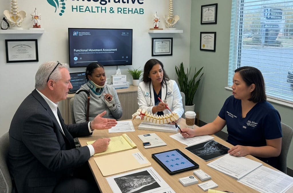
A cornerstone of our collaborative model is my partnership with Dr. Maria Guadalupe Cardenas, MD. Dr. Cardenas is Board Certified in Internal Medicine and serves as our esteemed Medical Director and Collaborative Physician. With over 40 years of invaluable experience, she provides essential medical oversight, ensuring our patients receive safe, comprehensive, and well-rounded care. This multidisciplinary structure allows us to blend the best of chiropractic and physical rehabilitation with the diagnostic and medical expertise of internal medicine. Our team works in synergy, designing treatment plans that address not just the symptoms but the underlying physiological dysfunction. Whether a patient is recovering from a personal injury, managing a chronic condition, or seeking to optimize their overall health, our integrated team provides a holistic, evidence-based pathway to recovery.
Navigating Low Back Pain with MLS Laser Therapy
One of the most common ailments we see is chronic low back pain. Today, we have a patient, John, who is experiencing persistent joint pain and stiffness in his lumbar spine, specifically around the L4-L5 facet joints, with some discomfort radiating down his right side. This is a classic presentation that responds exceptionally well to a targeted, multimodal approach.
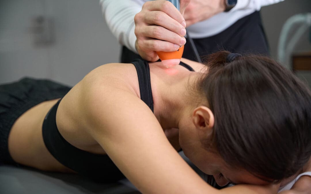
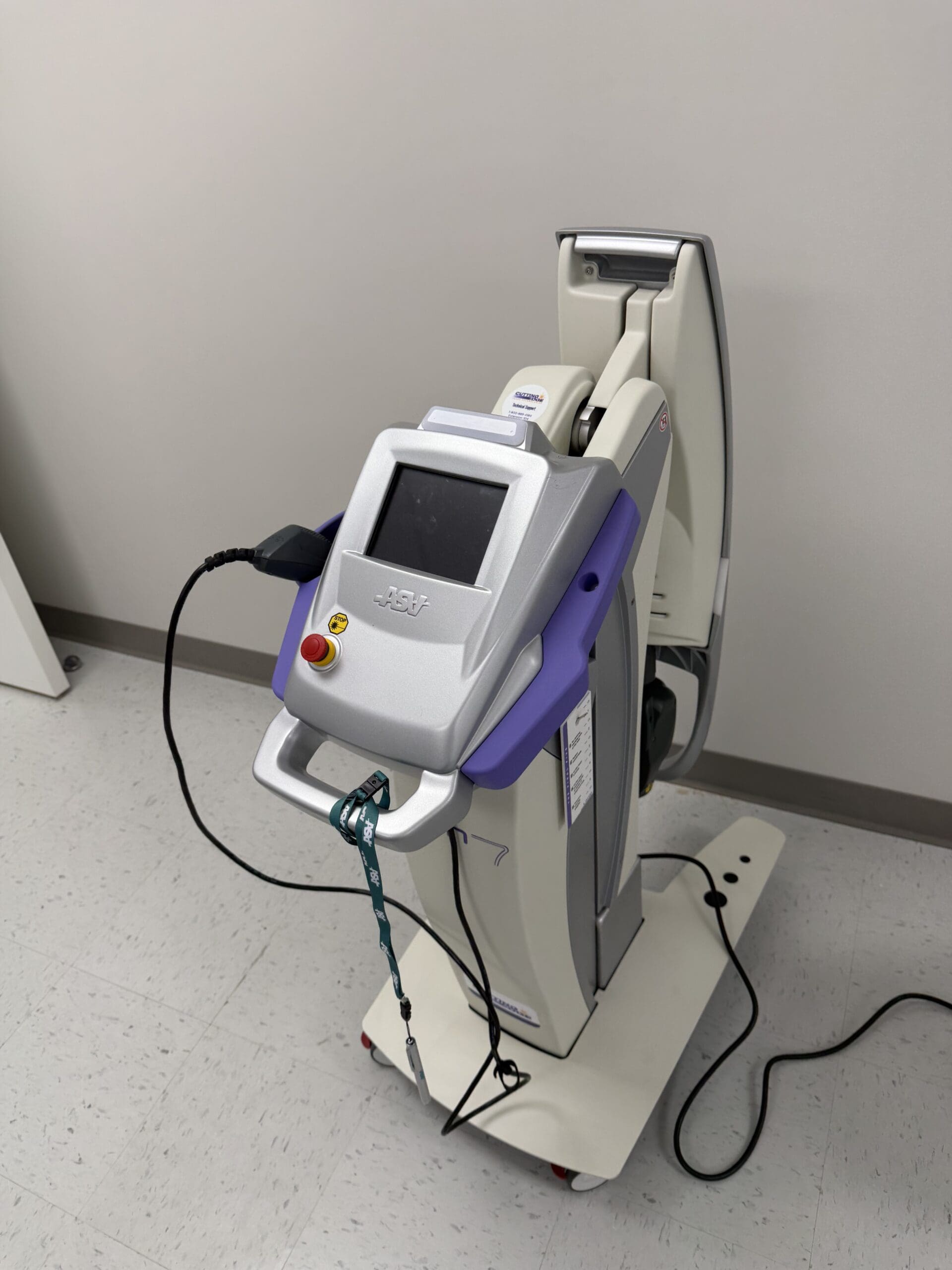
For John, we are utilizing the M6 Robotic MLS Laser. The first priority is always patient comfort. When using a robotic system, it’s critical that the patient remains still, as the laser is programmed to treat a precise area. We position the patient face down to allow direct access to the skin over the lumbar spine, as the laser energy must be delivered without the barrier of clothing.
The Clinical Multimodal Approach: More Than Just the “Spot of Pain”
Once John is comfortable, we begin the setup. The robotic laser interface is remarkably sophisticated yet user-friendly.
Targeting the Ailment: I select the “Joint Pain and Stiffness” protocol for the back.
Centering the Treatment: I zero out the X and Y axes on the control panel. This temporarily stops the robotic arm’s movement, allowing me to manually position the guiding red light directly over the primary source of John’s discomfort—the L4-L5 region he indicated.
Expanding the Field: This is where our clinical multimodal approach comes into play. Instead of just treating the single spot of pain, I expand the treatment area using the X and Y controls. This creates a larger therapeutic field that covers not only the symptomatic facet joints but also the surrounding connective tissue, muscles, and nerve roots. We aren’t just chasing pain; we are treating the entire functional unit to address the source of the dysfunction and support the interconnected biological systems.
The laser head is positioned at a precise distance from the skin—about six inches—using a provided ruler. This is crucial because the MLS laser beam is collimated, meaning the light rays are parallel. The focal point is engineered to be most effective at this distance, ensuring the therapeutic energy penetrates deep into the tissues rather than dissipating at the surface.
The Science of Healing: How MLS Laser Therapy Works
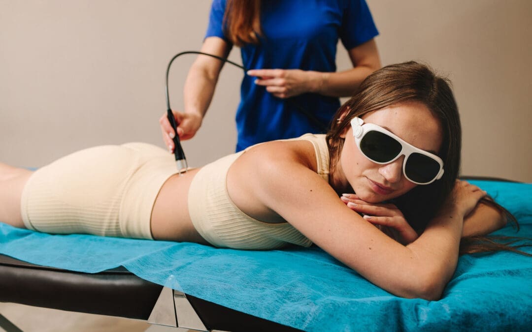
With the treatment underway—an eight-minute session for John’s low back—let’s dive into what’s happening at a cellular level. It’s common for patients to ask if they will feel anything. Most feel nothing at all, though some may notice a gentle warmth or tingling. This lack of intense heat is a hallmark of the MLS system’s advanced design.
The device combines two specific wavelengths of light: an 808-nanometer (nm) continuous-wave and a 905-nanometer (nm) pulsed-wave.
The 808 nm wavelength works more superficially to reduce inflammation and edema. It enhances blood circulation to the area, which helps clear out inflammatory byproducts and deliver oxygen and nutrients.
The 905 nm wavelength, delivered in powerful, short pulses, penetrates much deeper, reaching tissues such as muscle, nerve, and even the joint capsule. This pulsed energy is what provides the powerful analgesic (pain-relieving) effect.
These two wavelengths are synchronized, creating the patented “MLS pulse.” This enables delivery of very high peak power (up to 50 watts) in extremely short bursts (nanoseconds). This high-intensity “punch” of energy stimulates the cells without generating heat. A period of rest follows each pulse, allowing the tissue to absorb the energy efficiently. If a laser produces significant heat at the skin’s surface, it often means the energy isn’t being absorbed properly by the target tissues. The MLS system maintains tissue temperature at a constant level, ensuring optimal therapeutic delivery.
Seeing the Invisible: A Window into the Treatment
A fascinating demonstration of this technology involves using a smartphone camera. While the red aiming light is visible to the naked eye, the therapeutic infrared laser light is not. However, a camera’s sensor can detect it. If you were to look at John’s back through a phone camera during treatment, you would see a distinct triangle of light—this is the 808 nm wavelength at work, covering a significant area and illustrating how comprehensively we are treating the region.
Energy Density: The Key to Effective Dosing
A critical concept in laser therapy is energy density, measured in joules per centimeter squared (J/cm²). This is more important than the total number of joules delivered. Think of it like watering a plant: you need to provide the right amount of water for the pot’s size. Too little has no effect; too much drowns it. Similarly, our goal is to deliver a precise dose of light energy to the target tissue.
The World Association for Laser Therapy (WALT) and a large body of research support an optimal therapeutic window of 4-10 J/cm².
For John’s condition, the protocol is set to deliver approximately 6 J/cm². The laser’s software automatically calculates the treatment time required to achieve this density over the selected area. If I were to make the treatment area smaller or larger, the software would instantly recalibrate the time to ensure the correct dose is delivered.
This concept also relates to the Arndt-Schultz Law, a pharmacological principle stating that low doses stimulate, moderate doses inhibit, and high doses are toxic. With laser therapy, if you “overcook” an area with too much energy, you risk a bioinhibitory effect, in which the treatment becomes less effective or even counterproductive. The body’s cells can only absorb so much energy at once. This is why our protocols focus on precise energy density and, if more treatment is needed, we target different areas (e.g., an anterior and posterior approach for a knee) rather than just increasing the time on one spot.
Integrating Modalities for Superior Results
While the robotic laser treats the broader lumbar region, I can simultaneously use a handheld MLS laser applicator. This handpiece allows for more focused treatment on specific points, such as trigger points or “knots” in the muscle. I often use the “cooked meat” versus “raw meat” analogy that a physical therapist once taught me. Healthy, relaxed muscle feels like raw meat, while a tight, knotted trigger point feels firm, like cooked meat. The handheld applicator is perfect for treating these punctual spots.
The robot and the handpiece operate on two separate channels, allowing us to perform this dual treatment. This is a perfect example of our integrative philosophy in action:
Chiropractic Care: Before or after the laser session, I can perform specific chiropractic adjustments to restore proper motion to the L4-L5 facet joints and relieve mechanical stress.
Physical Rehabilitation: Our team can guide John through exercises to strengthen his core musculature and improve spinal stability.
MLS Laser Therapy: The laser works at the cellular level to reduce pain and inflammation that may be hindering his ability to engage in rehabilitation, thereby accelerating healing.
This combination addresses the structural, functional, and biochemical aspects of his condition simultaneously.
Advanced Applications: Augmenting Regenerative Medicine
The conversation around healing is increasingly turning toward orthobiologics, such as Platelet-Rich Plasma (PRP) injections. This is where MLS Laser Therapy shows even more remarkable potential. A common question arises: if PRP induces a beneficial pro-inflammatory phase to kickstart healing, won’t an anti-inflammatory laser treatment counteract it?
The answer is no. In fact, the laser augments the process. The data and our clinical observations show that using laser therapy in conjunction with PRP can improve outcomes by an estimated 15-20%.
Here is the progressive protocol we often recommend:
Pre-Injection Priming (2-3 treatments): In the weeks leading up to the PRP injection, we use the laser to “prepare the soil.” These sessions are designed to increase local blood circulation, reduce baseline chronic inflammation, and optimize the cellular environment, making the tissue more receptive to the growth factors in the PRP.
Day of Injection (1 treatment): A treatment on the day of the procedure can further enhance the effects.
Post-Injection Support (6+ treatments): Following the injection, a series of laser treatments helps manage pain and supports the regenerative cascade initiated by the PRP. The laser enhances mitochondrial function, which is critical for providing the cellular energy (ATP) needed for tissue repair.
The Cascade of Healing: From Acute Relief to Chronic Repair
How does a single modality address both acute pain and chronic conditions? The effects occur in a cascade.
Immediate Effect (Acute Phase): The initial pain relief often comes from the laser’s effect on small, unmyelinated nerve fibers (C-fibers) that transmit pain signals. The energy can temporarily block these signals, providing rapid relief. This is the analgesic effect.
Subsequent Effect (Inflammatory Modulation): Over the next few hours and days, the anti-inflammatory effect takes hold. The laser energy modulates the immune response, reducing pro-inflammatory cytokines and promoting the resolution of inflammation and edema.
Long-Term Effect (Biostimulation and Chronic Repair): With a series of treatments, we get to the core of cellular repair. Light energy is absorbed by cytochrome c oxidase in the mitochondria, the powerhouses of our cells. This significantly increases ATP (adenosine triphosphate) production, the body’s primary energy currency. This surge in available energy fuels all cellular repair processes, from protein synthesis to cell replication, promoting true, long-term tissue healing.
This mitochondrial boost is especially relevant in today’s world, where many common medications, such as statins, can impair mitochondrial function. By enhancing mitochondrial biogenesis and efficiency, laser therapy can help overcome these hurdles and optimize the body’s innate healing capacity. This is why we also discuss nutritional and lifestyle factors—such as CoQ10 supplementation to support mitochondrial function—as part of a truly comprehensive functional medicine approach.
Treatment Frequency and The Cumulative Effect
Healing is a process, not an event. The effects of MLS Laser Therapy are cumulative. We recommend a series of treatments to achieve lasting results.
Acute Conditions: Typically, a course of 6 treatments is effective.
Chronic Conditions: A more intensive course of 12 treatments is often needed.
Ideally, treatments are scheduled close together (e.g., Monday, Wednesday, Friday) to build therapeutic momentum. It is crucial for patients to complete the full course. Many start feeling significantly better after just 3-4 sessions and are tempted to stop. However, completing the entire protocol ensures deeper cellular repair, leading to more durable outcomes.
At Injury Medical Clinic, our mission is to empower your body’s own ability to heal. By integrating the best of chiropractic, medical oversight, and groundbreaking technologies like MLS Laser Therapy, we offer a path to recovery that is not only faster but also more complete.
World Association for Laser Therapy. (n.d.). WALT Recommended Treatment Doses for LLLT. WALT. Retrieved from https://waltza.co.za/wp-content/uploads/2012/08/Dose_table_780-860nm_for_Low_Level_Laser_Therapy_WALT-2010.pdf
Extreme Temperatures and Car Accident Risks in El Paso
In El Paso, Texas, summer heat often climbs above 100 degrees. This extreme heat does more than make you uncomfortable. It increases the risk of motor vehicle crashes and can worsen injuries. Scientific studies and safety data confirm that hot days and heat waves lead to more accidents and higher severity. On the road, heat creates a dangerous mix of tired drivers, stressed vehicles, and tough conditions.
This article walks you through why heat increases crash risks, how to prepare your vehicle and spot warning signs while driving, and what to do if you are in an accident. You will also learn about helpful integrative care options available right here in El Paso for faster, whole-person recovery.
Why Extreme Heat Leads to More Motor Vehicle Crashes
Research shows a clear link between high temperatures and more crashes. One review of studies found that hotter days are connected to rising numbers of fatal car crashes across the United States and other countries (Valentine, 2023). Another analysis noted a 3.4 percent rise in fatal crashes during heat waves (Adler, n.d.). In places like Texas, summer months often see the highest numbers of deadly wrecks.
Heat affects people, cars, and roads in several ways:
Your body struggles to stay cool. You sweat to cool down, but in extreme heat, you quickly lose water and important salts. This leads to dehydration. Dehydrated drivers often feel tired, have trouble focusing, and react more slowly to traffic lights or sudden stops.
Heat changes your mood and thinking. Many people become irritable or impatient when hot. This can lead to aggressive driving, tailgating, or risky decisions. Studies link heat to slower brain function and poorer judgment (Valentine, 2023; Adler, n.d.).
Your car turns into an oven. Sunlight passes through windows and traps heat inside. On a 100-degree day, the inside of a parked car can reach 130 to 150 degrees in a short time. Even with air conditioning, it takes time to cool down. Drivers in hot cabins feel distracted, sweaty, and less alert.
Vehicles face extra stress. Hot pavement and high temperatures can cause tire blowouts, especially on worn tires. Engines work harder and may overheat if coolant is low. Air conditioning systems strain to keep the cabin comfortable.
More traffic in summer. People drive more for vacations and outdoor plans. Higher traffic volume on hot, sunny days increases the likelihood of collisions (Adler, n.d.).
In El Paso’s desert climate, these factors combine often. Research cited by legal and safety sources shows that crash risks can rise by about 2.9 percent on heat-wave days, with even higher increases for crashes involving driver fatigue or distraction (Callahan Law, n.d.; Martinez, n.d.).
Preparing Your Vehicle for El Paso’s Hot Summers
A well-prepared car helps you avoid breakdowns and stay safer in extreme heat. Take these steps before and during summer:
Check tires carefully. Heat makes air inside tires expand, but worn tread or damage increases the risk of a blowout on hot roads. Check tire pressure when the tires are cool, usually in the morning. Look for cracks, bulges, or low tread. Replace tires that show wear.
Test and service the air conditioning. A strong AC keeps you cool and focused. If the air feels weak or takes too long to cool, have a mechanic check the system. Good cooling fights the greenhouse effect inside your car.
Inspect the cooling system. Make sure the radiator, hoses, and coolant levels are in good shape. Low coolant can cause engine overheating, leaving you stranded in dangerous heat.
Have the battery checked. Extreme heat shortens battery life and can cause sudden failure. Clean any corrosion from terminals and replace old batteries before problems start.
Use simple heat blockers. Keep a windshield sunshade handy. Park in shade or a garage whenever possible. These steps stop the inside of your car from reaching dangerous temperatures.
Carry basic supplies. Keep water bottles, a first-aid kit, a flashlight, and a phone charger in the car. If you break down, you can stay hydrated and call for help safely.
These simple actions reduce mechanical failures that, when combined with driver fatigue, cause crashes.
Spotting Heat-Related Fatigue While Driving
Even with a well-prepared car, long drives or heavy traffic in El Paso’s heat can tire you out quickly. Knowing the early signs lets you act before trouble starts. Common signs include:
Yawning often or feeling your eyelids grow heavy
Trouble staying focused on the road or missing exits and signs
Your vehicle drifting between lanes without you meaning to
Feeling more grumpy or frustrated with other drivers than usual
Headache, dry mouth, thirst, or general sluggishness
Slower reactions, such as braking late or not noticing hazards quickly
If you notice any of these, pull over to a safe spot right away. Drink water, sit in shade or cool air if possible, and rest. Some drivers find that calm music helps them stay relaxed (Martinez, n.d.). Do not try to push through severe tiredness. If you feel unsafe, let someone else drive or stop for the day. Your quick action can prevent a serious crash.
Regular Vehicle Maintenance to Lower Heat Dangers
Ongoing care keeps your car reliable when temperatures soar. Schedule a full inspection before summer begins. Ask a mechanic to check belts, hoses, fluids, and the air conditioning system. Change oil and filters on time so the engine runs cooler under heavy load. Monitor brake, transmission, and power steering fluid because heat makes these systems work harder. Replace wiper blades and ensure all lights work properly for better visibility in bright sunlight or dusty conditions.
Staying ahead on maintenance means fewer surprises and safer drives.
What to Do If You Are in a Motor Vehicle Accident
Even careful drivers can face crashes. In extreme heat, the stress on your body may make symptoms like headaches, back pain, or neck pain feel stronger or last longer. Getting the right care early supports better healing.
Integrative clinics offer a multifaceted approach. These clinics often bring together chiropractors, nurse practitioners, physical therapists, and medical doctors who work as a team. They address pain, movement, inflammation, and overall health instead of treating just one symptom.
How an Integrative Team Supports Recovery in El Paso
Many people involved in motor vehicle accidents deal with whiplash, spinal misalignments, soft tissue strains, headaches, back pain, or neck pain. These injuries happen from the sudden force of impact. An integrative and holistic approach can speed healing by combining treatments that support the whole body.
At Injury Medical Clinic PA (also known as Mission Plaza Injury Medical Clinic) in El Paso, Texas, the team uses this collaborative model. Dr. Alexander Jimenez, DC, APRN, FNP-BC, provides chiropractic care focused on spinal alignment and function. Through his extensive clinical experience treating patients in El Paso, shared on platforms such as dralexjimenez.com and his professional profiles, he has observed that recovery improves when care addresses both spinal issues and the body’s broader healing needs, often using advanced imaging and combined therapies.
Working with him is Dr. Maria Guadalupe Cardenas, MD, Board Certified in Internal Medicine. She brings over 40 years of experience as an internist, holds NPI #1164426749, and maintains Texas MD License #J2933. Dr. Cardenas serves as Medical Director and Collaborative Physician at the practice. This multidisciplinary setup is common in strong integrative and injury care clinics. The MD provides medical direction and oversight for complex cases, while the chiropractor delivers hands-on spinal care. Together, they create safe, coordinated plans.
The team integrates several services:
Chiropractic adjustments to gently realign the spine, relieve nerve pressure, reduce pain and inflammation, and restore mobility. This helps with common post-accident problems such as whiplash-related headaches and neck pain or lower back injuries.
Medical oversight and evaluation by Dr. Cardenas to assess overall health, manage inflammation or other factors, and guide the treatment path.
Functional medicine support, including nutrition and lifestyle guidance, to help the body repair tissues and regain energy.
Rehabilitation and physical therapy to build strength, improve flexibility, and prevent future issues.
Personal injury care that includes proper documentation and coordination for insurance or legal needs.
This combined approach often leads to faster relief, better mobility, and a lower risk of pain becoming chronic. It focuses on root causes rather than only covering symptoms. For anyone in the El Paso area experiencing headaches, back pain, or neck pain after a recent motor vehicle accident, the team can create a personalized recovery plan based on your specific injuries and health background. They may recommend imaging or referrals to other specialists when needed.
Patients frequently report improved comfort and function when care starts soon after an accident and includes this full-team support.
Moving Forward with Safety and Stronger Recovery
Extreme heat clearly raises the risks of motor vehicle crashes in El Paso, but preparation makes a difference. Checking your vehicle, watching for signs of fatigue, and keeping up with maintenance help protect you on the road. If an accident does occur, integrative care that blends chiropractic expertise, medical oversight, and functional support can help you heal more completely and quickly.
In El Paso, teams like the one at Injury Medical Clinic PA, with Dr. Alexander Jimenez and Dr. Maria Guadalupe Cardenas, offer this kind of coordinated, patient-centered care. They focus on restoring function and addressing the whole person so you can return to daily life with less pain and more confidence.
Drive safely, stay cool, and seek professional support when needed. Effective help is available close to home.
Welcome to our exploration of photobiomodulation therapy (PBMT), a revolutionary approach that harnesses the power of light to stimulate cellular healing. In this educational post, I will guide you through the intricate biological processes that make PBMT so effective. We will explore how specific wavelengths of light can penetrate tissues to activate mitochondria, modulate the immune response, and accelerate recovery. This journey will cover the fundamental science behind PBMT, from its effects on ATP production and cytokine modulation to its role in promoting angiogenesis and neurogenesis. Furthermore, we will examine the synergistic potential of combining PBMT with orthobiologics, such as Platelet-Rich Plasma (PRP), and demonstrate how this integrated approach can enhance healing outcomes. Drawing on the latest research, including fascinating studies from the veterinary world and our laboratory findings on tenocyte proliferation, we’ll demonstrate why light is not just a modality but a cornerstone of modern regenerative medicine. At Injury Medical Clinic, we integrate these advanced therapies within a collaborative framework, combining my expertise in chiropractic and functional medicine with the medical oversight of our Medical Director, Dr. Maria Guadalupe Cardenas, MD, to provide comprehensive, evidence-based care for our patients.
About Our Integrated Practice: A Collaborative Approach to Wellness
I, Dr. Alex Jimenez, am honored to share my passion for integrative and regenerative medicine with you. With a diverse background as a Doctor of Chiropractic (DC), Advanced Practice Registered Nurse (APRN), board-certified Family Nurse Practitioner (FNP-BC), and certifications in Functional Medicine (CFMP, IFMCP), Applied Traumatology (ATN), and Cranial Spinal Integration (CCST), my goal has always been to find the most effective, evidence-based paths to healing.
Here at Injury Medical Clinic PA in El Paso, Texas, we have built a unique, multidisciplinary practice. We believe that the best patient outcomes are achieved through a collaborative team approach. I am privileged to work alongside Dr. Maria Guadalupe Cardenas, MD, who serves as our Medical Director and Collaborative Physician. Dr. Cardenas is a highly respected, board-certified Internist with over 40 years of experience (NPI #1164426749, Texas MD License #J2933). Her extensive medical knowledge provides invaluable oversight and complements our services, ensuring our patients receive safe, comprehensive, and well-rounded care.
Our clinic integrates:
Advanced Chiropractic Care: Focused on spinal health, biomechanics, and nervous system function.
Physical Therapy & Rehabilitation: Tailored programs to restore movement, strength, and function.
Medical Oversight: Guided by Dr. Cardenas to ensure clinical safety and efficacy.
Functional Medicine: Investigating the root causes of chronic conditions.
Personal Injury Care: Specialized treatment for injuries sustained in accidents.
This model allows us to address health from multiple angles. While our core focus at elpasobackclinic.com is chiropractic and physical rehabilitation, we incorporate advanced modalities such as photobiomodulation to enhance the body’s innate healing capabilities, with all treatments guided by a solid medical and scientific foundation.
The Awakening: From Skepticism to Cellular Biology
I have been on this journey for nearly a decade, and for the first five years, discussing “laser” therapy in medical circles often felt like an uphill battle. It was a path paved with skepticism, much like the initial reception many of you in the biologics field have likely experienced. But today, I am thrilled to see the conversation shifting as the science catches up with the clinical results.
My evolution as a clinician mirrors this shift. For the first two decades of my career, I was a “mechanic,” using established tools to address specific conditions. Over the last ten years, however, I have become a “biologist,” focused on understanding and facilitating the body’s own healing processes at a cellular level. This is why I am so excited to share the science of photobiomodulation (PBMT) with you. It represents a profound shift from treating symptoms to enabling cellular recovery.
Understanding Photobiomodulation: The Science of Light and Life
The concept is beautifully simple, rooted in a phenomenon we all accept: photosynthesis. The sun’s light fuels life on Earth, and as a species that has evolved under this light for hundreds of thousands of years, our cells have developed a deep, genetic sensitivity to it. We readily accept that sunlight is necessary for Vitamin D synthesis, yet a significant gap remains in medical education regarding the broader therapeutic applications of light.
Photobiomodulation breaks down as:
Photo: Light
Bio: Life
Modulation: To affect or change
Light is energy, delivered in units called photons. These photons can transfer their energy to our cells, triggering a cascade of biological responses. This is the essence of PBMT.
The Cellular Engine: How PBMT Activates Mitochondria
The primary target of photobiomodulation within the cell is the mitochondria, our cellular powerhouses. Specifically, an enzyme in the mitochondrial respiratory chain, cytochrome c oxidase, acts as a photoacceptor. This means it is designed to absorb photons of light.
Here is the cascade of events that follows:
Activation: When light photons of the correct wavelength strike cytochrome C oxidase, the enzyme becomes more active.
Increased ATP Production: This heightened activity accelerates the Krebs cycle, leading to more efficient production of adenosine triphosphate (ATP), the primary energy currency of the cell. More ATP means more energy available for cellular repair, replication, and function.
Signaling Cascade: This process also triggers the release of key signaling molecules, including nitric oxide and reactive oxygen species (ROS) in controlled, beneficial amounts.
Gene Transcription: These signaling molecules then travel to the cell’s nucleus, initiating gene transcription. This is where the cell is instructed to produce specific proteins, including cytokines, which orchestrate the healing process.
Modulating the Immune Response: From Inflammation to Repair
When an injury occurs, the body initiates an inflammatory response characterized by the production of pro-inflammatory cytokines. PBMT helps guide the body out of this chronic or stalled inflammatory phase and into a reparative one by modulating the cytokine profile.
Anti-Inflammatory Effects: Research has clearly shown that PBMT, when used at the right wavelengths, can increase the production of interleukin-10 (IL-10), a potent anti-inflammatory cytokine.
Pro-Inflammatory Reduction: It has also been shown to reduce levels of pro-inflammatory cytokines, such as interleukin-6 (IL-6).
This shift moves the cellular environment from a state of chronic inflammation—such as that seen in a thickened, bulbous Achilles tendon—toward active healing and regeneration.
Building the Foundation for Healing: Angiogenesis, Neurogenesis, and Muscle Recovery
The benefits of PBMT extend beyond simple control of inflammation. The cellular signaling it initiates promotes the foundational elements of tissue repair.
Enhanced Blood Flow (Angiogenesis): PBMT has been shown to promote angiogenesis by stimulating the production of cytokines such as galectin-1. This improved microcirculation is crucial for delivering oxygen and nutrients to injured tissue and removing waste products. For anyone focused on healing, whether through chiropractic adjustments or post-surgical recovery, enhanced blood flow is paramount.
Nerve Repair (Neurogenesis): We can also document the repair of nerve cells. PBMT stimulates the production of proteins that encourage axonal growth, helping to repair damaged neurons. This is particularly relevant in our practice for treating neuropathies and nerve entrapment syndromes like carpal tunnel.
Muscle and Tissue Recovery: Electron microscopy studies have provided clear evidence that PBMT improves muscle cell development and increases myoglobin production, which enhances oxygenation. It also activates fibroblasts, the cells responsible for producing collagen and building the structural framework for new tissue.
In essence, PBMT orchestrates a symphony of healing: it modulates the immune system, increases blood flow, repairs nerves, and rebuilds tissue.
The Therapeutic Window: Why Wavelength Matters
Not all light is created equal. The electromagnetic spectrum ranges from deadly short-wavelength gamma rays to long-wavelength radio waves that pass harmlessly through us. The therapeutic potential of light lies within a specific “therapeutic window,” approximately from 600 nanometers (red light) to 1200 nanometers (near-infrared light).
The primary challenge is getting the photons to the target tissue. Three main obstacles absorb light energy before it can penetrate deeply:
Skin (Melanin)
Blood (Hemoglobin)
Water
While red light is effective for superficial tissues (penetrating 3-4 millimeters), treating deeper musculoskeletal structures requires wavelengths in the near-infrared spectrum, which can penetrate more effectively.
In my practice, we leverage this science daily. For acute injuries, such as those our Division 1 athletes sustain, PBMT significantly reduces recovery time. Post-operatively, it minimizes swelling and bruising and improves incision healing. And for the chronic inflammatory conditions we see so often, it provides the cellular energy needed to break the cycle of pain and dysfunction.
Synergy in Action: Combining PBMT and Orthobiologics
This is where the conversation becomes truly exciting. We know that orthobiologics, such as Platelet-Rich Plasma (PRP), deliver a potent cocktail of growth factors and anti-inflammatory proteins. They are essentially sending a “message” to the cells, instructing them to heal.
Now, imagine providing the “fuel” for that message.
By combining PRP with PBMT, we are doing just that. The PRP provides the blueprint for repair, and the PBMT provides the cellular energy (ATP) needed to carry out those instructions. We turn on the mitochondrial engine, allowing the cells to fully utilize the growth factors and signaling proteins delivered by the biologic treatment. We are creating a synergistic effect where the whole is greater than the sum of its parts.
Evidence from Our Four-Legged Friends: A Canine Study
When exploring emerging therapies, I often look to veterinary medicine. Animals, particularly dogs, do not have confounding factors such as secondary gain or placebo effects associated with complex human emotions. A treatment either works or it does not.
An outstanding randomized controlled trial on canines with knee osteoarthritis provides compelling evidence for this synergy.
Study Design: Each dog served as its own control. The dogs first received PBMT alone. After a washout period, they received a PRP injection alone. Finally, after another washout period, they received a combination of PRP and PBMT.
Results: The outcomes, measured by owner-reported functional improvements (like climbing stairs or getting into a car), were significantly better with the combined therapy than with either treatment alone.
This study strongly suggests that combining light energy with biologics creates a more robust and effective healing response.
Our Own Research: Proving Cellular Proliferation
To further validate these concepts, we embarked on our own research. My son, Zachary, led a study at the Mass General Brigham Enable BioSkills Lab to investigate the direct effects of PBMT on human tendon cells.
We treated human tenocytes (tendon cells) with our laser therapy. The results were remarkable: we demonstrated a 20% dose-dependent increase in tenocyte proliferation with PBMT alone. We were able to literally watch the cells multiply under the influence of light.
We are now conducting additional qPCR and ELISA testing to analyze gene expression and protein levels, which will give us an even deeper understanding of the pathways being activated. This work confirms that PBMT is not a passive modality; it is an active biological stimulus that directly promotes cellular regeneration.
The Future of Medicine is Biology
We are moving away from an era of purely symptomatic treatments and toward a future of true disease modification. The goal is to intervene earlier and more effectively, harnessing the body’s innate biological wisdom to heal from within. Photobiomodulation is a cornerstone of this new paradigm. It has been validated by major health organizations, including its mention in the CDC’s revised opioid guidelines as a non-pharmacological option for pain.
I have seen the profound impact of this therapy in my clinic and in the research lab. It works. The synergy between photobiomodulation and other regenerative therapies, all within an integrated care model that prioritizes chiropractic and physical rehabilitation, represents the future of orthopedic and musculoskeletal health. It has been a pleasure to share this journey with you.
Delayed Car Accident Pain and Integrative Recovery
Many people feel fine right after a small car bump or fender bender. They drive away thinking everything is okay. Then, hours or even days later, pain, stiffness, or odd symptoms appear. This happens more often than most expect. Delayed symptoms after minor auto accidents are common because the body initially hides problems. Understanding why this occurs and what to do next can make a big difference in how well and how fast you recover.
Why Symptoms Often Appear Hours or Days Later
During a car accident, even a minor one, your body goes into a high-alert mode. It releases adrenaline to give you energy and focus. At the same time, it pumps out endorphins. These natural chemicals act like pain blockers. They help you stay calm and move if needed. Muscles also tense up and brace for impact. This response can mask damage to ligaments, discs, nerves, or soft tissues.
Once the adrenaline and endorphins fade, usually within 24 to 72 hours, swelling and inflammation begin to show. Hidden strains or small tears start to bother you. In some cases, symptoms wait even longer—weeks after the crash. This delay occurs because other parts of the body compensate at first. Or swelling builds slowly in deeper tissues. Low-speed collisions can still cause real problems because the body may not brace the same way as in bigger crashes. The result is neck pain, backaches, or nerve pain that seems to come out of nowhere.
Ignoring these signs can let small issues turn into bigger ones. Scar tissue may form, movement patterns change, and chronic discomfort can settle in. That is why paying attention early matters.
Common Warning Signs to Watch After a Minor Crash
Delayed symptoms vary from person to person. Some feel them the next day. Others notice changes a week or more later. Here are frequent ones to track:
Headaches that stick around or get worse: These can start from neck strain or small head movements during impact.
Neck or back stiffness and pain: Whiplash often shows up this way, with tightness that makes turning or bending hard.
Numbness, tingling, or radiating pain: This may travel into the shoulders, arms, or legs and may point to nerve irritation or pressure.
Unusual fatigue or low energy: Your body uses extra resources to heal, leaving you drained.
Brain fog, irritability, or trouble focusing: These cognitive changes can follow even mild impacts and affect daily tasks.
Dizziness, balance problems, or vertigo: Inner ear or neck issues sometimes appear later.
Other possible signs include shoulder or hip discomfort, sleep trouble, or mood shifts. If any new symptom starts after an accident, write down when it began, how strong it feels, and what makes it better or worse. This record helps healthcare providers connect it to the event.
Why See a Healthcare Professional Right Away
Even if the crash seemed small and you felt okay at the scene, get checked soon. A healthcare professional or nearby urgent care can spot hidden issues before they grow. They document the link between your symptoms and the accident. This step supports insurance claims and guides proper care. Early evaluation often leads to simpler, non-invasive help that works better than waiting until pain becomes constant.
Seek emergency medical help right away if you notice:
Sudden weakness in arms or legs
Severe vertigo or spinning sensations
Pain that quickly gets much worse
Confusion, vision changes, or slurred speech
Chest pain, shortness of breath, or abdominal swelling
These can signal more serious problems that need immediate attention. For most delayed symptoms from minor accidents, though, a prompt visit to a knowledgeable clinic sets the stage for steady healing.
How Integrative Chiropractic Care Supports the Body’s Natural Healing
Your body has a built-in healing process that works at the cellular level. After injury, it sends signals to reduce inflammation, repair damaged tissue, and rebuild strength. An integrative chiropractic clinic helps this natural cascade along. They combine hands-on biomechanical work with targeted regenerative therapies. The goal is to remove roadblocks so healing happens smoothly and completely.
Chiropractic adjustments gently move spinal joints back into better alignment. This relieves pressure on nerves and improves overall movement. Myofascial release loosens tight bands of tissue around muscles that often form after an accident. These tight spots create compensations—extra strain on other areas as the body tries to avoid pain. By restoring normal motion early, the clinic reduces the chance that old compensations become new long-term problems.
Regenerative Injections and Chiropractic Adjustments: A Strong Team Approach
When used together, regenerative biological injections and chiropractic care give a well-rounded path to recovery. Regenerative injections, such as platelet-rich plasma (PRP), work at the cellular level. A small amount of your blood is processed to concentrate platelets. These platelets release growth factors and signaling proteins. The factors tell local cells to multiply, build new collagen, improve blood supply, and shift from ongoing irritation to active repair. This supports healing of ligaments, tendons, muscles, and joints damaged in the crash.
Chiropractic adjustments and soft tissue work then correct the bigger picture. They restore spinal alignment and smooth movement patterns. Without this step, even repaired tissues can face ongoing stress from poor posture or guarded motions. The injections handle the microscopic repair work. The adjustments ensure the entire structure supports the repair and prevents reinjury. Patients often notice improved mobility, reduced pain, and a faster return to normal activities when both parts work in sequence.
This combined method is well-suited to delayed symptoms. It addresses both the hidden cellular damage and the mechanical changes that develop after the initial shock wears off. Many people find they heal more completely and with fewer setbacks than with either approach alone.
Expert Multidisciplinary Care in El Paso
In El Paso, Texas, Injury Medical Clinic PA—also known as El Paso Back Clinic—offers this kind of integrative care for people dealing with auto accident injuries. Dr. Alexander Jimenez, DC, APRN, FNP-BC, leads the team. He is a chiropractor and board-certified family nurse practitioner with advanced training in functional medicine, spinal trauma, and musculoskeletal care. His clinical observations show that many patients with symptoms that appear days or weeks after minor crashes improve significantly when care targets both alignment and early tissue repair. He notes that addressing compensation and supporting cellular healing help prevent chronic pain and keep people moving well long term.
Working alongside him is Dr. Maria Guadalupe Cardenas, MD. She is Board Certified in Internal Medicine with over 44 years of experience. Dr. Cardenas serves as Medical Director and Collaborative Physician at the clinic (NPI #1164426749, Texas MD License #J2933). Her role brings medical oversight to the practice. She helps ensure adherence to safety protocols, coordinates care for complex health needs, and supports the integration of chiropractic services with broader internal medicine perspectives. This includes attention to chronic conditions, preventive strategies, nutrition, and referrals when needed.
The setup is a common multidisciplinary model in integrative injury clinics. Chiropractic care from Dr. Jimenez focuses on biomechanical correction and rehabilitation. Medical direction from Dr. Cardenas provides an internal medicine lens for whole-person health. The team also incorporates functional medicine principles, personal injury documentation, and regenerative options. Together, they create personalized plans that respect each patient’s unique situation after a car accident. This collaboration helps people recover function while addressing any underlying factors that could slow healing.
Moving Forward After Delayed Symptoms Appear
If you have noticed new stiffness, headaches, nerve feelings, or fatigue following a minor auto accident—recent or even from months ago—consider reaching out for a full evaluation. A clinic experienced with these patterns can assess your spine, soft tissues, and overall function. They can then build a plan that supports your body’s healing steps without jumping straight to heavy medications or surgery.
Keep notes on your symptoms and how they affect daily life. Save records from any visits. These details help the care team connect the dots and may support insurance or legal processes if needed. Recovery does not have to mean living with ongoing discomfort. With the right combination of expert adjustments, regenerative support, and medical guidance, many people regain comfort and mobility.
Delayed symptoms after minor car accidents do not have to control your days. Understanding the timeline, recognizing the signs, and choosing care that works with your body’s natural processes can lead to real improvement. Teams that blend chiropractic precision with regenerative therapies and medical oversight offer a clear path forward—one focused on lasting function and feeling like yourself again.
In this educational post, I will take you on a journey through the cutting-edge landscape of regenerative and integrative medicine for treating common musculoskeletal conditions. Drawing on the latest evidence-based research and my clinical experience, we will explore which injuries respond best to advanced orthobiologic therapies such as Platelet-Rich Plasma (PRP) and microfragmented adipose tissue. We will explore a systematic, algorithm-based approach for patient selection, focusing on conditions such as partial rotator cuff tears, tendinopathies like tennis elbow, and mild-to-moderate osteoarthritis. Furthermore, I will introduce a groundbreaking study that uses machine learning to identify key biomarkers—such as uric acid and lipoprotein(a)—that predict patients’ treatment response. Finally, I will explain how our unique multidisciplinary practice in El Paso, Texas, integrates advanced medical oversight with chiropractic care, physical therapy, and functional medicine to create a comprehensive and personalized healing environment for our patients.
A New Era of Collaboration in Patient Care
I am thrilled to announce a significant enhancement to our patient care model here at Injury Medical Clinic. We are honored to welcome Dr. Maria Guadalupe Cardenas, MD, to our team as our Medical Director and Collaborative Physician. Dr. Cardenas is a highly respected, board-certified Internist with over four decades of clinical experience (NPI #1164426749, Texas MD License #J2933).
This collaboration represents a powerful fusion of expertise. Our clinic has always been at the forefront of providing exceptional chiropractic care, physical therapy, and rehabilitation, particularly for those suffering from personal injuries. With Dr. Cardenas providing medical oversight, we can now offer an even more robust and integrated treatment paradigm. This multidisciplinary setup allows us to manage complex cases by combining my expertise in chiropractic, functional, and regenerative medicine with her profound knowledge of internal medicine. This ensures that every aspect of a patient’s health—from musculoskeletal alignment and function to underlying systemic factors—is addressed, creating a truly holistic path to recovery.
The Foundation of Our Approach: Evidence-Based Integrative Care
When I established my practice in El Paso, TX, this environment ingrained in me the necessity of grounding every clinical decision in solid, evidence-based research. We developed a structured protocol to identify which conditions were most appropriate for orthobiologic treatments. This required a deep dive into the scientific literature to ensure we were offering therapies with proven efficacy.
This commitment to evidence is the cornerstone of our practice in El Paso. We specialize in treatments that bridge the gap between conservative care and invasive surgery. Our focus is on harnessing the body’s innate healing capabilities, supported by advanced diagnostics and targeted interventions.
Identifying the Right Conditions for Orthobiologic Therapies
Through rigorous review of studies and extensive clinical experience, we have identified a specific cohort of conditions that respond well to integrative and regenerative treatments. It is crucial to be precise in our diagnosis and patient selection to achieve the best possible outcomes.
Here are some of the primary conditions we treat:
Shoulder: Low-grade, partial-thickness rotator cuff tears and mild-to-moderate glenohumeral arthritis. For arthritis, it is vital to consider the Walsh classification (e.g., A1, A2, B1) to ensure that the joint architecture is stable and that the “golf ball” (humeral head) isn’t falling off the “tee” (glenoid).
Elbow: Lateral epicondylitis (tennis elbow) and medial epicondylitis (golfer’s elbow), as well as proximal partial tears of the ulnar collateral ligament (UCL).
Hand/Wrist: Mild-to-moderate carpometacarpal (CMC) arthritis. A landmark study from my professor at the Mayo Clinic validated the use of biologics for this condition.
Hip: Femoroacetabular Impingement (FAI) of grade two or less, where the labrum is not shredded, and there are no large pincer or cam deformities. We also achieve great results with gluteus medius and hamstring tendinopathy, especially focal mid-portion tears.
Foot/Ankle: Plantar fasciitis.
Knee: Classically, mild-to-moderate knee osteoarthritis and very small meniscal tears.
Interestingly, recent literature has shown promise in the use of PRP post-operatively. Some forward-thinking surgeons now refer patients for a PRP injection between 0 and 6 weeks after a rotator cuff repair to potentially enhance healing.
A Deeper Look at Tendinopathy: Diagnosis and Treatment Strategy
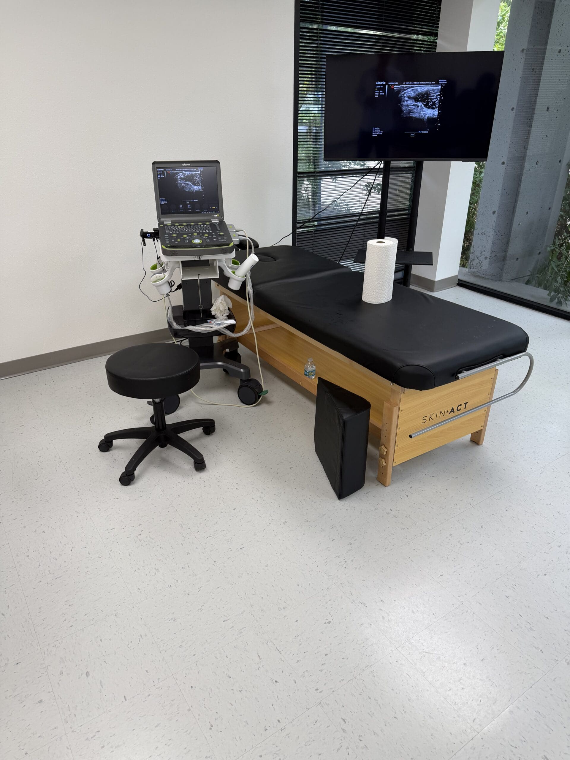
Let’s examine a common case: tennis elbow, or a partial-thickness tear of the common extensor tendon. Using musculoskeletal ultrasound, we can visualize the injury with incredible detail. I look at the tendon in both long-axis and short-axis views to measure the tear’s precise length and width.
A key to my treatment success has been the technique of tenotomy with fenestration. This involves using a needle to meticulously break up the scarred, degenerative tissue throughout the entire length and width of the tear. Many practitioners might inject only into one spot, but I have found that ensuring the biologic agent is delivered throughout the full extent of the damaged area significantly improves results. We are essentially creating micro-trauma to stimulate a new, robust healing cascade and delivering the growth factors right where they are needed most. The study by Gosens et al. (2011) provides strong support for using PRP to treat chronic tennis elbow, and it is a paper I often share with my colleagues to explain the rationale for this approach.
Consider the case of a 31-year-old weightlifter with patellar tendinopathy. His ultrasound revealed a complex picture: early-stage arthritis with a knee effusion (fluid), a large partial-thickness tear of the patellar tendon, heterogeneous echogenicity changes (indicating tendinosis), and even a large calcium deposit. The critical question becomes: what is the primary pain generator? Is it the joint cartilage, the degenerated tendon, or the calcification?
After a thorough discussion about the risks and benefits, and correlating his physical exam findings with the imaging, I decided to treat the tendon tear with PRP. My decision was influenced by research, such as the work of Jason Dragoo, who demonstrated the efficacy of leukocyte-rich PRP for tendinopathy. For a tear of this significant size, PRP provides a powerful concentration of growth factors to orchestrate cellular repair and tissue regeneration. In these challenging cases, pinpointing the source of pain is paramount.
The Nuances of Treating Rotator Cuff Tears
Rotator cuff tears present another layer of complexity. An MRI might show a partial-thickness tear (less than 50% of the tendon’s thickness) and also an interstitial tear (a split within the tendon fibers), along with surrounding edema (fluid). My approach is often to treat both. I will perform a guided injection into the subacromial bursa to reduce inflammation and another directly into the interstitial tear itself.
Using ultrasound guidance is non-negotiable. I can watch the needle in real-time as it passes through the deltoid muscle and subacromial bursa to precisely target the tear on the superficial facet of the greater tuberosity. I use a small amount of fluid to hydrodissect the tissue planes, which confirms I am in the correct location and helps distribute the biologic throughout the length of the tear.
It’s important to clarify terminology. A partial-thickness tear involves only a portion of the tendon’s depth. A full-thickness tear goes all the way through, but this can be a partial-width tear (affecting only part of the tendon’s footprint) or a full-thickness, full-width tear (a complete rupture). Orthobiologics are most effective for partial-thickness and full-thickness, partial-width tears, not complete ruptures, which typically require surgery.
Choosing the Right Tool: PRP vs. Adipose Tissue
When a patient presents with a more severe injury, we must consider more robust therapies. This is where my treatment algorithm helps guide the decision-making process.
For low-grade partial-thickness tears (less than 50%): I will consider PRP, sometimes augmented with dextrose prolotherapy (P2G), to stimulate a healing response.
For high-grade partial-thickness tears (greater than 50%): I will consider using microfragmented adipose tissue.
Why adipose? Adipose tissue is not just fat; it is a rich source of mesenchymal stem cells (MSCs) and other perivascular cells that create a biological scaffold. This scaffold provides a structural framework and a sustained-release reservoir of signaling molecules that guide tissue repair over a longer period. This is particularly beneficial in larger defects where a simple injection of PRP might not be sufficient to bridge the gap. For moderate-to-severe arthritis (Kellgren-Lawrence grade 3-4), I also lean towards adipose tissue or bone marrow aspirate concentrate (BMAC) for their more potent anti-inflammatory and regenerative capabilities.
For patients with neuralgia or nerve entrapment, I have found that hydrodissection—using fluid to carefully separate the nerve from surrounding fibrotic tissue—can provide significant relief by freeing the nerve and reducing compression.
An Algorithmic Approach to Treating Knee Osteoarthritis
To standardize care and optimize outcomes, I have developed a treatment algorithm for patients with knee osteoarthritis (OA). This systematic process ensures we address all contributing factors:
Assess Systemic Health: First, I investigate for underlying systemic diseases (like autoimmune conditions) or factors that impair healing. We must address the whole person, not just the knee.
Evaluate Functional Markers: Next, I consider a functional medicine workup. What are their hormone levels? Is there evidence of gut dysbiosis or microbiome imbalance? These factors create the systemic environment in which the knee must heal.
Grade the Arthritis: Using X-rays and MRIs, I determine the severity. Is it grade 3 or 4 arthritis? Is there significant subchondral bone edema (a sign of stress and inflammation in the bone beneath the cartilage)?
Select the Treatment:
If the patient has mild-to-moderate OA (grade 1-2) without the above complicating factors, PRP is my first-line orthobiologic treatment.
If they have severe OA (grade 3-4) or significant bone edema, I will discuss microfragmented adipose tissue or BMAC.
Monitor and Adjust: Healing is a process. PRP typically causes increased soreness for about three days, with functional improvements beginning around weeks three to six. By twelve weeks, we should have a clear indication if we are on the right track. If the patient has achieved at least 60% improvement, we continue with our supportive care plan. If not, we re-evaluate and adjust the strategy.
The Future is Now: Machine Learning and Personalized Medicine
A groundbreaking study published in April 2026 in BMC Musculoskeletal Disorders is already changing how I think about patient selection. Researchers in China used a machine learning algorithm to predict clinical response to PRP for knee osteoarthritis. They analyzed a vast dataset including patient demographics, BMI, lab markers, and pain scores.
The algorithm aimed to identify the factors that were most predictive of a high response rate (defined as increasing the success rate from 65% to 85%). The results were fascinating. While we often focus on the “special recipe” of the PRP itself, the study found that three biomarkers were most important in predicting success:
Osmotic Pressure (Joint Swelling): This was self-explanatory. My clinical experience confirms that patients with recurrent, large effusions do not respond as well. The inflammatory environment dilutes the biologic and impedes healing.
Lipoprotein(a) [Lp(a)]: A marker for cardiovascular risk, elevated Lp(a) is also strongly associated with inflammation.
Uric Acid: Commonly known for its role in gout, high uric acid is a powerful pro-inflammatory marker.
This study reinforces the critical link between systemic metabolic health and local musculoskeletal healing. It’s making me consider routinely checking uric acid and Lp(a) levels in my patients. Perhaps by addressing these metabolic imbalances first—through diet, lifestyle, and targeted supplementation, a core principle of functional medicine—we can turn potential non-responders into high-responders. It highlights the importance of our integrative model, in which chiropractic adjustments and physical therapy optimize biomechanics, while functional and internal medicine address the underlying biochemistry.
This is the future of medicine: personalized, predictive, and integrative. By combining advanced orthobiologics, sophisticated diagnostics, and a deep understanding of the body as an interconnected system, we can offer our patients in El Paso a truly transformative level of care.
References
Gosens, T., Peerbooms, J. C., van Laar, W., & den Oudsten, B. L. (2011). Ongoing positive effect of platelet-rich plasma versus corticosteroid injection in lateral epicondylitis: a double-blind randomized controlled trial with 2-year follow-up. The American Journal of Sports Medicine, 39(6), 1200–1208. https://doi.org/10.1177/0363546510397173
How Integrative Chiropractic Clinics Help Personal Injury Cases After Car Accidents
After a car crash, many people feel pain right away or notice it days later. Whiplash, back pain, neck stiffness, and soft tissue injuries often show up slowly. When a personal injury attorney recommends an integrative chiropractic clinic, it’s for a clear reason. They want their client to receive care that is safe, well-documented, and easy to explain in a settlement or in court. Good clinics give timely treatment and keep detailed records that show exactly how the crash hurt the person and how treatment is helping.
Attorneys look for providers who are credible and follow the rules. They want clinics that can withstand scrutiny from insurance companies. An integrative team that combines hands-on chiropractic care, medical oversight, and advanced healing options provides a complete picture of injuries and recovery. This approach helps from the first days of sharp pain through long-term tissue repair.
What Personal Injury Attorneys Look For in a Recommended Clinic
Personal injury attorneys carefully choose clinics for their motor vehicle accident clients. They need proof that the care is real, necessary, and properly recorded. Here are the main things they check:
Credibility and experience — The providers must know how car crash injuries affect muscles, ligaments, nerves, and the spine. They should have worked with many personal injury cases before.
Strong, clear documentation — Every visit needs detailed notes on what hurts, how it limits daily life, and how the person is improving. These records must link the injuries directly to the crash.
Compliance with state rules — In Texas, clinics must follow regulations for chiropractors, nurse practitioners, and medical doctors working together. Proper oversight keeps everything legal and defensible.
Timely communication — Attorneys want quick reports, often within days, so they can keep the case moving and answer insurance questions fast.
Comprehensive care in one place — A team that handles many types of treatment reduces the need to send the client to many different offices. This creates smoother records and better healing.
When these pieces are in place, the clinic helps build a stronger case. Insurance companies take the injuries more seriously when the records are complete and professional (Kaizo Health, 2025; Gain Servicing, n.d.).
The Power of an Integrative Team for Motor Vehicle Accident Recovery
An integrative clinic uses multiple tools together rather than just one type of care. Chiropractic adjustments help the spine and joints move better. Medical oversight by a doctor assesses overall health and guides any further steps. Regenerative and rehabilitation therapies then support the body’s own healing.
This multi-layered plan works for both sudden pain and deeper tissue damage. It gives the body what it needs at each stage of recovery.
Here are some of the therapies an integrative team often provides and how they help:
Chiropractic care and rehabilitation — Gentle spinal adjustments and exercises restore movement, reduce muscle tightness, and improve posture after the crash. Care starts with a full exam that measures range of motion and checks how the injuries affect walking, working, or sleeping.
Ultrasound and shockwave therapy — These non-invasive treatments deliver sound waves or gentle pulses to sore areas. They increase blood flow, calm inflammation, and speed soft tissue repair without drugs or surgery.
Spinal decompression and traction — Special tables gently stretch the spine. This takes pressure off pinched nerves and bulging discs, often giving quick relief from sciatica or neck pain that travels down the arms or legs.
Regenerative options such as PRP, PRF, MFAT, and epidural spinal injections — PRP (platelet-rich plasma) uses a small amount of the patient’s own blood, spun to concentrate healing cells, then injected into damaged areas. Similar ideas apply to PRF and MFAT treatments that support tissue repair. Epidural injections, done with imaging guidance, can calm irritated spinal nerves when pain is severe. These steps are used when basic care needs extra help to heal deeper injuries.
By combining these treatments, the clinic addresses pain today while also working on long-term repair. The goal is to help the person return to normal activities with a lower risk of ongoing problems (Injury Medical Clinic PA, n.d.).
How Good Documentation Builds a Strong Medical-Legal Case
Insurance companies often try to claim that injuries are minor or unrelated to the crash. Detailed records from an integrative clinic make that argument much harder.
Strong documentation usually includes:
A clear story of the accident and the symptoms that followed
Objective measurements such as range of motion, strength tests, and imaging results
Notes on how the injuries affect daily activities like driving, working, or caring for family
A treatment plan that explains why each therapy is needed
Regular progress notes that show improvement or remaining limits
A final summary when care ends, including any lasting effects
When records are this complete and shared quickly with the attorney, they create a believable timeline. They show the crash caused real harm and that the person made honest efforts to get better. This kind of evidence supports fair settlement talks and stands up if the case goes further (Integrated Health & Injury Center, 2026; Align Med, n.d.; Chiropractic Economics, n.d.).
Many reputable clinics also work with attorneys on a lien basis. The client gets care now, and the clinic is paid from the final settlement. This removes money stress so healing can stay the focus.
A Leading Integrative Team in El Paso, Texas
One example of this approach is found at Injury Medical Clinic PA in El Paso. Dr. Alexander Jimenez, DC, APRN, FNP-BC, leads the chiropractic, functional medicine, and personal injury care. He has decades of experience treating car crash injuries. His clinical observations show that looking at the whole person — spine, nerves, muscles, and the body’s healing process — leads to better results. He focuses on identifying the root cause of pain and using natural methods first, while keeping very careful records for the attorneys.
Working alongside him is Dr. Maria Guadalupe Cardenas, MD, a board-certified internal medicine physician with more than 40 years of experience. Her NPI is 1164426749, and her Texas medical license is J2933. She serves as Medical Director and Collaborative Physician at the clinic.
This setup is common in strong integrative injury clinics. Dr. Jimenez and his team provide hands-on chiropractic adjustments, rehabilitation exercises, and many regenerative and decompression therapies. Dr. Cardenas gives medical oversight. She reviews overall health, helps guide any advanced procedures requiring medical direction, and ensures the entire plan remains within Texas regulations. The two doctors and their staff coordinate closely. This means the patient receives seamless care that covers both the musculoskeletal injuries from the crash and any related internal or functional health needs.
The result is a complete record from multiple professional viewpoints. Chiropractic notes show progress in spinal and soft tissue. Medical oversight adds another layer of credibility and safety. Functional medicine examines nutrition, inflammation, and lifestyle factors that can slow or accelerate healing. All of this happens in one coordinated location, which attorneys appreciate because it creates consistent, easy-to-follow documentation (Injury Medical Clinic PA, n.d.; Injury Medical Clinic PA, 2026).
Why This Approach Supports Better Settlements and Real Recovery
When an attorney recommends an integrative chiropractic clinic, they are thinking about both healing and the legal case. The client experiences faster pain relief and improved function with combined therapies. At the same time, the detailed records show the true impact of the crash and the real work being done to recover.
This combination often leads to:
Quicker identification of hidden injuries before they become long-term problems
Clear proof that treatment was necessary and helpful
Stronger position when negotiating with insurance companies
Reduced chance that the case will be undervalued or delayed
People who receive this kind of coordinated care often report less ongoing pain and better ability to return to work and daily life. The medical-legal strength of the records gives attorneys solid ground to fight for fair compensation that covers medical bills, lost wages, and the real effects on quality of life.
Choosing the right clinic after a car accident can make a meaningful difference. An integrative team that blends chiropractic expertise, medical direction, and advanced healing options provides both the care and the documentation that personal injury attorneys need to build a strong case.
Functional Orthopedics for Spine and Joint Health: The Unit Approach to Integrative Care
Abstract
Hello, I’m Dr. Alex Jimenez. In this educational post, we will journey beyond traditional pain management to explore a comprehensive, patient-centered model for treating musculoskeletal conditions. I will introduce the concept of Interventional and Functional Orthopedics, a philosophy that goes beyond simply treating a “pain generator” to address the body’s entire functional unit. We will delve into the latest evidence-based research from leading experts, examining how treating intra-articular (inside the joint), extra-articular (outside the joint), and even intraosseous (inside the bone) structures can lead to superior, long-term outcomes. This discussion will highlight the critical interplay between structure and function, from the microscopic level of cellular health in the subchondral bone to the macroscopic mechanics of how your hip and ankle affect your knee. I’ll also explain how our unique, multidisciplinary practice at Injury Medical Clinic PA integrates cutting-edge chiropractic care, advanced rehabilitation, and medical oversight to restore not just comfort, but true, lasting function.
Our Integrated Approach: A Collaboration for Your Health
At Injury Medical Clinic PA, we believe that the future of healthcare lies in collaboration. That’s why I am proud to announce a significant development for our practice and our community here in El Paso, Texas. I, Dr. Alex Jimenez, am thrilled to be working alongside Dr. Maria Guadalupe Cardenas, MD, who has joined our team as the Medical Director and Collaborative Physician.
Dr. Cardenas is a highly respected internist, Board Certified in Internal Medicine, with an impressive career spanning over 40 years (NPI #1164426749, Texas MD License #J2933). Her extensive experience and deep understanding of internal medicine provide an invaluable layer of medical oversight and diagnostic expertise to our practice.
This multidisciplinary setup allows us to offer a truly integrative model of care. Here’s how our team works together for you:
Medical Direction (Dr. Cardenas): Provides comprehensive medical evaluations, oversees patient care plans, and manages any underlying medical conditions that could be contributing to musculoskeletal pain. While our focus remains on non-surgical solutions, her expertise ensures that all aspects of your health are considered.
Chiropractic & Functional Neurology (Dr. Jimenez): I focus on the body’s biomechanical and neurological integrity. Through precise chiropractic adjustments, spinal decompression, and advanced soft tissue therapies, we correct structural misalignments that are often the root cause of pain and dysfunction.
Functional Medicine & Rehabilitation: We dive deep to understand the “why” behind your condition. This includes advanced diagnostics, nutritional counseling, and personalized rehabilitation programs designed to strengthen weaknesses, improve mobility, and restore proper movement patterns.
Personal Injury Care: Our integrated team is uniquely equipped to manage the complex needs of patients injured in accidents, providing comprehensive documentation and a coordinated treatment plan that addresses everything from acute spinal injury to long-term rehabilitation.
By combining the structural focus of chiropractic care with the medical oversight of an experienced internist, we ensure a safe, effective, and holistic journey back to health. Our focus at elpasobackclinic.com remains centered on chiropractic and physical rehabilitation, but this collaboration allows us to address the whole person in a way that sets a new standard for patient care.
Beyond the Pain Point: Understanding Interventional Orthopedics
For years, the standard approach to joint pain was to identify the single “thing” causing the pain and treat it. This might mean an injection into a knee joint or therapy focused solely on a sore shoulder. But I ask, is that enough? What if the pain is just a symptom of a much larger, more complex issue?
This is where the concept of Interventional Orthopedics comes in. It’s a philosophy that shifts our focus from just treating the pain to understanding and treating the entire system. It means we’re not just “chasing the pain.” Instead, we use advanced imaging guidance, such as musculoskeletal ultrasound and fluoroscopy, to precisely target and treat the specific anatomical structures involved in a person’s unique condition. We look at the whole picture.
But how do we know what to target? How do we build a treatment plan that goes beyond the obvious? This brings us to a philosophy I’ve developed based on my background and clinical experience: Functional Orthopedics.
Functional Orthopedics: The “Why” Behind the “What”
You likely haven’t heard the term Functional Orthopedics before, because it’s a concept I’ve coined to describe my approach. However, the principles behind it are timeless and deeply rooted in well-established medical philosophies. It draws heavily from my training as an osteopathic physician and my background in Physical Medicine and Rehabilitation (PM&R).
The core tenets are:
The Body is a Unit: No part of the body works in isolation. The foot is connected to the knee, the knee to the hip, the hip to the spine. A problem in one area will inevitably affect others.
Structure and Function are Interrelated: The way your body is built (structure) dictates how it moves (function), and vice versa. Poor movement patterns can lead to structural damage, and structural problems will compromise function.
The Body Has Self-Healing Mechanisms: Our bodies possess an incredible, innate ability to heal. Our role as clinicians is to identify and remove the barriers to this process and provide the necessary support to facilitate it.
Rational Treatment is Based on These Principles: A truly effective treatment plan must honor these truths.
Functional Orthopedics applies these principles by looking for the root causes of a condition. Imagine a tree. The leaves and branches might be the symptoms—the knee pain, the back ache—but the real problem may lie in the roots and the soil. We need to examine all factors that may be involved in optimizing the patient’s biological environment for healing. A crucial part of this is the Functional Unit Approach.
The Functional Unit Approach: Treating the System, Not Just the Joint
The idea of a “functional unit” originated in the surgical literature, specifically in the context of the functional spinal unit. Surgeons recognized that when dealing with the spine, you couldn’t just look at a single vertebra or disc. You had to consider the adjacent vertebrae, the disc between them, the ligaments holding them together, the facet joints that guide their movement, and the muscles that power them. All these components work together as a single unit.
We are now applying this powerful concept to the world of orthopedics and regenerative medicine. Recent research is validating this comprehensive approach.
Studies on the Spine: Pioneering research has investigated the use of orthobiologics such as Platelet-Rich Plasma (PRP) and Bone Marrow Aspirate Concentrate (BMAC) in the spine. Instead of just injecting one area, researchers treated the entire functional unit: the epidural space, facet joints, stabilizing ligaments, and paraspinal muscles. The results showed more significant and longer-lasting benefits compared to single-target treatments.
Expanding to the Knee: This principle isn’t limited to the spine. A landmark study looked at patients with knee osteoarthritis. One group received a standard intra-articular (inside the joint) injection. The other group received injections both intra-articularly and into the extra-articular structures—the surrounding ligaments and tendons that stabilize and support the knee. While both groups improved, the group that received the comprehensive treatment reported significantly better outcomes.
This marks a major paradigm shift. For conditions like knee osteoarthritis, we should not just be injecting the joint space. We must also assess and treat the supporting cast of characters—the ligaments, tendons, and muscles that make up the knee’s functional unit. But does it stop there?
The Critical Role of Subchondral Bone: Digging Deeper
For decades, we were taught—and we taught our patients—that osteoarthritis is a disease of cartilage. You’ve likely heard someone say, “My cartilage is gone,” as if that’s the end of the story. While cartilage loss is a feature of osteoarthritis, we now recognize that it does not always equate to pain. The plot thickens when the damage goes deeper.
When cartilage wears away, the underlying bone, known as the subchondral bone, becomes exposed to abnormal stress. This bone is not a dead, inert scaffold; it is a living, dynamic tissue rich with blood vessels, nerves, and even a reservoir of stem cells (pericytes) crucial for healing.
Dr. Philippe Hernigou, a true pioneer in regenerative medicine, conducted groundbreaking research comparing the stem cell populations in bone marrow. He found that as knee osteoarthritis worsens with age, the concentration of healing cells in the subchondral bone of the knee declines dramatically, whereas the concentration at a distant site, such as the pelvis (PSIS), remains relatively stable. This tells us that the local healing environment within the arthritic joint becomes depleted. The bone itself is sick.
This has led to a revolutionary treatment strategy: intraosseous injections, or injections directly into the subchondral bone.
Evidence for Intraosseous PRP: A recent meta-analysis and a consensus statement we just published for the American Academy of Physical Medicine and Rehabilitation (AAPM&R) have recognized the significant merit of injecting PRP directly into the bone for knee osteoarthritis, particularly in more advanced cases.
Compelling Data on Bone Marrow: The most robust data, in my opinion, comes from two sister studies on intraosseous bone marrow aspirate concentrate (BMAC).
In the first study, patients had one knee that had already been replaced and a second knee with severe osteoarthritis. The arthritic knee was treated with an intraosseous BMAC injection. With an average follow-up of 15 years, an astounding 80% of these patients avoided a knee replacement on the treated side. Furthermore, they overwhelmingly preferred their “bone marrow knee” to their artificial one.
The second study involved patients with severe osteoarthritis in both knees who wanted to avoid surgery. One knee received an intra-articular BMAC injection, while the other received an intraosseous BMAC injection. While both knees improved, the knees treated with the intraosseous injection had a significantly lower rate of eventually needing a knee replacement.
The message is clear: for moderate-to-severe osteoarthritis, the most effective approach must address the entire functional unit—the intra-articular space, the extra-articular soft tissues, and the underlying subchondral bone.
The Art of Diagnosis: How We Decide What to Treat
So, how do we put this all together in the clinic? How do we analyze the complex interplay of forces and decide which structures to treat? This is where a thorough physical examination and a deep understanding of biomechanics become indispensable. It is not just a matter of “poking to see where it hurts.”
Let’s use the knee as an example:
Varus Stress (Bow-Legged): If a patient presents with a bow-legged posture, the medial (inner) part of their knee is under compressive stress. This might lead to medial knee osteoarthritis or a medial meniscus tear. In addition to treating these compressed structures, we must ask: what is happening on the other side? The lateral collateral ligament (LCL) on the outside of the knee is likely being chronically stretched and weakened. To restore stability to the entire functional unit, we must also address this laxity in the LCL.
Valgus Stress (Knock-Knees): Conversely, in a patient with knock-knees, the lateral (outer) part of the joint is compressed. But we also need to examine the medial structures, such as the medial collateral ligament (MCL), which may be overstretched and require support.
Patellofemoral Maltracking: If the kneecap (patella) is being pulled laterally (to the outside), causing pain and cartilage wear, it’s not enough to just treat the cartilage. We must investigate why it’s maltracking. Often, the medial patellofemoral ligament (MPFL), which acts as a tether to prevent lateral movement, becomes lax. Treating and tightening this ligament is key to correcting the underlying mechanical problem.
Looking Proximal and Distal: The Buck Doesn’t Stop at the Knee
Here is the final piece of the puzzle, and it’s one I implore every patient and clinician to consider. If someone develops knee pain, like a meniscus tear or patellofemoral pain, without a specific traumatic injury, does the problem really originate in the knee?
Or should we be looking elsewhere?
The Hip and Glutes: The gluteal muscles, particularly the gluteus medius, are critical for pelvic and knee stability. Weakness in these muscles is a very common driver of knee pain and faulty movement patterns. As a clinician, I always strength-test these muscles.
The Ankle and Foot: How a person’s foot strikes the ground reverberates up the entire kinetic chain. Poor foot mechanics, such as overpronation, can cause the tibia to rotate internally, placing abnormal stress on the knee.
The Lumbar Spine: Is there a subclinical radiculopathy? A subtle nerve irritation in the lower back could be causing weakness in the muscles that control the leg, leading to instability and pain downstream at the knee. We must test for this.
True, long-term success comes not from just treating the joint itself but from identifying and correcting these dysfunctions throughout the kinetic chain. This is what it means to look at the patient as a whole. This is the essence of integrative chiropractic care and functional rehabilitation. By correcting spinal and pelvic alignment, restoring proper nerve function, and strengthening weak links in the chain, we don’t just put a bandage on the problem—we rebuild the foundation for lasting health.
This journey back to our roots in physical diagnosis, combined with the exciting advancements in orthobiologics, allows us to provide truly transformative care. It’s about creating not just a “pain generator” treatment plan, but a “health and function generator” plan for life.
IFM's Find A Practitioner tool is the largest referral network in Functional Medicine, created to help patients locate Functional Medicine practitioners anywhere in the world. IFM Certified Practitioners are listed first in the search results, given their extensive education in Functional Medicine