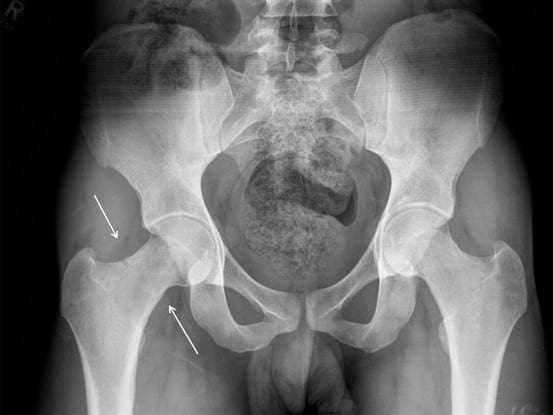
The hip is a ball-and-socket joint composed of the head of the thigh bone, or femur,�which acts as the ball and fits into the round socket of the hip bone, or acetabulum. The neck of the femur is located under the ball of the hip joint. Stress fractures to the femoral neck can entirely or partially detach the femoral head from the rest of the femur.
Femoral neck stress fractures can be either displaced, where the bone is transferred out of its normal position, or non-displaced, where the bone remains stable. These fractures may interrupt blood flow to the portion of the broken bone. In recovery, the blood supply prevents severely displaced femoral neck stress fractures from healing correctly.
Causes and Symptoms of Femoral Neck Stress Fractures
Femoral neck stress fractures can result due to: a small slip-and-fall accident or twisting of the hip in older adults, osteoporosis, a high-impact�injury, such as from an automobile accident, and�sudden strenuous physical activity or changes in physical activity in younger individuals unaccustomed to the events, including from sports injuries.�
The symptoms of femoral neck stress fractures generally include: pain and discomfort, radiating pain which extends to the knee, inability to bear weight on the affected lower extremity, shortening or sideways rotation of the leg, increased pain in the hip during the rotation of the leg, and inflammation on the side of the hip with the femoral neck stress fractures.
Diagnosis and Treatment of Femoral Neck Stress Fractures
A healthcare professional will diagnose femoral neck stress fractures based on the causes and symptoms of the health issue, followed by clinical evaluation. Many doctors order x-rays to diagnose femoral neck stress fractures. The doctor may also order�magnetic resonance imaging, or MRI, and computer tomography, or CT, scanning for a better diagnosis.
Treatment for femoral neck stress fractures depends on the patient’s age as well as on the extent of the broken bone. Treatment for femoral neck stress fractures may include�bed rest for several days followed by a physical rehabilitation program. A healthcare professional may prescribe drugs and/or medications to relieve pain, prevent blood clots and treat infection.
Many femoral neck stress fractures are treated through surgical interventions. Surgery for femoral neck stress fractures involves hip pinning if the bone is minimally displaced and the patient has�enough bone density. The surgeon performs this by making a small incision and then inserting several screws to stabilize the bones which are broken.
Hip hemiarthroplasty or partial hip replacement is utilized for displaced fractures where the surgeon will replace the�femoral head with a metal implant. The socket is not replaced in a partial hip replacement procedure. For total hip replacement, the surgeon will replace the socket of the hip joint, as well as the femoral head, with artificial metallic implants.
�
Femoral neck stress fractures are hip injuries which occur just below the femoral head, or the ball-and-socket hip joint. This area of the thigh bone, or femur, is known as the femoral neck. Femoral neck stress fractures happen when the ball is disconnected from the rest of the femur, or thigh bone. Treatment for this health issue includes rest and physical rehabilitation.
Dr. Alex Jimenez D.C., C.C.S.T.
Conclusion
Femoral neck stress fractures occur�in the hip area below the ball-and-socket joint of the hip. A healthcare professional will suggest treatment based on the severity of the femoral neck stress fractures and the patient’s age.�The scope of our information is limited to chiropractic as well as to spinal injuries and conditions. To discuss the subject matter, please feel free to ask Dr. Jimenez or contact us at�915-850-0900�.
Curated by Dr. Alex Jimenez
Additional Topics: Chiropractic for Athletes with Back Pain
Back pain�is one of the most prevalent causes of disability and missed days at work worldwide. Back pain is the second most common reason for doctor office visits, outnumbered only by upper-respiratory infections. Approximately 80 percent of the population will experience back pain at least once throughout their life. The spine is a complex structure made up of bones, joints, ligaments, and muscles, among other soft tissues. Because of this, injuries and/or aggravated conditions, such as�herniated discs, can eventually lead to symptoms of back pain. Sports injuries or automobile accident injuries are often the most frequent cause of back pain, however, sometimes the simplest of movements can have painful results. Fortunately, alternative treatment options, such as chiropractic care, can help ease back pain through the use of spinal adjustments and manual manipulations, ultimately improving pain relief.
If you�ve ever had a rib slip out of place, you know well the extreme pain it can cause. Every breath can be excruciating. Movement and laughing can also be very painful. It can be located in the back, side, or front on of the ribcage. It is often confused with other conditions such as gastroesophageal reflux disease, a heart condition, pleurisy, or heartburn. The area is usually very tender, and sometimes the area will swell, and a lump will form over the joint. Chiropractic care has been proven to be a very effective treatment for this painful condition.
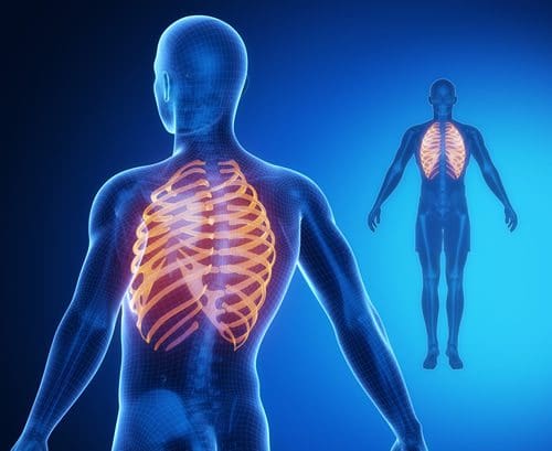
Structure of the Rib cage
Many people believe that the ribcage is a fixed skeletal structure that houses and protects the heart, lungs, and other internal organs. That is only partly true.
The ribcage is somewhat flexible. Note how the chest expands when inhaling. This is because each rib is attached to the spine by three joints in the back, and to the breastbone by one joint in the front. These joints are small but do allow some movement or flexing so that the ribs do not impair breathing. Instead, they rise and fall with each breath.
However, these joints can become inflamed, and that is where the problems start. Because breathing is an involuntary response � and necessary for life � it is impossible to avoid movement in these joints. When there is inflammation in one or several, it can be unbearable.
Causes of a Rib Subluxation
There is any number of reasons for a dislocated rib. Some experience it by doing simple, everyday things like putting dishes in the dishwasher or putting the milk in the refrigerator. Some of the more common causes include:
Extreme sneezing or coughing � Excessive or severe coughing such as is associated with bronchitis or pneumonia puts a great deal of strain on the ribcage. However, even coughing due to a common cold can add enough stress to cause the rib to dislocate. Sneezing very hard can also cause it. Often the illness associated with coughing and sneezing can make a person more susceptible to rib dislocation due to the weakened state of the muscles.
Excessive vomiting � Much like sneezing or coughing, vomiting can also cause this condition. While it does not necessarily involve the lungs, the convulsive action of vomiting can cause a rib to �pop.�
Exercise � Working out can cause the ribs to move out of position, particularly if the person has poor or improper form, or if they do a lot of work with their arms extended in front of them. This is especially true when weights are involved. The muscles involved in the movement may not be strong enough to handle the added weight and movement combination, causing the rib to move out of place.
Improper Posture � Poor posture puts stress on the body, including the spine which, in turn, puts pressure on the posterior portion of the ribcage. Over time, this can cause ribs to dislocate.
Pregnancy � As a woman�s body changes toward the end of her pregnancy, her weight shifts to the front. This can create a continual downward pull on her rib cage, increasing her risk of rib dislocation.
Pain or discomfort in the area of the chest or back.
Swelling and bruising in the affected area.
The formation of a lump over the injured rib.
Extreme pain and difficulty when breathing, trying to sit up, or while straining.
Painful sneezing and coughing.
Pain when moving or walking.
Difficulty breathing.
Numbness or paralysis in nearby or surrounding ribs.
Tenderness in the affected area.
Treatments for a Dislocated Rib
Chiropractic care is considered one of the best, most effective treatments for dislocated or subluxated ribs. Once the chiropractor has determined that the rib is out of place, he or she will often begin by using various techniques that will �loosen� the area, making the muscles more pliable.
They may do this by using stretching, massage, or a vibration tool. They will then apply gentle but firm pressure to �pop� the rib back into place. In some cases stabilization may be used after to keep the area protected, allowing it to heal. The treatment is usually far less painful than the condition, and some patients report not experiencing any pain at all.
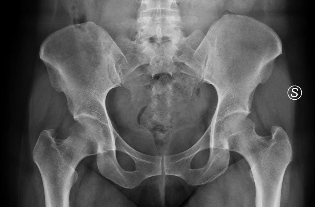
Pain along the pelvis and groin region is known as osteitis pubis. Osteitis pubis develops through the inflammation of the pubic symphysis, or the joints of the major pelvic bones found at the front of the pelvis.
The pubic symphysis is a thin joint which generally provides very minimal motion. The joint retains the two sides of the pelvis together in the front, where they connect�at the sacrum in the rear side of the pelvis.
Osteitis Pubis Symptoms
Osteitis pubis is commonly characterized by pain in the front of the pelvis. Other causes of pelvic pain, such as a strain or a sprain, are frequently confused and diagnosed as osteitis pubis. While many patients report painful symptoms on one side, the�pain�typically occurs in the middle of the pelvis. Other symptoms of osteitis pubis include limping and weakness.
Osteitis Pubis Causes
For some patients, the pubic symphysis itself can become irritated and inflamed, causing the well-known symptoms of osteitis pubis. Other common causes of osteitis pubis comprise of: sports injuries, particularly from football, hockey, and soccer; pregnancy; gynecologic or abdominal surgical interventions; and trauma or injury from accidents.
�
Osteitis pubis is known as the inflammation of the pubis symphysis which causes various degrees of lower abdominal, pelvic, and groin pain. Symptoms of osteitis pubis include pain and discomfort in the region of the pelvis when engaging in physical activities, and loss of flexibility. A variety of causes, including sports injuries, can cause osteitis pubis. Fortunately, rest alone can help treat this painful health issue.
Dr. Alex Jimenez D.C., C.C.S.T.
Osteitis Pubis Diagnosis
Diagnosis of osteitis pubis generally involves x-rays which demonstrate an irregular pubic symphysis with sclerotic, or thick, bone borders as a result of chronic inflammation. An MRI test is generally not required, however, it will help demonstrate the inflammation of the bone and the joint.
Additional tests may be performed to ensure there’s no infection in the bone which could also be causing symptoms similar to osteitis pubis. This complication is more of a concern for those patients who have had recent surgery or for those who are more prone to suffer from infections.
Osteitis Pubis Management
The most recommended treatment for osteitis pubis is rest. Since inflammation is the problem, the human body often only requires the joint to rest in order to heal correctly. Other treatment, however, consists of:
Rest
An essential treatment for osteitis pubis is rest as this will permit the intense inflammation in the pelvis and groin to subside. For many patients, rest alone is the only treatment necessary for their�osteitis pubis. If the pain is severe, crutches or a cane may provide additional assistance.
Ice and Heat
Ice packs and heating pads are among the most commonly used remedies for inflammation. Make sure to follow the instructions of your healthcare professional before utilizing ice and heat for your osteitis pubis symptoms.
Chiropractic Care
Chiropractic care is a well-known, alternative treatment option for osteitis pubis. A doctor of chiropractic, or chiropractor, will utilize a variety of treatment methods and techniques, to help restore strength, mobility, and flexibility while rest is needed to subside the painful symptoms. Chiropractic care can also help correct any spinal misalignments which may be causing additional pain and discomfort for the patient.
Drugs and/or Medications
Nonsteroidal anti-inflammatory drugs and/or medications, commonly referred to as NSAIDs, are frequent prescriptions provided for patients with hip pain brought on by problems like arthritis, bursitis, and tendonitis.
Treatment of osteitis pubis may take some time to completely relieve the painful symptoms. The use of drugs and/or medications is demonstrated to be better than the other treatment options listed above, although attempts to heal osteitis pubis with cortisone injections have been tested.
Surgical interventions are generally not necessary for patients with osteitis pubis.�The scope of our information is limited to chiropractic as well as to spinal injuries and conditions. To discuss the subject matter, please feel free to ask Dr. Jimenez or contact us at�915-850-0900�.
Curated by Dr. Alex Jimenez
Additional Topics: Acute Back Pain
Back pain�is the most prevalent cause of disability worldwide and the second most common reason for doctor office visits, outnumbered only by upper-respiratory infections. Approximately 80 percent of the population will experience back pain at least once throughout their life. The spine is a complex structure made up of bones, joints, ligaments, and muscles, among other soft tissues. Because of this, injuries and/or aggravated conditions, such as�herniated discs, can eventually lead to symptoms of back pain. Sports injuries or automobile accident injuries are often the most frequent cause of back pain, however, sometimes the simplest of movements can have painful results. Fortunately, alternative treatment options, such as chiropractic care, can help ease back pain through the use of spinal adjustments and manual manipulations, ultimately improving pain relief.
Ankylosing Spondylitis is a type of arthritis that typically begins during adolescence or in a person�s early twenties and occurs more often in men than in women. However, once experiences onset, they are affected for the rest of their lives. It is estimated that between 0.2% and 0.5% of individuals in the United States suffers from ankylosing spondylitis. �It can cause significant pain, discomfort, and immobility. While there is no cure for the condition, the symptoms can be treated, bringing some degree of comfort and mobility.
What is Ankylosing Spondylitis?
Ankylosing Spondylitis, or AS, is a type of arthritis that causes inflammation in the spine. While the vertebrae are primarily involved, it can also affect other joints as well, including the hips, shoulders, heels, ribs, and the small joints of the feet and hands.
In some cases, the heart, lungs, and even eyes can be involved. If left untreated, the condition can progress, causing chronic pain that can be severe as the spinal inflammation increases. More advanced cases can cause the spine to grow new bone formations so that it is immobile, or fixed, sometimes resulting in kyphosis, which is a bowed or forward-stooped posture.
What Causes Ankylosing Spondylitis?
While genetics is believed to be a key player in the development of Ankylosing Spondylitis, the exact cause has not yet been determined. The majority of people who have AS also carry a specific gene that has been linked to the condition.
This gene produces HLA-B27, a protein or genetic marker, that more than 95% of Caucasians with ankylosing spondylitis have. However, some people don�t have this protein who develop AS and many people do carry this marker yet never develop the condition.
Researchers theorize that there may be other genes that may be involved, as well as environmental factors that trigger the gene activation, such as a bacterial infection, causing people who are susceptible to AS to activate it. Scientists have identified more than 60 genes that are believed to be associated with AS with only about 30% that are linked to HLA-B27 regarding overall risk. Other genes that have been identified as key to AS include IL-23, IL-17, IL-12, and ERAP.
It is also believed that AS can be triggered when the intestinal defenses break down, allowing certain bacteria into the bloodstream. This can, in turn, cause an immune response.
How is Ankylosing Spondylitis Treated?
AS cannot be cured, but the symptoms can be treated to relieve stiffness and pain as well as delay or prevent spinal deformity and other complications. The damage that it does to the joints is irreversible, so it is best if treatment is started before that occurs. There are several ways that AS is treated:
Medication � Nonsteroidal anti-inflammatory drugs (NSAIDs) like indomethacin (Indocin) and naproxen (Naprosyn) are commonly used to treat the symptoms of AS. They can be useful in relieving pain, inflammation, and stiffness but may cause some side effects, including gastrointestinal bleeding. This makes long-term use impractical and even unsafe. If NSAIDs do not help, other medications may be prescribed, including:
Golimumab (Simponi; Simponi Aria)
Certolizumab pegol (Cimzia)
Adalimumab (Humira)
Etanercept (Enbrel)
Infliximab (Remicade)
Physical therapy – PT is often recommended to help with flexibility, strength, and pain relief. It can help with posture and prevent some of the more debilitating symptoms.
Surgery � Most people with AS do not require surgery, but it may be recommended if there is severe joint damage or pain. In some cases, it can cause significant damage to hip joints, and they will need to be replaced.
Chiropractic � Many patients with AS have with outstanding results with chiropractic treatment. It is non-invasive and does not have the unpleasant side effects that many medications have.
Chiropractic Treatment for Ankylosing Spondylitis
Chiropractors strongly recommend chiropractic treatment for the non-acute inflammatory stage of AS. Once the condition has progressed to acute joint disease, there is a very high risk of injury or damage to the connective tissue. Adjustments and exercise are used to relieve symptoms, but some of the traditional spinal manipulation treatments are not performed.
A chiropractor will also make recommendations to the patient regarding lifestyle changes that can help with symptoms, such as stopping smoking. Tobacco use can increase inflammation and damage connective tissue. They may also advise increasing their intake of omega three fatty acids in their diet. Regular chiropractic care can help patients manage symptoms and prevent disease progression, improving their quality of life.
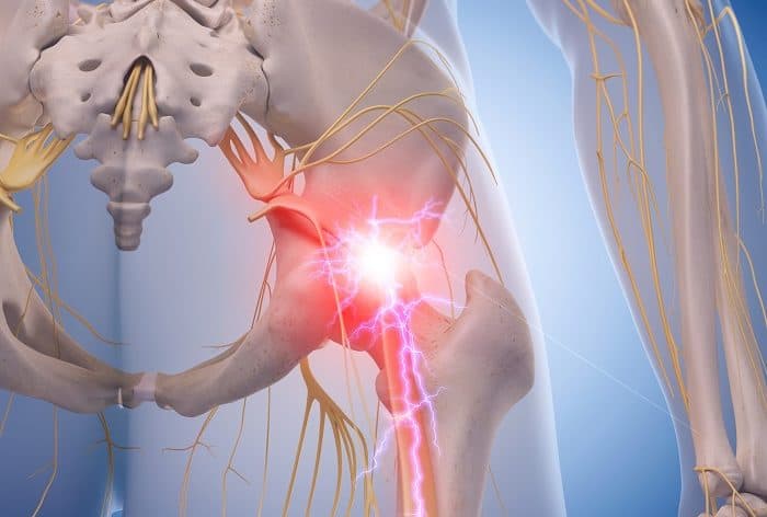
Sciatica is a collection of symptoms in the low back, which radiate down one or both legs. Sciatica is generally caused by the compression or irritation of the sciatic nerve, the largest nerve in the human body. One of the most common health issues that cause sciatic nerve pain is called piriformis syndrome. The piriformis muscle stretches from the front of the sacrum, the triangle-shaped bone between the hipbones on the pelvis.
The piriformis muscle extends to the top of the femur around the sciatic nerve. The femur, as previously mentioned, is the large bone in the upper leg. The piriformis muscle functions by helping the thigh move from side to side. A piriformis muscle spasm, or any other type of injury and/or condition along the piriformis muscle, can place pressure on the sciatic nerve and cause pain and discomfort. The result is piriformis�syndrome.
Piriformis Syndrome Causes and Symptoms
Sciatic nerve pain,�or sciatica, is one of the most prevalent�symptoms of piriformis syndrome. The pain and discomfort, however, may be felt in another part of the body. This is known as referred pain. Other common symptoms of piriformis syndrome include tingling sensations and numbness; tenderness;�difficulty sitting along with�pain while sitting and pain in the buttocks and thighs with physical activities.
The piriformis muscle can easily become damaged or injured from periods of inactivity or an excessive amount of exercise. Some common causes of piriformis syndrome include overuse; repetitive movements involving the legs; sitting for lengthy periods of time; lifting heavy objects; and extensive stair climbing. Sports injuries or automobile accident injuries can also harm the piriformis muscle and cause it to compress the sciatic nerve.�
�
Piriformis Syndrome Diagnosis
A doctor appointment for diagnosis of piriformis syndrome may include a review of the patient’s health history, their symptoms, and other probable causes of their pain and discomfort. If you recall straining a muscle during physical activity, be sure to share that information with your doctor. The�doctor may also perform a physical exam. The patient will participate in a series of range of movements to determine the cause of symptoms.
Some imaging tests may also be essential to help rule out other causes of piriformis syndrome. A CT scan or an MRI scan may help the healthcare professional determine whether even a herniated disc or arthritis is causing the patient’s pain and discomfort. An ultrasound of the piriformis muscle may also be helpful in diagnosing the problem if it seems that piriformis syndrome is causing the patient’s overall symptoms.
�
Piriformis syndrome is a health issue associated with the compression or impingement of the sciatic nerve around the piriformis muscle. Symptoms may include pain and discomfort, tingling sensations and numbness along the low back, or sciatica. Chiropractic care is a well-known alternative treatment option which can help reduce the compression of the sciatic nerve and improve piriformis syndrome.
Dr. Alex Jimenez D.C., C.C.S.T.
Piriformis Syndrome Treatment
Piriformis syndrome may often not need any treatment to�relieve its symptoms. Just avoiding the physical activities which caused the pain and discomfort to manifest and rest can help improve the health issue. If symptoms do persist, however, alternating between ice and heat can help decrease pain. Apply ice for 15 to 20 minutes then use a heating pad on the affected area. Try that every couple of hours to help relieve symptoms.
Over-the-counter painkillers�may also help decrease pain and discomfort. The symptoms associated with piriformis syndrome can go away with no additional treatment, however, if it doesn’t, the patient might benefit from alternative treatment options, such as chiropractic care or physical therapy. Chiropractic care is a treatment approach which utilizes spinal adjustments and manual manipulations to treat a variety of injuries and/or conditions.
A chiropractor,�or doctor of chiropractic, may also provide piriformis syndrome relief through the use of transcutaneous electrical nerve stimulator, or TENS, treatment. A TENS device is a handheld unit which sends electrical charges directly to the affected region of the piriformis muscle. The nerves are then stimulated by the electric energy, which interferes with pain signals being transmitted to the brain.
The chiropractor or physical therapist may also recommend a series of lifestyle modifications, including physical activity guidance and nutritional advice. Various stretches and exercises can help improve the strength, flexibility, and mobility of the�piriformis muscle. In severe cases of piriformis syndrome, corticosteroid injections or even surgical interventions may be required to help alleviate the symptoms.�The scope of our information is limited to chiropractic as well as to spinal injuries and conditions. To discuss the subject matter, please feel free to ask Dr. Jimenez or contact us at�915-850-0900�.
Curated by Dr. Alex Jimenez
Additional Topics: Chiropractic for Athletes with Back Pain
Back pain�is one of the most prevalent causes of disability and missed days at work worldwide. Back pain is the second most common reason for doctor office visits, outnumbered only by upper-respiratory infections. Approximately 80 percent of the population will experience back pain at least once throughout their life. The spine is a complex structure made up of bones, joints, ligaments, and muscles, among other soft tissues. Because of this, injuries and/or aggravated conditions, such as�herniated discs, can eventually lead to symptoms of back pain. Sports injuries or automobile accident injuries are often the most frequent cause of back pain, however, sometimes the simplest of movements can have painful results. Fortunately, alternative treatment options, such as chiropractic care, can help ease back pain through the use of spinal adjustments and manual manipulations, ultimately improving pain relief.
Roberto Varela was always actively involved with chores at home before he started to experience neck, shoulder and leg pain. Due to his symptoms, Mr. Varela had difficulties engaging in regular physical activities, such as driving. However, after being recommended by his wife, he first received chiropractic care with Dr. Alex Jimenez, and Roberto Varela experienced tremendous relief from his neck, shoulder and leg pain, regaining his quality of life. Mr. Varela highly recommends Dr. Alex Jimenez and his staff for their services.
Shoulder Pain Treatment
Shoulder pain or leg pain is common; however, sometimes these problems don’t originate in the location of the symptoms. Shoulder and leg pain may also occur due to health issues in the neck or cervical spine. A variety of injuries and conditions can have their roots in improper posture, sports injuries or automobile accident injuries, causing misalignments, or subluxations, in the cervical spine or neck. Many healthcare professionals will discuss how damage to the cervical spine can be an underlying cause for shoulder pain and leg pain, among other symptoms.
We are blessed to present to you�El Paso�s Premier Wellness & Injury Care Clinic.
As El Paso�s Chiropractic Rehabilitation Clinic & Integrated Medicine Center,�we passionately are focused on treating patients after frustrating injuries and chronic pain syndromes. We focus on improving your ability through flexibility, mobility and agility programs tailored for all age groups and disabilities.
If you have enjoyed this video and we have helped you in any way, please feel free to subscribe and recommend�us.
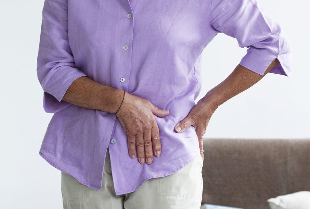
The hips are some of the most flexible structures in the human body, providing the necessary amount of strength and stability needed to support the human body when walking, running or jumping. However, the hip joint can also be vulnerable to damage or injury, resulting in debilitating hip pain. Trochanteric bursitis is hip pain brought on by the inflammation of the fluid-filled sac, or bursa, found on the outer border of the hip.
Trochanteric Bursitis Overview
There are about 160 bursae located around the entire body. Bursae act as a sort of “cushion” between soft tissues and bones, preventing bones from rubbing against tendons, ligaments, and muscles. Trochanteric bursitis can affect any of the bursae inside the human body. Trochanteric bursitis affects the outer part of the thighbone, or the femur, at the edge of the hip. This bony point is best known as the greater trochanter.
Another bursa, called the iliopsoas bursa, can be found on the inside of the hip. Inflammation of the iliopsoas bursa also triggers pain in the groin. Bursitis is considered to be one of the top causes of hip pain. Repetitive physical activities, such as climbing stairs, or even surgical interventions to the hip may cause inflammation in the bursa. Many doctors commonly refer to trochanteric�bursitis as greater trochanteric pain syndrome.
Signs and Symptoms of Trochanteric Bursitis
The main characteristic of trochanteric bursitis involves pain in the outer area of the hip or pain when laying on the affected side of the hip. The painful signs and symptoms will also generally become worse through certain physical activities, such as walking or climbing stairs. Pain may also�radiate down the�thigh and into the feet, or it may disperse. Pain can be sharp and fade into an ache, accompanied by swelling in the legs.
Causes of Trochanteric Bursitis
Common causes of trochanteric bursitis include�slip-and-fall accidents, strong blows to the hip, or lying on one side of the body for an extended period of time. Sports injuries involving�overuse from repetitive physical activities like running, bicycling, or climbing stairs, a ripped tendon or even standing may cause trochanteric�bursitis. Health issues, such as�bone spurs in the hip or thighbone, may consequently cause trochanteric bursitis.�
A variety of conditions and disorders may also lead to trochanteric bursitis, including spine problems, such as scoliosis or arthritis of the lumbar spine, even rheumatoid arthritis, and gout as well as thyroid disease. Moreover, legs of two different lengths,�hip surgery or prosthetic implants can create problems in the hips. Trochanteric bursitis is most common in middle-aged or elderly people and it is most prevalent in women than men.
�
Trochanteric Bursitis Treatment and Chiropractic Care
Avoiding the physical activities which caused trochanteric bursitis will allow time for the body to heal. After seeing a healthcare professional for diagnosis, the doctor may often recommend nonsteroidal anti-inflammatory drugs, or NSAIDs to help control pain and inflammation. The recommended amount should be used to avoid side effects. Some doctors may also use steroid injections to control pain and inflammation.
Many healthcare professionals may also recommend alternative treatment options,�such as chiropractic care and physical therapy to help improve trochanteric bursitis signs and symptoms. A chiropractor may utilize spinal adjustments�and manual manipulations to reduce pressure from the spine while a physical therapist may teach the patient exercises to maintain strength. A cane or crutches can also take the weight off a patient’s hip.
If pain relievers or alternative treatment options, such as chiropractic care or physical therapy, do not work for the patient, the healthcare professional might recommend surgery to remove the bursa. This procedure can be accomplished through very small incisions with a camera. Other treatment approaches should be considered before following through with surgery.� The scope of our information is limited to chiropractic as well as to spinal injuries and conditions. To discuss the subject matter, please feel free to ask Dr. Jimenez or contact us at�915-850-0900�.
Curated by Dr. Alex Jimenez
�
�
Additional Topics: Acute Back Pain
Back pain�is one of the most prevalent causes of disability and missed days at work worldwide. Back pain is the second most common reason for doctor office visits, outnumbered only by upper-respiratory infections. Approximately 80 percent of the population will experience back pain at least once throughout their life. The spine is a complex structure made up of bones, joints, ligaments, and muscles, among other soft tissues. Because of this, injuries and/or aggravated conditions, such as�herniated discs, can eventually lead to symptoms of back pain. Sports injuries or automobile accident injuries are often the most frequent cause of back pain, however, sometimes the simplest of movements can have painful results. Fortunately, alternative treatment options, such as chiropractic care, can help ease back pain through the use of spinal adjustments and manual manipulations, ultimately improving pain relief.
IFM's Find A Practitioner tool is the largest referral network in Functional Medicine, created to help patients locate Functional Medicine practitioners anywhere in the world. IFM Certified Practitioners are listed first in the search results, given their extensive education in Functional Medicine