The term excitotoxicity was first employed to demonstrate the capability of L-glutamate, in addition to structurally-associated amino acids, to destroy nerve cells, a process which has been suggested to occur in acute and chronic health issues of the central nervous system (CNS). Excitotoxicity is caused by the excess stimulation of iGluRs into a characteristic loss of cell bodies and dendrites as well as post-synaptic structures. There is a substantial degree of variation in the sensitivity of nerve cells compared to the variety of iGluRs which is associated with the specific receptors demonstrated on the nerve cells and their metabolisms. The susceptibility of neurons to excitotoxicity can be affected with age. �
Acute excitotoxic nerve cell death is believed to occur in reaction to a number of severe insults, including cerebral ischemia, traumatic brain injury (TBI), hypoglycemia, and status epilepticus. However, what about neurodegenerative diseases, such as Alzheimer’s disease? Does chronic excitotoxicity also occur? Could exposure of nerve cells to low but above-average concentrations of L-glutamate, or even glutamatergic neurotransmission through a variety of molecules be involved as previously mentioned, within an extended time period also significantly result in neural cell death? The purpose of the article below is to demonstrate the concepts of acute and chronic glutamate toxicity on the health and wellness of the brain. �
Acute and Chronic Glutamate Toxicity
Excitotoxicity was initially studied in animals, however, so as to comprehend the mechanisms underlying this procedure, cell culture models were developed. The basic cell culture model of acute excitotoxicity involves the treatment of principal neurons in accordance with L-glutamate or particular iGluRs for a brief time interval (min) and then analyzing downstream events in the time point which is most relevant for the research study. By way of instance, cell death is frequently determined after 24 hours. While these types of research studies are proven to be quite useful for understanding the pathways involved in acute excitotoxicity, it has demonstrated to be far more difficult to evaluate chronic excitotoxicity in culture partially because it is not completely clear how to specify “chronic” in the context of cell culture. Does consistent imply a minimal dose supplied for 24 hours instead of a maximum dose supplied for 5 to 10 minutes or is it more complicated than that? �
Among the few research studies which tried to come up with a model of chronic excitotoxicity, it was revealed that it is indeed more complicated with acute and chronic excitotoxicity appearing to be different processes. In this research study, the researchers utilized pure cultures of primary cortical neurons developed from day 14 mouse embryos and treated them after seven and 14 days in culture (DIV). For constant excitotoxicity, the neurons were exposed to L-glutamate or NMDA for 24 hours and for severe excitotoxicity for 10 minutes. In both circumstances, cell death was measured after 24 hours. Surprisingly, the EC50s in their toxicity of L-glutamate were lower for acute toxicity, particularly in the 7 DIV cultures, when compared with the EC50s for chronic toxicity. Additionally, it was discovered that a high cell culture density increased the cells’ sensitivity into excitotoxicity that was acute but not chronic. Further research studies indicated that the lower sensitivity of these neurons to L-glutamate in the chronic excitotoxicity paradigm was due to the stimulation of mGluR1, associated with earlier data on the neuroprotective effects of mGluR1 stimulation, among other important processes. �
Further Research Studies for Glutamate Toxicity
An alternative approach for understanding chronic glutamate toxicity used organotypic spinal cord cultures in conjunction with L-glutamate uptake inhibitors. These spinal cord cultures, which had been prepared from 8-day-old rat pups, were kept in culture for up to 3 months. Persistent inhibition of L-glutamate uptake utilizing two varieties of uptake inhibitors caused a consistent increase of L-glutamate in the cell culture medium and time period as well as a concentration of dependent motor neuron cell death. The highest concentration of uptake inhibitor increased extracellular L-glutamate levels at least 25-fold and began to kill the cells within 1 week whereas a five-fold lower concentration raised extracellular L-glutamate levels eight-fold and cell death only began after 2 to 3 weeks of treatment. The toxicity was obstructed with non-NMDA but not NMDA receptors as well as by inhibitors of L-glutamate synthesis or release. These research studies ultimately indicate that moderately increased L-glutamate concentrations can also induce toxicity as well as a variety of other health issues. �
In vivo approaches to studying excitotoxicity have relied on an approach analogous to that utilized with the spinal cord cultures. In the wide variety of the research studies, a single or multiple EAATs were transiently or permanently genetically eliminated and the effects on brain function were evaluated. During the first few research studies, which utilized rats, chronic intraventricular administration of antisense RNA was utilized to eliminate every one of the 3 primary EAATs (EAAT1, EAAT2, and EAAT3). The loss of either of the glial L-glutamate transporters (EAAT1 and EAAT2) but not the neuronal transporter (EAAT3) caused large increases in extracellular L-glutamate concentrations in the striatum following 7 days as demonstrated by microdialysis (EAAT2, 32-fold increase; EAAT1, 13-fold increase). Treatment with the EAAT1 or EAAT2 antisense oligonucleotides caused a progressive motor impairment whereas epilepsy was produced by the EAAT3 antisense oligonucleotide. The loss of any of the 3 transporters demonstrated clear evidence of neuronal damage in the striatum and hippocampus after 7 days of treatment although the effects of the EAAT1 and EAAT2 antisense oligonucleotides were far more dramatic, consistent with the substantial increases in extracellular L-glutamate brought about by treatment. �
Particularly different results were demonstrated with homozygous mice deficient in EAAT2 or EAAT1. Mice deficient in EAAT2 demonstrated sudden and normally deadly seizures with 50 percent dead by 6 weeks of age. Approximately 30 percent of these mice demonstrated selective degeneration in the CA1 area at 4 to� 8 weeks of age. L-glutamate amounts in the CA1 region of the hippocampus measured by microdialysis were three-fold greater in the mutant mice as compared with the wild type mice. In contrast, heterozygous EAAT2 knock-out mice have an average lifespan and do not reveal hippocampal CA1 atrophy. However, they exhibit several behavioral abnormalities suggestive of moderate glutaminergic hyperactivity. While mice deficient in EAAT1, that is expressed in cerebellar astrocytes, didn’t reveal changes in cerebellar arrangement or obvious indicators of cerebellar impairment, such as ataxic gait, they had not been able to adapt to difficult motor tasks like rapidly running the rotorod. When taken collectively, these results imply that disruptions in homeostasis which are glutamatergic have a greater impact when they occur in the animal rather than when they are found from conception. �
Other Health Issues in Glutamate Toxicity
Tuberous sclerosis complex (TSC) is a multi-system genetic disease caused by the mutation of both TSC1 or TSC2 genes, where it is characterized by severe neurodegenerative diseases. Mice with inactivation of the TSC1 gene in glia have a less than 75 percent reduction in the expression and function of EAAT1 and EAAT2 as well as to cause seizures. At 4 weeks of age, prior to the development of seizures in these mice, there was a 50 percent increase in extracellular L-glutamate in the hippocampus of the mutant mice, as determined by microdialysis, which correlated with increases in markers of cell death in neurons in both hippocampus and cortex. Utilizing slices from mice that were 2 to 4 week old, impairments in long-term potentiation were determined, which translated into deficits when mice were analyzed for contextual and spatial memory in the Morris water maze and fear conditioning assays. Further research studies are still necessary for outcome measures. �
In the majority of the research studies described above, there was a large increase in extracellular L-glutamate that, when analyzed, caused adverse effects on the role of specific neuronal populations. To ascertain the long-term effects of more moderate increases in extracellular glutamate, further research studies created transgenic (Tg) mice with extra copies of this gene for Glud1, especially in neurons. Mitochondrial 2-oxoglutarate from Glud1 is transported into the cytoplasm of nerve terminals in which it’s converted back into L-glutamate and kept in synaptic vesicles thus leading to the pool of synaptically releasable L-glutamate. Nine-month-old Glud1 Tg mice demonstrated a 10 percent boost in L-glutamate in the hippocampus and striatum relative to wild type mice as determined to utilize magnetic resonance spectroscopy. In addition, 50 percent caused increased L-glutamate release in the striatum. At 12 to 20 months of age, the Glud1 Tg mice revealed significant decreases in the numbers of neurons in the CA1 area of the hippocampus and granule cell layer of the dentate gyrus in addition to an age-dependent loss of the two dendrites and dendritic spines in the hippocampus. There was also a drop in long-term potentiation after high frequency stimulation in hippocampal slices in the mice when compared with the wild type mice. Evaluation of the transcriptome of those Glud1 Tg mice in comparison with wild type mice indicated that long-term moderate increases in cerebral L-glutamate ultimately caused both rapid aging in the level of gene expression combined with compensatory reactions which protected against pressure and/or promoted recovery, among other capabilities. �
Conclusion
Brain function and nerve cell survival can be affected by excitotoxicity. The results appear to be highly dependent on the degree of L-glutamate increase, however, even a 10 percent growth appears to influence nerve cell survival, particularly in the context of aging indicating that chronic excitotoxicity may be associated with neurodegenerative diseases. �
Several toxins which connect to iGluRs and that have also been demonstrated to cause excitotoxicity in cell culture may cause slowly growing neurological health issues in both animals and humans. Surprisingly, each toxin appears to target a particular type of neuron, an effect which may be associated with the pharmacokinetics and ADME properties of the toxins, which have not been analyzed to any great extent. The data from these types of toxins supports the idea that excitotoxicity may play a fundamental role in neurodegenerative diseases as well as in other health issues which exist in humans. �
Because iGluRs are demonstrated both from the synapse and in extra-synaptic locations, there has been a great deal of effort devoted to discovering if the region of the receptors impacts the toxicity of molecules. An influential research study with primary neuronal cultures indicated that synaptic and extrasynaptic NMDA receptors have counteracting effects on cell survival with neural cell death being primarily controlled by extrasynaptic NMDA receptors. Nonetheless, these outcome measures have not been reproduced in brain slices or in vivo. Furthermore, many more recent research studies utilizing the exact same primary neuronal culture preparation protocol as the prior research study found either no difference between synaptic and extrasynaptic NMDA receptors in boosting excitotoxicity or discovered that both receptors were needed for cell death. Finally, a variety of research studies that supported the idea that extrasynaptic NMDA receptors promote excitotoxicity relied on the NMDA receptor inhibitor memantine that was originally believed to specifically act on extrasynaptic NMDA receptors. However, more recent research studies demonstrate that memantine can inhibit both synaptic and extrasynaptic NMDA receptors. These results strongly imply that synaptic and extrasynaptic NMDA receptors may contribute to excitotoxicity but the contribution of each depends on the experimental and/or pathological conditions. �
Glutamate is the primary excitatory neurotransmitter in the brain. Although it plays a fundamental role in the overall structure and function of the central nervous system, excessive amounts of glutamate can ultimately cause excitotoxicity which may lead to a variety of health issues, such as Alzheimer’s disease and other types of neurodegenerative diseases. Acute and chronic excitotoxicity treatment currently focuses on decreasing or restricting glutamate receptors or extracellular glutamate. The article above summarizes the available research studies for glutamate toxicity in neurodegenerative diseases. – Dr. Alex Jimenez D.C., C.C.S.T. Insight
Excitotoxicity demonstrates the capability of L-glutamate, as well as structurally-associated amino acids, processes which have been suggested to occur in acute and chronic excitotoxicity. Excitotoxicity is caused by the excess stimulation of iGluRs in cell bodies and dendrites as well as post-synaptic structures. There is a substantial degree of variation in nerve cells compared to iGluRs associated with the receptors demonstrated on the nerve cells and their metabolisms. The scope of our information is limited to chiropractic, musculoskeletal and nervous health issues as well as functional medicine articles, topics, and discussions. We use functional health protocols to treat injuries or chronic disorders of the musculoskeletal system. To further discuss the subject matter above, please feel free to ask Dr. Alex Jimenez or contact us at 915-850-0900 . �
Curated by Dr. Alex Jimenez �
References �
Lewerenz, Jan, and Pamela Maher. �Chronic Glutamate Toxicity in Neurodegenerative Diseases-What Is the Evidence?� Frontiers in Neuroscience, Frontiers Media S.A., 16 Dec. 2015, www.ncbi.nlm.nih.gov/pmc/articles/PMC4679930/.
Additional Topic Discussion: Chronic Pain
Sudden pain is a natural response of the nervous system which helps to demonstrate possible injury. By way of instance, pain signals travel from an injured region through the nerves and spinal cord to the brain. Pain is generally less severe as the injury heals, however, chronic pain is different than the average type of pain. With chronic pain, the human body will continue sending pain signals to the brain, regardless if the injury has healed. Chronic pain can last for several weeks to even several years. Chronic pain can tremendously affect a patient’s mobility and it can reduce flexibility, strength, and endurance.
Neural Zoomer Plus for Neurological Disease
�
Dr. Alex Jimenez utilizes a series of tests to help evaluate neurological diseases. The Neural ZoomerTM Plus is an array of neurological autoantibodies which offers specific antibody-to-antigen recognition. The Vibrant Neural ZoomerTM Plus is designed to assess an individual�s reactivity to 48 neurological antigens with connections to a variety of neurologically related diseases. The Vibrant Neural ZoomerTM Plus aims to reduce neurological conditions by empowering patients and physicians with a vital resource for early risk detection and an enhanced focus on personalized primary prevention. �
Formulas for Methylation Support
XYMOGEN�s Exclusive Professional Formulas are available through select licensed health care professionals. The internet sale and discounting of XYMOGEN formulas are strictly prohibited.
Proudly,�Dr. Alexander Jimenez makes XYMOGEN formulas available only to patients under our care.
Please call our office in order for us to assign a doctor consultation for immediate access.
If you are a patient of Injury Medical & Chiropractic�Clinic, you may inquire about XYMOGEN by calling 915-850-0900.
�
For your convenience and review of the XYMOGEN products please review the following link.*XYMOGEN-Catalog-Download �
* All of the above XYMOGEN policies remain strictly in force.
L-glutamate is one of the main excitatory neurotransmitters in the human brain and it plays an essential role in practically all activities of the nervous system. In the following article, we will discuss the general principles of L-glutamate signaling in the brain. Then, we will demonstrate this scheme by describing the different pools of extracellular glutamate, including the synaptic, the perisynaptic, and the extrasynaptic, resulting from vesicular and non-vesicular sources or abnormally located glutamate receptors outside of synapses as well as discuss their possible physiological functions in the human brain. �
Glutamate Signaling in the Brain
According to research studies, the human brain has about a 6 to 7 ?mol/g wet weight of L-glutamate. L-glutamate, together with glutamine, is one of the most abundant free amino acids in the central nervous system (CNS). More than five decades ago, several research studies demonstrated that L-glutamate has an excitatory response on nerve cells. Since then, its role as an excitatory neurotransmitter as well as its cerebral metabolism has been evaluated in numerous research studies. �
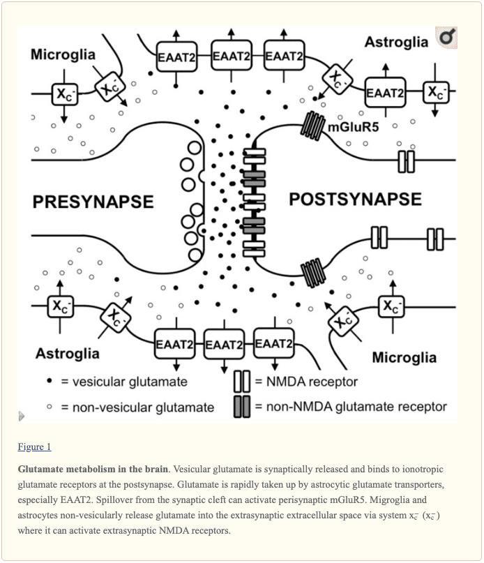
L-glutamate is commonly found throughout synaptic vesicles in the presynaptic terminal through the process of vesicular glutamate transporters. Additionally, several of the L-glutamate in the vesicles may develop by a vesicle-associated aspartate amino-transferase from 2-oxoglutarate utilizing L-aspartate as the amino group donor. During the depolarization of the presynaptic membrane, L-glutamate is released into the synaptic cleft and connects to ionotropic glutamate receptors, known as iGluRs, at the postsynaptic membrane, as shown in Figure 1. According to research studies, iGluRs are characterized as ligand-gated ion channels which include receptors of the ?-amino-3-hydroxy-5-methyl-4-isoxazole propionic acid (AMPA), kainate, and N-methyl-D-aspartic acid (NMDA) types. While AMPA and kainate receptors primarily regulate and maintain sodium influx, NMDA receptors actually have a high calcium conductivity. Moreover, the activation of NMDA receptors plays a fundamental role in synaptic plasticity and learning. In contrast to the other iGluRs, the activity of NMDA receptors is ultimately restricted by an Mg+2 block at the regular membrane potential, however, the ion channel is immediately unblocked by membrane depolarization which eliminates Mg+2 from the pore. Furthermore, NMDA receptors are tetramers that have two NR1 subunits and two NR2 or NR3 subunits, according to several research studies. �
Additionally to iGluRs, there are also eight isoforms of metabotropic glutamate receptors (mGluRs) which belong to the family of G-protein-coupled receptors, where they don’t develop ion channels but instead signal through a variety of second messenger systems. L-glutamate-associated depolarization causes a postsynaptic excitatory potential which eases the development of an action potential at the axon hillock. The glutamatergic synapse is activated by astrocytic processes that demonstrate high levels of excitatory amino acid transporters (EAATs). There are five different EAATs, EAAT1 to 5, of which EAAT1 and 2 are the primary astrocytic EAATs, whereas EAAT3 shows a predominantly neuronal expression. Approximately 90 percent of the L-glutamate transport is regulated and maintained by EAAT2 such as GLT-1 in rodent models. These transporters then co-transport 2 or 3 molecules of Na+ and a proton with each molecule of L-glutamate or L-aspartate together with the counter-transport of a K+ ion. Therefore, by utilizing the electrochemical gradient of these ions throughout the plasma membrane as an energy source, the transporters are able to safely and effectively accumulate L-glutamate and L-aspartate in cells against their sudden intra- to extracellular concentration gradients. This allows the brain to control a very low extracellular L-glutamate concentration in the low micromolar range. It is generally believed that L-glutamate taken up by astrocytes is turned to glutamine by the enzyme glutamine synthetase, the glutamine is then released, taken up by neurons and turned to L-glutamate, where it is ultimately utilized once again for neurotransmission. �
Extrasynaptic Glutamate in the Brain
Aside from the essential role of L-glutamate as the primary excitatory neurotransmitter released from glutamatergic presynapses, as previously mentioned above, it has become evident that L-glutamate receptors outside the synaptic cleft also play an essential role in brain physiology. In the cerebellum, it was demonstrated by evaluating AMPA receptor-mediated currents in Bergmann glia that synaptically released L-glutamate concentrations can reach extrasynaptic concentrations of up to 190 ?M while concentrations in the synaptic cleft can exceed 1 mM. Moreover, several mGluRs have been shown to demonstrate a different localization in proximity to the postsynaptic density which would allow them to immediately recognize L-glutamate escaping from the synaptic cleft, as shown in Figure 1. However, current research studies have demonstrated that iGluRs, especially of the NMDA type, are also found at extrasynaptic regions in the neuronal cell membrane. Utilizing light and electron microscopy, other research studies also demonstrated that extrasynaptic NMDA receptors gather at different regions of close contact in the dendritic shaft with axons, axon terminals, or astrocytic processes. The proportion of extrasynaptic NMDA receptors was estimated to be as high as 36 percent of the dendritic NMDA receptor pool in rat hippocampal slices. Although extrasynaptic NMDA receptors were associated with similar scaffolding proteins as synaptic NMDA receptors, an in vitro research study suggested that extrasynaptic and synaptic NMDA receptors may ultimately activate different downstream signaling pathways with a variety of results, including the suppression of CREB activity by extrasynaptic NMDA receptor activation as well as activation by synaptic NMDA receptors. Furthermore, NMDA receptors localized extrasynaptically on dendritic shafts connect extrasynaptic L-glutamate as well as regulate and maintain Ca2+ influx during the elimination of the Mg+2 block by dendrite depolarization throughout the backfiring of action potentials. Research studies demonstrated that L-glutamate release from astrocytes can activate slow inward currents through extrasynaptic NMDAR receptors in CA1 neurons which can also be ultimately synchronized. The mechanisms through which glial cells release L-glutamate as well as how the extrasynaptic L-glutamate concentrations are controlled are vital towards understanding how the activity of extrasynaptic NMDA receptors is controlled. �
Different mechanisms through which astrocytes can release L-glutamate have been suggested, including vesicular L-glutamate release and non-vesicular release through anion channels as well as connexin hemichannels and release through the cystine/glutamate antiporter system x?c. Several research studies strongly suggest that vesicular release from astrocytes plays a minor role because the Ca+2-associated release of L-glutamate was still present in astrocytes created from dominant-negative SNARE mice where vesicular release can be blocked by doxycycline withdrawal. System x?c is a cystine/glutamate antiporter which is characterized as heterodimeric amino acid transporters, made up of xCT as the specific subunit and 4F2hc as the promiscuous heavy chain. This transporter is demonstrated in the brain, especially in astroglial and microglial cells, as shown in Figure 1. The fact that extrasynaptic L-glutamate levels in different regions of the human brain are downregulated by approximately 60 percent to 70 percent in xCT knock out mice, research studies demonstrated that system x?c releases L-glutamate into the extrasynaptic space and suggests that this transporter is essential in the regulation of extrasynaptic L-glutamate levels. This is further supported by the observation that when measured by in vivo microdialysis, the increase in extrasynaptic L-glutamate developed by EAAT inhibitors is neutralized by blocking system x?c while blocking neuronal vesicular L-glutamate release is ineffective. Further research studies are still required. �
Taken together, glutamatergic neurotransmissions don’t simply happen through classical excitatory synapses but also through extrasynaptic L-glutamate receptors, as shown in Figure 1. Finally, the levels of extrasynaptic L-glutamate are determined, at least partially, by glial non-vesicular L-glutamate release, as also shown in Figure 1. However, the regulation of extrasynaptic L-glutamate levels, as well as its temporal-spatial dynamics and its effect on neuronal function, neurodegeneration, and behavior, are far from being fully understood by researchers, healthcare professionals, and patients. �
Glutamate, together with aspartate, is one of the main excitatory neurotransmitters in the human brain. Although it plays a fundamental role in the overall structure and function of the nervous system, excessive amounts of glutamate can ultimately cause excitotoxicity which may lead to a variety of health issues, such as Alzheimer’s disease and other types of neurological diseases. The following article describes the role of glutamate in the human brain. – Dr. Alex Jimenez D.C., C.C.S.T. Insight
L-glutamate is one of the main excitatory neurotransmitters in the human brain and it plays an essential role in practically all activities of the nervous system. In the article above, we discussed the general principles of L-glutamate signaling in the brain. Then, we demonstrated this scheme by describing the different pools of extracellular glutamate, including the synaptic, the perisynaptic, and the extrasynaptic, resulting from vesicular and non-vesicular sources or abnormally located glutamate receptors outside of synapses as well as discussed their possible physiological functions in the human brain. The scope of our information is limited to chiropractic, musculoskeletal and nervous health issues as well as functional medicine articles, topics, and discussions. We use functional health protocols to treat injuries or chronic disorders of the musculoskeletal system. To further discuss the subject matter above, please feel free to ask Dr. Alex Jimenez or contact us at 915-850-0900 . �
Curated by Dr. Alex Jimenez �
References �
Lewerenz, Jan, and Pamela Maher. �Chronic Glutamate Toxicity in Neurodegenerative Diseases-What Is the Evidence?� Frontiers in Neuroscience, Frontiers Media S.A., 16 Dec. 2015, www.ncbi.nlm.nih.gov/pmc/articles/PMC4679930/.
Additional Topic Discussion: Chronic Pain
Sudden pain is a natural response of the nervous system which helps to demonstrate possible injury. By way of instance, pain signals travel from an injured region through the nerves and spinal cord to the brain. Pain is generally less severe as the injury heals, however, chronic pain is different than the average type of pain. With chronic pain, the human body will continue sending pain signals to the brain, regardless if the injury has healed. Chronic pain can last for several weeks to even several years. Chronic pain can tremendously affect a patient’s mobility and it can reduce flexibility, strength, and endurance.
Neural Zoomer Plus for Neurological Disease
�
Dr. Alex Jimenez utilizes a series of tests to help evaluate neurological diseases. The Neural ZoomerTM Plus is an array of neurological autoantibodies which offers specific antibody-to-antigen recognition. The Vibrant Neural ZoomerTM Plus is designed to assess an individual�s reactivity to 48 neurological antigens with connections to a variety of neurologically related diseases. The Vibrant Neural ZoomerTM Plus aims to reduce neurological conditions by empowering patients and physicians with a vital resource for early risk detection and an enhanced focus on personalized primary prevention. �
Formulas for Methylation Support
XYMOGEN�s Exclusive Professional Formulas are available through select licensed health care professionals. The internet sale and discounting of XYMOGEN formulas are strictly prohibited.
Proudly,�Dr. Alexander Jimenez makes XYMOGEN formulas available only to patients under our care.
Please call our office in order for us to assign a doctor consultation for immediate access.
If you are a patient of Injury Medical & Chiropractic�Clinic, you may inquire about XYMOGEN by calling 915-850-0900.
�
For your convenience and review of the XYMOGEN products please review the following link.*XYMOGEN-Catalog-Download �
* All of the above XYMOGEN policies remain strictly in force.
For people who love drinking diet sodas, recent research studies have found that diet drinks can increase the risk of stroke and dementia. Although diet drinks have been previously advertised as a much more healthier, low-calorie alternative than regular carbonated drinks, a closer look at the results of these recent research studies ultimately suggests otherwise. �
One research study, consisting of 2,888 participants, ages 45 and older, in the Framingham Heart Study, asked for diet entries to be filled out up to three times within a seven-year period. According to the research study, participants who said they drank one diet soda a day were roughly twice as likely to have a stroke within the next decade than individuals who didn’t drink diet soda. Drinking regular, sugar-sweetened carbonated drinks did not seem to increase the risk of stroke. �
However, these types of research studies have only been able to prove an association between diet drinks, stroke, and dementia. “Also, only 97 people (about 3 percent) had strokes during the follow-up, which means that only two or even three of those strokes may be associated to drinking diet soda,” stated Dr. Kathryn Rexrode, an associate professor of medicine at Harvard-affiliated Brigham and Women’s Hospital which co-authored a research study on soda intake and stroke risk. �
Risk of Stroke Associated with Diet Drinks
The research study found a slightly increased risk of stroke in people who drank more than one soda per day, whether or not it contained any type of artificial sweetener. Although the research study didn’t particularly show a considerable increase in stroke risk, that doesn’t necessarily suggest that they’re a better option than diet sodas. Research studies have shown that drinking carbonated drinks may lead to weight gain, diabetes, high blood pressure, heart disease, and stroke, ” she stated. �
As a matter of fact, researchers believe that one possible explanation as to why regular, sugar-sweetened carbonated drinks weren’t associated with stroke in the recent research study is a phenomenon known as the survival bias. In this instance, it would mean that individuals who drink a lot of carbonated drinks may have died from health issues such as heart disease. �
Conversely, diet drinks may be associated with an increased risk of stroke due to a variety of health issues known as reverse causation. In an attempt to be healthier, individuals who are overweight or have diabetes may be more inclined to select diet drinks over regular drinks. Their increased risk of stroke may come from their health issues rather than their drink option. “We may ultimately only be measuring the residual effect of weight gain, obesity, and diabetes,” says Dr. Rexrode. �
Artificial Sweeteners and Stroke
� Although researchers need further evidence to determine why artificial sweeteners may increase stroke risk, there are other reasons as to why these should be avoided. Research studies show that artificial sweeteners can make individuals crave sugary, high-calorie meals, therefore, decreasing the artificial sweetener’s purpose of cutting your total calorie consumption. �
Moreover, many researchers believe that people who use these artificial sweeteners, which can be many times sweeter than sugar, can come to find naturally sweet foods, such as fruits, to be less appealing and less-sweet foods, such as vegetables, to be entirely unpalatable. Furthermore, individuals may be missing out on the many nutrients found in fresh, natural foods. �
“I encourage my patients to stop drinking soda and other sugar-sweetened carbonated drinks regularly to prevent empty calories,” she says. “However, if someone says that they can’t do without soda in the morning to wake up, I will encourage them to switch to diet soda.” Water is a much better choice, however. “There are plenty of ways to make it more attractive, both visually and taste-wise.” She adds. Try flavoring sparkling or flat water or add crushed mint, cucumber, or frozen fruit. �
Risk of Dementia Associated with Diet Drinks
In another research study, people who drank diet soda were associated with an increased risk of developing dementia. “The research study can’t prove a connection between drinking habits and health issues, however, it does strongly suggest an association,” Stated Dr. Matthew Pase, neurology fellow at Boston University School of Medicine and contributing author. �
The initial research study evaluated food questionnaires, MRI scans, and cognitive tests of approximately 4,000 people ages 30 and up. Researchers found that individuals who consumed over three diet sodas per week were more likely to have memory problems, a reduced brain volume, and a smaller hippocampus, an area of the brain used in memory and learning. In the research study, drinking a minimum of one diet soda per day was also associated with a reduced brain volume. �
During a second research study, the researchers tracked two different groups of adults for ten years. According to the research study, out of almost 3,000 adults over age 45, approximately 97 adults suffered a stroke during that time and from almost 1,500 adults over age 60, approximately 81 adults developed Alzheimer’s disease or another type of dementia. �
Past research studies have connected diet drinks to an increased risk of weight gain and stroke. Researchers believe that artificial sweeteners may ultimately affect the human body in many different ways, such as by transforming gut bacteria and tricking the brain into craving more calories. This is the first-time diet sodas have been associated with dementia. Because people with diabetes drink more diet soda, researchers believe that the health issue may partly explain the rise in dementia, although not completely. When people with diabetes were excluded from the research study, the association stayed. �
As stated by the United States Department of Agriculture, Americans consumed 11 million metric tons of sugar in 2016, much of it in the form of sugary, sweetened carbonated drinks. Because it would have been difficult to measure total sugar consumption from all type of different food sources, the research study focused on sugary, sweetened carbonated drinks. �
A growing number of research studies suggest that diet drinks may not be a safe alternative to sugary, sweetened drinks. Even small causal effects can have much bigger consequences on health, given the popularity of both diet and regular sodas. The research study concluded that both glucose and artificially sweetened soft drinks “may be hard on the brain.” �
Diet soda is basically a mixture of carbonated water, natural or artificial sweetener, colors, flavors, and other food additives. Although diet drinks generally have very few to no calories, these essentially have no significant nutritional value. Many research studies have demonstrated that drinking diet soda is associated with an increased risk of stroke and dementia. Researchers have also found that diet drinks can cause a variety of other health issues. It’s essential for to avoid drinking too much diet soda. – Dr. Alex Jimenez D.C., C.C.S.T. Insight
Recent research studies have found that diet drinks are associated with an increased risk of stroke and dementia. Although diet drinks are advertised as a much more healthier, low-calorie alternative than regular carbonated drinks, a closer look at the results of these recent research studies ultimately suggests otherwise. The scope of our information is limited to chiropractic, musculoskeletal and nervous health issues as well as functional medicine articles, topics, and discussions. To further discuss the subject matter above, please feel free to ask Dr. Alex Jimenez or contact us at 915-850-0900 . �
Curated by Dr. Alex Jimenez �
References �
Corliss, Julie. �Does Drinking Diet Soda Raise the Risk of a Stroke?� Harvard Health Blog, 31 July 2017, www.health.harvard.edu/blog/drinking-diet-soda-raise-risk-stroke-2017073112109.
MacMillan, Amanda. �A Daily Diet Soda Habit May Be Linked to Dementia.� Health.com, 21 Apr. 2017, www.health.com/alzheimers/diet-soda-linked-to-dementia-stroke.
Additional Topic Discussion: Chronic Pain
Sudden pain is a natural response of the nervous system which helps to demonstrate possible injury. By way of instance, pain signals travel from an injured region through the nerves and spinal cord to the brain. Pain is generally less severe as the injury heals, however, chronic pain is different than the average type of pain. With chronic pain, the human body will continue sending pain signals to the brain, regardless if the injury has healed. Chronic pain can last for several weeks to even several years. Chronic pain can tremendously affect a patient’s mobility and it can reduce flexibility, strength, and endurance.
Neural Zoomer Plus for Neurological Disease
Dr. Alex Jimenez utilizes a series of tests to help evaluate neurological diseases. The Neural ZoomerTM Plus is an array of neurological autoantibodies which offers specific antibody-to-antigen recognition. The Vibrant Neural ZoomerTM Plus is designed to assess an individual�s reactivity to 48 neurological antigens with connections to a variety of neurologically related diseases. The Vibrant Neural ZoomerTM Plus aims to reduce neurological conditions by empowering patients and physicians with a vital resource for early risk detection and an enhanced focus on personalized primary prevention. �
Formulas for Methylation Support
XYMOGEN�s Exclusive Professional Formulas are available through select licensed health care professionals. The internet sale and discounting of XYMOGEN formulas are strictly prohibited.
Proudly,�Dr. Alexander Jimenez makes XYMOGEN formulas available only to patients under our care.
Please call our office in order for us to assign a doctor consultation for immediate access.
If you are a patient of Injury Medical & Chiropractic�Clinic, you may inquire about XYMOGEN by calling 915-850-0900.
�
For your convenience and review of the XYMOGEN products please review the following link.*XYMOGEN-Catalog-Download�
* All of the above XYMOGEN policies remain strictly in force.
Excitotoxicity is a pathological mechanism seen in a variety of health issues where an excessive synaptic excitation causes neuronal death and is also believed to be caused by the extracellular accumulation of the excitatory neurotransmitter glutamate, which triggers and connects ionotropic N-methyl-D-aspartate glutamatergic receptors (NMDARs) in the brain. Generally, NMDARs regulate and maintain calcium in cells to help manage physiological mechanisms like synaptic plasticity and memory, however, excessive stimulation can ultimately increase intracellular calcium which triggers cell death signaling to activate apoptosis. This pathological mechanism has been suggested in a variety of health issues, such as traumatic brain injury (TBI) and Alzheimer’s disease (AD), where it is extensively examined to understand health issues and treatment approaches. In a stroke, excitotoxicity has been shown to be the main pathological mechanism where neuronal damage happens and it is considered to be a well-known goal for many recent attempts at developing stroke therapeutics. �
Stroke is an acute brain health issue which causes neuronal damage which has currently no safe and effective neuroprotective treatment approaches. Immediately following a stroke, the brain tissue loses blood perfusion and the center of the infarct deteriorates quickly. This then causes milder ischemia and many brain cells or neurons will result in delayed death which can take up to several hours or even days. Research studies show that the mechanism of cell death is mainly NMDA receptor-dependent excitotoxicity. In ischemic areas, extracellular glutamate levels increase while preventing glutamate release, synaptic activity, or NMDAR activation which was capable of limiting cell death in a variety of stroke models. Thus, preventing excitotoxicity is an important treatment approach for reducing brain damage and improving patient outcome measures following a stroke, and this has definitely encouraged extensive efforts towards developing NMDA receptor-based stroke treatment approaches over the last two decades. Unfortunately, these have largely met with rather disappointing results. Several research studies have failed to find the expected efficiency of NMDAR for decreasing brain injuries. The reasons behind the basic research study results and clinical trials are still unknown, however, several reasons have been suggested. These include, but are not limited to, the inability to utilize the correct doses necessary for neuroprotection due to their side-effects, the inability to use the drugs within their neuroprotective windows, poor experimental designs, and heterogeneity in the patient population. However, as we will briefly summarize in the following article, improvement in our understanding of the physiological and pathological mechanisms of NMDAR activation as well as the different pathways connected to different NMDAR subtypes, has allowed researchers to develop new treatment approaches which improve therapeutic windows and increase specificity for death signaling pathways, achieving neuroprotection without interrupting other essential signaling pathways downstream of the NMDAR receptor. �
Neuroprotectants Targeting NMDAR Subtypes
NMDAR subtypes have different purposes in excitotoxicity and physiology. The NMDAR is a receptor which generally has two GluN1, also known as NR1, subunits as well as two subunits from the GluN2 subfamily (GluN2A-2D, also known as NR2A-2D). In the cortex, the major subpopulations of NMDARs are GluN2A- or GluN2A and 2B-containing receptors. GluN2A-containing receptors are found in synapses whereas GluN2B-containing receptors are found on extrasynaptic membranes. GluN2A- and GluN2B-containing receptors are different from each other because they regulate and manage plasticity, favoring either long-term potentiation (GluN2A) or depression (GluN2B) through a variety of electrophysiological and pharmacological properties as well as signaling proteins. In addition, these receptors play a fundamental role in promoting cell survival (GluN2A) or death (GluN2B) after excitotoxic stimulation. Because GluN2A-containing receptors are mainly focused on synapses while GluN2B-containing receptors are focused to both synaptic and extrasynaptic membranes, when excitotoxic conditions cause glutamate to extend beyond synapses, GluN2B-mediated death signaling becomes stronger in comparison to survival signaling which ultimately results in death. Through a stroke, by way of instance, NMDARs are less likely to favor cell survival and can instead cause detrimental effects by preventing considerable normal physiological purposes. Selfotel, a non-specific NMDAR blocker, was neuroprotective against stroke in vitro and in vivo, however, it ultimately failed to be neuroprotective against stroke in clinical trials by causing a variety of intolerable side-effects. �
Treatment strategies to reduce undesirable side-effects, including glycine site antagonists and NMDAR subtype-specific improvements, was to target the allosteric glycine binding regions on the GluN1 subunits with licostinel and gavestinel instead of directly blocking the receptor. These drug candidates performed well in preclinical examinations, however, they also failed as a result of low efficiency despite minimal side-effect profiles. The negative side-effects were perhaps due to a missed window of time following a stroke that shows which receptor blockers are safe and effective in preventing death. �
Better treatment methods and techniques for reducing unwanted side-effects of NMDAR are to utilize the differences between their variations. By way of instance, the GluN2B-specific inhibitor traxoprodil is neuroprotective in stroke research studies and minimal side-effects, however, it has also failed in clinical trials. Similar to the glycine region antagonists, it possibly needs to be properly regulated and managed to function efficiently. GluN2A agonists should promote cell survival signaling which could allow recovery following a stroke as well as cell survival to prevent passing signaling. As a matter of fact, activation of GluN2A-containing receptors utilizing increased doses of glycine was neuroprotective in an animal model of stroke but further research studies must examine GluN2A activation as a treatment approach in human participants. �
While NMDAR antagonists and modulators are safe and effective at attenuating excitotoxicity in experimental versions, their shortcoming is the challenge in implementing treatment approaches early to coincide with the summit of excitotoxic glutamate release. Stroke patients frequently have no chance of receiving these treatment approaches in time. However, the health issue can be avoided if receptor blockers can be utilized in at-risk populations. One research study has shown that low doses of prophylactic memantine, an NMDAR non-competitive antagonist with few side-effects, can considerably decrease brain injury and functional deficits following a stroke. Whether any medications are tolerable, safe, and effective when taken this way remains to be demonstrated but innovative solutions may nevertheless address how to deliver those drugs. �
One factor apart from those of the failed clinical trials is the interplay of NMDARs in cell survival which may be completely misunderstood. In the last few decades, there has been accumulating evidence that synaptic NMDARs may also cause cell death and GluN2A, as well as GluN2B, do not necessarily have dichotomous functions in excitotoxicity. Further research studies may be required to demonstrate more nuanced receptor inhibitor strategies and to solve this controversy. �
Neuroprotectants Targeting Cell Death Signaling
A treatment approach for NMDAR inhibitors is to focus on the most downstream events for cell death which happen over a much longer time period following receptor activation. A variety of cell death pathways following activation have been determined and several groups have provided proof-of-principle evidence that these pathways can be regulated and managed with the utilization of peptides to ultimately protect brain cells or neurons without any side-effects. �
The oldest reported and most explored peptide strategy in stroke goals is nitrous oxide synthase (nNOS)-mediated cell death. NNOS connects to postsynaptic protein 95 (PSD95) which then connects to the C-terminal tail of the GluN2B subunit. NOS is a calcium-activated enzyme which activates the development of nitric oxide (NO) and its own status in the receptor complex which associates it in proximity to the focused stream of calcium entering activated GluN2B. In a stroke, the excessive calcium influx activates GluN2B-coupled nNOS. An interference peptide is utilized to disconnect the complex to prevent NO development. The peptide, Tat-NR2B9c, is made up of an HIV-1 Tat-derived cell penetration sequence which allows passage through the blood-brain barrier and cell membranes, connected to a copy of the region on the GluN2B for PSD95. The peptide and GluN2B disconnect PSD95, therefore, decoupling nNOS in the local considerable levels of calcium without interrupting the function of the receptor from different pathways. Utilization results in considerable protection against tissue and functional damage with no side-effects in vitro and in vivo after a single dose given before or after ischemia in vivo. The peptide has lately succeeded in Phase II clinical trial where it decreased iatrogenic infarcts during intracranial aneurysm treatment. This is the first time a research study has demonstrated efficiency in humans which also shows authenticity that targeting downstream cell death can be helpful against excitotoxic/ischemic neuronal injuries. �
While the utilization of peptides in a clinical setting is safe and effective, a similar efficiency has been achieved with small molecule drugs which act on the exact same goal and function like the peptides in a laboratory setting. To mimic Tat-NR2B9c, two small molecules, IC87201 and ZL006 have been individually demonstrated to compete at the identical GluN2B-specific connecting region without affecting the connection of PSD95 to other proteins. Additionally, ZL006 imitates the peptide’s neuroprotection without causing any considerable adverse side-effects. By identifying the goals and the specific regions, research studies can simulate small molecule drugs and accelerate their discovery towards excitotoxicity and stroke. �
Other GluN2B-specific pathways have been demonstrated in a similar manner and are showing promise in the stages of development. One such pathway which is triggered following GluN2B activation is the potentiation and recruiting of GluN2B in the cell membrane by death-associated protein kinase 1 (DAPK1). DAPK1 is a protein which connects to calmodulin to activate apoptosis but it is phosphorylated in an inactive form which is incapable of associating cell death and calmodulin. Following excitotoxicity, calcineurin activation dephosphorylates and triggers DAPK1, contributing to cell death. Furthermore, active DAPK1 can connect to and phosphorylate the C-terminal tail of receptors, excitotoxicity, and their function, aggravating calcium influx. A Tat-linked interference peptide which has the C-tail phosphorylation region which is GluN2B managed to block the interaction of active DAPK1 with GluN2B and promote excitotoxicity. Once the peptide was utilized in mice, dubbed Tat-NR2B-CT, it improved the outcome following ischemia. However, Tat-NR2B-CT was only efficient at preventing activity and runaway insertion instead of the downstream apoptotic of DAPK1 signaling. Researchers were also able to connect and guide DAPK1 towards lysosomes by including a sequence in the close of the hindrance peptide to create a degradation peptide. The result has been a serious and temporary fall in busy DAPK1 levels with a corresponding decrease in infarction when administering the peptide hours after ischemia, according to several research studies. �
The c-Jun N-terminal kinase 3 (JNK) acts upon many pathways and is a mediator for cell death in excitotoxicity. JNK interacting protein (JIP) connects and prevents JNK activity through a JNK binding domain (JBD) which spans over 20 residues. When these residues are connected to Tat as from the Tat-JBD20 interrupted peptide, they are capable of limiting JNK activity and preventing cell death in stroke models when administered before or after ischemia. The Tat-JBD20 peptide has also been shown utilizing D-amino acids instead of L-amino acids to withstand degradation by endogenous proteases. Doing so tremendously increases the peptide’s half-life and doesn’t negatively affect its binding affinity and selectivity, demonstrating that this alteration may be utilized for several interference peptides to boost efficiency and bioavailability. �
New targets are always being discovered. While currently, no new stroke treatment approaches are being utilized, a great deal of progress has been made by targeting the processes which occur during stroke towards creating treatment approaches. With the debut of the achievement of degradation and interruption peptides targeting GluN2B-specific passing signaling events, there’s hope that new treatments are on the horizon for health issues which have excitotoxicity. �
Excitotoxicity is the pathological mechanism by which brain cells or neurons are ultimately damaged or eliminated by excessive stimulation from neurotransmitters, including glutamate and other similar substances. This ultimately occurs when the NMDA receptor and the AMPA receptor are overactivated by excitatory neurotransmitter glutamate receptors. This can cause a variety of processes which can damage cell structures, including components of the cytoskeleton, membrane, and DNA. Regulating and managing excitotoxicity can help maintain overall well-being. – Dr. Alex Jimenez D.C., C.C.S.T. Insight
Excitotoxicity is a pathological mechanism where an excessive synaptic excitation causes neuronal death and is also believed to be caused by the extracellular accumulation of the excitatory neurotransmitter glutamate, which triggers and connects ionotropic N-methyl-D-aspartate glutamatergic receptors (NMDARs) in the brain. This pathological mechanism has been suggested in a variety of health issues, such as traumatic brain injury (TBI) and Alzheimer’s disease (AD), where it is extensively examined to understand health issues and treatment approaches. The scope of our information is limited to chiropractic, musculoskeletal and nervous health issues as well as functional medicine articles, topics, and discussions. To further discuss the subject matter above, please feel free to ask Dr. Alex Jimenez or contact us at 915-850-0900 . �
Curated by Dr. Alex Jimenez �
References �
Li, Victor, and Yu Tian Wang. �Molecular Mechanisms of NMDA Receptor-Mediated Excitotoxicity: Implications for Neuroprotective Therapeutics for Stroke.� Neural Regeneration Research, Medknow Publications & Media Pvt Ltd, Nov. 2016, www.ncbi.nlm.nih.gov/pmc/articles/PMC5204222/.
Additional Topic Discussion: Chronic Pain
Sudden pain is a natural response of the nervous system which helps to demonstrate possible injury. By way of instance, pain signals travel from an injured region through the nerves and spinal cord to the brain. Pain is generally less severe as the injury heals, however, chronic pain is different than the average type of pain. With chronic pain, the human body will continue sending pain signals to the brain, regardless if the injury has healed. Chronic pain can last for several weeks to even several years. Chronic pain can tremendously affect a patient’s mobility and it can reduce flexibility, strength, and endurance.
Neural Zoomer Plus for Neurological Disease
�
Dr. Alex Jimenez utilizes a series of tests to help evaluate neurological diseases. The Neural ZoomerTM Plus is an array of neurological autoantibodies which offers specific antibody-to-antigen recognition. The Vibrant Neural ZoomerTM Plus is designed to assess an individual�s reactivity to 48 neurological antigens with connections to a variety of neurologically related diseases. The Vibrant Neural ZoomerTM Plus aims to reduce neurological conditions by empowering patients and physicians with a vital resource for early risk detection and an enhanced focus on personalized primary prevention. �
Formulas for Methylation Support
XYMOGEN�s Exclusive Professional Formulas are available through select licensed health care professionals. The internet sale and discounting of XYMOGEN formulas are strictly prohibited.
Proudly,�Dr. Alexander Jimenez makes XYMOGEN formulas available only to patients under our care.
Please call our office in order for us to assign a doctor consultation for immediate access.
If you are a patient of Injury Medical & Chiropractic�Clinic, you may inquire about XYMOGEN by calling 915-850-0900.
�
For your convenience and review of the XYMOGEN products please review the following link.*XYMOGEN-Catalog-Download �
* All of the above XYMOGEN policies remain strictly in force.
Many healthcare professionals believe that peripheral neuropathy, which affects the peripheral nerves or the nerves which connect from the brain and spinal cord to the upper and lower extremities, can be permanent or irreversible. However, healthcare professionals like Dr. John Coppola and Dr. Valerie Monteiro have demonstrated that peripheral neuropathy can be treated through the utilization of a variety of treatment methods and techniques.
Dr. Coppola and Dr. Monteiro describe that because peripheral neuropathy can manifest due to a variety of health issues, such as diabetes, treating the underlying cause of a patient’s peripheral neuropathy can help treat their symptoms. The 5 critical keys for defeating peripheral neuropathy are ultimately described to help promote overall health and wellness. Dr. Alex Jimenez, a chiropractor in El Paso, Tx, can help ease symptoms associated with peripheral neuropathy. Dr. Alex Jimenez is the non-surgical choice for peripheral neuropathy.
Peripheral Neuropathy Relief & Treatment | El Paso, TX (2019)
Neuropathy is a medical term used to describe a collection of general diseases or malfunctions which affect the nerves. The causes of neuropathy, or nerve damage, can vary greatly among each individual and these may be caused by a number of different diseases, injuries, infections, and even vitamin deficiency states. However, neuropathy can most commonly affect the nerves that control the motor and sensory nerves. Because the human body is composed of many different kinds of nerves which perform different functions, nerve damage is classified into several types.
Neuropathy can also be classified according to the location of the nerves being affected and according to the disease-causing it. For instance, neuropathy caused by diabetes is called diabetic neuropathy. Furthermore, depending on which nerves are affected will depend on the symptoms that will manifest as a result. Below we will discuss several specific types of neuropathies clinically treated by chiropractors, physical therapists and physical medicine doctors alike, as well as briefly describing their causes and their symptoms.
Peripheral neuropathy, which is often simply referred to as �neuropathy,� is a state that happens when your nerves become damaged or injured, oftentimes simply disrupted. It�s estimated that neuropathy affects roughly 2.4 percent of the general populace and approximately 8 percent of people older than age 55. However, this quote doesn�t include people affected by neuropathy caused by physical trauma to the nerves.
Types
Neuropathy can affect any of the three types of peripheral nerves:
Sensory nerves, which transmit messages from the sensory organs, eyes, nose to the brain
Motor nerves, which track the conscious movement of the muscles
Autonomic nerves, which regulate the involuntary functions of the body
Sometimes, neuropathy will only impact one nerve. This is medically referred to as mononeuropathy and instances of it include:
Ulnar neuropathy, which affects the elbow
Radial neuropathy, which affects the arms
Peroneal neuropathy, which affects the knees
Femoral neuropathy, which affects the thighs
Cervical neuropathy, which affects the neck
Sometimes, two or more isolated nerves in separate regions of the body can become damaged, injured or disrupted, resulting in mono neuritis multiplex neuropathy. Most often, however, multiple peripheral nerves malfunction at the same time, a condition called polyneuropathy. According to the National Institute for Neurological Disorders and Stroke, or the NINDS, there are over 100 kinds of peripheral neuropathies.
Causes
Neuropathies are often inherited from birth or they develop later in life. The most frequent inherited neuropathy is the neurological disease Charcot-Marie-Tooth disease, which affects 1 in 2,500 people in the USA. Although healthcare professionals are sometimes not able to pinpoint the exact reason for an acquired neuropathy, medically referred to as idiopathic neuropathy, there are many known causes for them, including systemic diseases, physical trauma, infectious diseases, and autoimmune disorders.
A systemic disease is one which affects the whole body. The most frequent systemic cause behind peripheral neuropathy is diabetes, which can lead to chronically high blood glucose levels that harm nerves.
Other systemic issues can cause neuropathy, including:
Kidney disorders, which permit high levels of nerve-damaging toxic chemicals to flow in the blood
Toxins from exposure to heavy metals, including arsenic, lead, mercury, and thallium
Certain drugs and/or medications, including anti-cancer medications, anticonvulsants, antivirals, and antibiotics
Chemical imbalances because of liver ailments
Hormonal diseases, including hyperthyroidism, which disturbs metabolic processes, potentially inducing cells and body parts to exert pressure on the nerves
Deficiencies in vitamins, such as E, B1 (thiamine), B6 (pyridoxine), B12, and niacin, that can be vital for healthy nerves
Alcohol abuse, which induces vitamin deficiencies and might also directly harm nerves
Cancers and tumors that exert damaging pressure on nerve fibers and pathways
Chronic inflammation, which can damage protective tissues around nerves, which makes them more vulnerable to compression or vulnerable to getting inflamed and swollen
Blood diseases and blood vessel damage, which may damage or injure nerve tissue by decreasing the available oxygen supply
Signs and Symptoms
Depending on the reason and unique to each patient, signs, and symptoms of neuropathy can include:
Such signs and symptoms are dependent on whether autonomic, sensory, or motor nerves, as well as a combination of them, are ultimately affected. Autonomic nerve damage can influence physiological functions like blood pressure or create gastrointestinal problems and issues. Damage or dysfunction in the sensory nerves may impact sensations and sense of equilibrium or balance, while harm to motor nerves may affect movement and reflexes. When both sensory and motor nerves are involved, the condition is known as sensorimotor polyneuropathy.
Complications
Peripheral�neuropathy�may result in several complications, as a result of disease or its symptoms. Numbness from the ailment can allow you to be less vulnerable to temperatures and pain, making you more likely to suffer from burns and serious wounds. The lack of sensations in the feet, for instance, can make you more prone to developing infections from minor traumatic accidents, particularly for diabetics, who heal more slowly than other people, including foot ulcers and gangrene.
Furthermore, muscle atrophy may cause you to develop particular physical disfigurements, such as pes cavus, a condition marked by an abnormally high foot arch, and claw-like deformities in the feet and palms.
Neuropathy Treatment
The first step in neuropathy treatment should be finding the root cause that’s causing the neuropathy.
Treatment of diseases such as:
Diabetes
Guillain-Barre syndrome
Rheumatoid arthritis
Sarcoidosis
Other underlying diseases
Prevents continued nerve damage and in some cases heals the damaged nerves.
If you are unaware of any underlying disease that is causing the peripheral neuropathy, make sure to let your doctor know of abnormal symptoms you may be experiencing.
Medication
Peripheral neuropathy can be treated with various medications.
The first type used to treat mild symptoms are:
Over-the-counter pain medications
In more severe cases:
Opiates
Narcotic medications
Anti-seizure medications
A doctor may prescribe a lidocaine patch or anti-depressants, as well to relieve symptoms.
Patients should thoroughly discuss medication for neuropathy treatment with a doctor before proceeding.
Physical Therapy
Physical therapy can benefit symptoms in neuropathy treatment.
A therapist will teach the patient exercises and stretches to help improve symptoms and increase muscle strength/control.
A therapist may also recommend braces or splints to improve mobility.
Patient’s should attend all physical therapy sessions to gain the maximum benefits.
Acids
Supplements like:
Essential acids called ALA (alpha-Lipoic acid)
GLA (gamma-linolenic acid) and omega-3 fatty acids
These can have a beneficial effect on diabetic peripheral neuropathy.
L-Carnitine
L-carnitine is a substance that the body makes and stores in the:
Liver
Brain
There have been reports that certain diabetics with neuropathy symptoms could regain regular sensation in the limbs when they increased their consumption of carnitine called acetyl-L-carnitine.
Red meat
Peanut butter
Dairy products
Are good dietary sources of this nutrient.
Supplements are also available at health food stores and pharmacies and health/wellness clinics.
Vitamins/Minerals
Vitamin deficiencies can result in peripheral neuropathy in some people.
Therefore there needs to be a replenishing of vitamins:
B
B12
E
Can help to decrease symptoms.
Recommended dosages are 300mg daily of vitamin E.
Doses of the different B vitamins differ, but one option for patients is to take a daily B-complex supplement.
Herbal Supplements
Herbal remedies are an alternative to explore.
St. John’s Wort, is a herbal supplement that can be taken orally and can reduce the pain.
Topical creams that have capsaicin, which is an anti-inflammatory found in chili peppers, can reduce the burning sensation.
Traditional Chinese Medicine TCM
Acupuncture can be an effective way to manage peripheral neuropathy.
Acupuncture uses pressure points throughout the body to realign the body’s energy, called the qi or chi.
Also, movement therapy is a way to manage the condition.
Tai chi and yoga can also help:
Align the body
Mind
Encourage relaxation
Distract from the pain
Even if the neuropathy treatment is only temporary, it can still help.
We are blessed to present to you�El Paso�s Premier Wellness & Injury Care Clinic.
Neuropathy can be caused by a variety of injuries and/or aggravated conditions, often manifesting into a plethora of associated signs and symptoms. While every type of neuropathy, such as diabetic neuropathy or autoimmune disease-associated neuropathy, develops its own unique group of signs and symptoms, many patients will often report common complaints. Individuals with neuropathy generally describe their pain as stabbing, burning or tingling in character.
If you experience unusual or abnormal tingling or burning sensations, weakness and/or pain in your hands and feet, it�s essential to seek immediate medical attention in order to receive a proper diagnosis of the cause of your specific signs and symptoms. Early diagnosis may help prevent further nerve injury. Visit http://www.neuropathycure.org�for more details.
The most common causes of TBI which result in ER visits include slip-and-fall accidents, blows to the head, and automobile accidents. Abrupt forces which jolt the brain violently within the skull, such as shock waves from explosions, which can also cause TBI. Traumatic brain injury can also result from bullet wounds or other injuries which penetrate the skull and brain. �
Doctors characterize traumatic brain injury as mild, moderate, or severe depending on whether the injury causes unconsciousness, how long it lasts, and other symptoms. Although most traumatic brain injuries are characterized as mild because they’re not considered life-threatening, even a mild TBI can have serious and long-lasting effects if left untreated. � Resulting from an impact to the head which interrupts brain function, TBI is a threat to cognitive health in two ways: �
The effects of traumatic brain injury, which may be long-lasting or even permanent, can include unconsciousness, inability to recall the event, confusion, difficulty learning new information, trouble speaking, unsteadiness, lack of coordination, and health issues associated with vision or hearing, among other common symptoms.
TBI may increase the risk of developing Alzheimer’s disease or dementia, years after the injury takes place.
According to the Centers for Disease Control and Prevention (CDC), approximately 2.8 million TBI-associated ER visits, hospitalizations, and deaths occurred in 2013, the latest year for which information is available. The purpose of the following article is to discuss traumatic brain injury (TBI) and its connection with Alzheimer’s disease and other health issues. �
Traumatic Brain Injury Causes
Slip-and-fall accidents are the most common cause of traumatic brain injury, where falls pose a potentially serious risk factor for older adults. According to a CDC special report evaluating data from several federal agencies, approximately 56,000 seniors are hospitalized every year as a result of head injuries sustained in falls. A serious TBI from a slip-and-fall accident may ultimately result in long-term cognitive changes and reduced ability to function as well as overall mood changes. �
About 775,000 older adults have traumatic brain injury-related disability. Measures to reduce the risk of falls include: �
Using a walker or other assistive device to compensate for mobility problems, muscle weakness or poor balance.
Having your vision checked regularly and using glasses or contact lenses that correct for changes.
Working with your doctor to watch for medication side effects or interactions among drugs you�re taking.
Avoiding household hazards, such as clutter, loose rugs or poor lighting.
Automobile accidents are another common cause of traumatic brain injury (TBI). People can reduce the risk of being involved in an auto accident by keeping their vehicle in good condition, following the rules of the road, and buckling their seat belt. Wearing a helmet and when biking, inline skating, or playing contact sports can also help protect the head from TBI. �
TBI Symptoms
The severity of symptoms for traumatic brain injuries largely depends on whether the injury is mild, moderate, or severe. Mild traumatic brain injury (TBI), also known as a concussion, can either not cause unconsciousness or can cause unconsciousness which lasts for 30 minutes or less. Mild traumatic brain injury (TBI) symptoms may include: �
Inability to remember the traumatic event immediately before or up to 24 hours after
Confusion and disorientation
Difficulty learning new information
Headache
Dizziness
Blurry vision
Nausea and vomiting
Ringing in the ears
Trouble speaking coherently
Mood changes or changes in sleeping patterns
These symptoms will commonly manifest at the time of the TBI or soon after, however, these may sometimes not develop till several days or even weeks following the traumatic event. Mild TBI symptoms are generally temporary and these will clear up within hours, days, or weeks following the traumatic even, however, they can occasionally last several months or longer. �
Moderate traumatic brain injury can cause unconsciousness which lasts more than 30 minutes but less than 24 hours and severe traumatic brain injury can cause unconsciousness for more than 24 hours. Symptoms of moderate and severe traumatic brain injury are similar to those of mild traumatic brain injury but these are more serious and longer-lasting. �
In all types of TBI, cognitive changes are the most common symptoms. The ability to learn and remember new information is also frequently affected. Other commonly affected cognitive skills include the ability to pay attention, organize thoughts, plan effective strategies for completing tasks and activities, and/or make sound judgments. More severe changes in cognitive skills may develop years after the traumatic event where the person may appear to have recovered from the previous TBI. �
TBI Diagnosis
Evaluations performed by healthcare professionals to help diagnose traumatic brain injury (TBI) generally include: �
Questions about the traumatic event
Analysis of the person’s level of consciousness and confusion
Neurological tests to analyze memory and thinking, vision, hearing, touch, balance, and reflexes
Let your doctor know if you are taking any drugs and/or medications, especially blood thinners, because they can increase the chance of complications. Also, inform your healthcare professional if you drink alcohol or take illicit drugs. �
Depending on the cause of the TBI and the severity of symptoms, brain imaging with computed tomography (CT) may be necessary to determine if there�s swelling or bleeding in the brain. If you experience a traumatic brain injury, it should be noted in your permanent medical record and mentioned whenever familiarizing a new doctor with your medical history. �
Traumatic Brain Injury Treatment
The most serious traumatic brain injuries commonly require specialized hospital care and can also need several months of rehabilitation. Most traumatic brain injuries are mild and can be treated with either a short hospital stay for observation or at-home monitoring followed by outpatient rehabilitation, if necessary. Treatment of dementia in a person with a history of traumatic brain injuries varies depending on the type of dementia diagnosed. Treatment strategies for Alzheimer’s disease or another type of dementia are ultimately the same for people with and without a history of traumatic brain injury. �
Alzheimer’s disease and other types of dementia which may occur as a long-term result of traumatic brain injury (TBI) are progressive health issues which worsen over time. As with all types of dementia, they can affect a person’s quality of life, shorten lifespan, and complicate the effort to manage other health issues effectively. However, because other types of dementia, such as CTE, are considerably new for researchers and healthcare professionals, clinical guidelines for diagnosis and treatment do not exist. Several research studies are underway to gain further insight into the patterns of TBI and Alzheimer’s disease which may be implicated in CTE and to develop strategies for prevention, diagnosis, and treatment. �
As previously mentioned in the article above, Alzheimer�s disease and other types of dementia which may occur as a long-term result of traumatic brain injury (TBI) are progressive health issues which may ultimately worsen over time. As with all types of dementia, these can affect quality of life, shorten life span, and complicate the effort to manage other health issues effectively. It’s essential for patients and healthcare professionals to diagnose and treat a traumatic brain injury to prevent further health issues in the future, including Alzheimer’s disease and dementia. – Dr. Alex Jimenez D.C., C.C.S.T. Insight
According to research studies, TBI is ultimately associated with Alzheimer�s disease and other types of dementia. Doctors commonly characterize traumatic brain injury as mild, moderate, or severe depending on whether the previous traumatic event causes unconsciousness, how long it lasts, and other well-known symptoms. The scope of our information is limited to chiropractic, musculoskeletal and nervous health issues as well as functional medicine articles, topics, and discussions. To further discuss the subject matter above, please feel free to ask Dr. Alex Jimenez or contact us at 915-850-0900 . �
Curated by Dr. Alex Jimenez �
Additional Topic Discussion: Chronic Pain
Sudden pain is a natural response of the nervous system which helps to demonstrate possible injury. By way of instance, pain signals travel from an injured region through the nerves and spinal cord to the brain. Pain is generally less severe as the injury heals, however, chronic pain is different than the average type of pain. With chronic pain, the human body will continue sending pain signals to the brain, regardless if the injury has healed. Chronic pain can last for several weeks to even several years. Chronic pain can tremendously affect a patient’s mobility and it can reduce flexibility, strength, and endurance.
Neural Zoomer Plus for Neurological Disease
Dr. Alex Jimenez utilizes a series of tests to help evaluate neurological diseases. The Neural ZoomerTM Plus is an array of neurological autoantibodies which offers specific antibody-to-antigen recognition. The Vibrant Neural ZoomerTM Plus is designed to assess an individual�s reactivity to 48 neurological antigens with connections to a variety of neurologically related diseases. The Vibrant Neural ZoomerTM Plus aims to reduce neurological conditions by empowering patients and physicians with a vital resource for early risk detection and an enhanced focus on personalized primary prevention. �
Formulas for Methylation Support
XYMOGEN�s Exclusive Professional Formulas are available through select licensed health care professionals. The internet sale and discounting of XYMOGEN formulas are strictly prohibited.
Proudly,�Dr. Alexander Jimenez makes XYMOGEN formulas available only to patients under our care.
Please call our office in order for us to assign a doctor consultation for immediate access.
If you are a patient of Injury Medical & Chiropractic�Clinic, you may inquire about XYMOGEN by calling 915-850-0900.
�
For your convenience and review of the XYMOGEN products please review the following link.*XYMOGEN-Catalog-Download �
* All of the above XYMOGEN policies remain strictly in force.
Traumatic brain injury (TBI) is one of the most common causes of disability and death among the general population, especially in young adults. Additionally, TBI is associated with a variety of neurodegenerative diseases, such as Alzheimer�s disease (AD) and Parkinson�s disease (PD). It is essential for patients and healthcare professionals to understand the pathophysiological mechanisms of traumatic brain injury and neurodegenerative diseases to diagnose factors which may ultimately cause neurodegeneration associated with TBI as well as determine possible treatment approaches. �
Oxidative stress, neuroinflammation, and glutamatergic excitotoxicity have previously been associated with TBI and neurodegenerative diseases. As a matter of fact, oxidative stress is believed to be an essential pathological mechanism which connects TBI to neurodegenerative diseases. Research studies have demonstrated that reactive oxygen species and their subsequent byproducts may play a role as novel fluid markers for the identification and monitoring of cellular damage. These reactive oxygen species can also serve as a suitable treatment approach to ultimately help reduce the risk of neurodegenerative diseases and promote quality of life for people suffering from TBI and other health issues. �
Pathogenesis of TBI and Neurodegenerative Diseases
Several research studies have demonstrated the development of neurodegenerative diseases following TBI. Previous research studies have also shown a three times higher prevalence of PD following TBI. Likewise, the prevalence of AD has also been shown to be higher following TBI. Moreover, traumatic brain injury has been demonstrated to be a risk factor for ALS with several research studies demonstrating an increased risk of neurological diseases in professional Italian soccer players. A case-control research study of ALS patients in the United States also found an increased risk of ALS with repeated TBI. However, it currently appears unlikely that a single occurrence of TBI could considerably affect the risk of ALS. Additionally, chronic traumatic encephalitis (CTE), a tau pathology, has been demonstrated in NFL players and professional athletes which suffer from repeated TBI. Because of the prevalence of neurodegenerative diseases and other health issues appears to increase after TBI, it is relevant to discuss the pathogenesis of TBI and neurodegenerative diseases. �
In several research studies, TBI patients and TBI animal models have been shown to demonstrate characteristic pathological mechanisms in key proteins, indicating the disruption of axonal transport due to axonal injury. The accumulated proteins which result in protein neuropathy include A?, ?-synuclein, and tau protein. These abnormal proteins are specifically interesting because it is well-known that A? protein aggregation is an essential pathological factor of AD, ?-synuclein protein aggregation is an important characteristic of PD, and tau protein aggregation is fundamental in the pathogenesis of CTE and AD. Surprisingly, these protein neuropathological changes occur in all three proteins through oxidative stress-associated free radicals and reactive aldehydes which are commonly increased following TBI. Additionally, the reactive aldehyde byproducts of lipid peroxidation have been demonstrated to result in further lipid peroxidation. Provided that these pathological proteins can also cause the development of free radicals through excitotoxicity or changes in mitochondrial ion balance. Because reactive aldehydes can cause further lipid peroxidation and protein carbonylation, it is possible that oxidative stress also plays a key role in a self-propagating cycle of lipid peroxidation, protein carbonylation, and neurodegenerative protein aggregation. Further research studies are still necessary to determine these outcome measures. �
TBI patients and TBI animal models have also demonstrated behavioral signs and symptoms, such as post-TBI dementia which resembles AD, post-TBI motor deficits which offer evidence of post-TBI brain tissue damage in the region of the hippocampus thus, resembling brain tissue damage in AD, and damage in the basal ganglia thus, resembling the brain tissue damage which occurs in PD. Functional magnetic resonance imaging (fMRI) research studies have also shown transient and persistent neuropathological functional changes in the brain of TBI patients which may contribute to the development of chronic neurodegenerative diseases. These changes observed in post-injury patients suggest that TBI could cause the initial tissue damage which resembles or results in processes in the pathophysiology of neurodegenerative diseases. �
Based on the essential role which oxidative stress plays in post-TBI secondary injury and in the pathophysiology of neurodegenerative diseases, it is possible that oxidative stress is a key process in connecting TBI to the increased prevalence of neurodegenerative diseases. Furthermore, oxidative stress may serve as a therapeutic, diagnostic, or prognostic marker in evaluating the risks of long term neurological diseases following TBI which can help determine a proper treatment approach. �
Treatment of TBI and Neurological Diseases
Considering the considerable risks caused by TBI, it is clear that there is a need for effective methods and techniques for early diagnosis and treatment of TBI patients to ultimately reduce the prevalence of post-TBI neurological sequelae. Currently, the diagnosis of TBI is primarily based on the patient’s provided history and clinical observations. Several clinical systems have been developed for the evaluation of mTBI, which is the most common type of clinical TBI, including the Sports Concussion Assessment Tool and Military Acute Concussion Evaluation. However, these assessments are made to be utilized immediately after injury and, as such, quickly decreasing in sensitivity with delayed evaluation. Moreover, the Glasgow Coma Scale has been utilized for decades and allows for both quick and constant communication of the patient’s condition nevertheless, the currently accepted threshold score of 13 may not be adequate to exclude visible abnormalities on computed tomography imaging which require neurosurgical intervention. Due to these outcome measures in current diagnostic methods and techniques, civilian and military work-groups have recommended the development of fluid or imaging-based biomarkers for the diagnosis of mTBI to ultimately determine the most appropriate treatment approach. �
Several substances and proteins have been suggested to play an essential role as fluid biomarkers, including glial fibrillary acidic protein (GFAP), calcium-binding protein S100B, and tau protein. In most cases, the presence of these biomarkers demonstrates a blood-brain barrier disruption within the central nervous system. These proteins have been demonstrated to be acutely increased following TBI in human participants, however, these currently face challenges of low specificity, poor correlation with the development of post-concussive symptoms, and poor correlation with imaging abnormalities. �
Provided the key role of oxidative stress and neuroinflammation in secondary neuronal injury and neurodegeneration, it is possible that the results of these processes may also serve as suitable biomarkers. As previously mentioned, plasma levels of several oxidative stress and inflammation-associated markers have been demonstrated to be increased in serum up to 42 days following multiple blast injuries and as early as one day following a single injury. Furthermore, lipid peroxidation products, such as acrolein and 4-hydroxynonenal, have also been demonstrated to be associated not only in TBI secondary injury but also in other types of neuronal health issues, such as spinal cord injury and ischemia-reperfusion injury. Provided that these peroxidation products are not only a cause of damage but also able to cause the modification of biomacromolecules where it is possible that measured increases may be able to demonstrate not only present damage but also continued secondary injury. Treatment of oxidative stress could help as a possible prophylactic treatment to decrease the risk of post-TBI neurodegeneration. Direct supplementation with endogenous antioxidants, such as glutathione and superoxide dismutase, has not demonstrated considerable benefits because these do not easily cross the blood-brain barrier. However, the glutathione precursor N-acetylcysteine has demonstrated several acute benefits in both animal and human research studies. Additionally, focusing on substances of the oxidative cascade, such as reactive aldehydes, has been demonstrated as a possible treatment due to the more lengthened half-lives of these substances when compared to ROS. However, despite the lengthened increase of inflammatory and oxidative byproducts, trials of antioxidant therapies have generally favored acute treatment, often within hours of the TBI, suggesting that acute treatment is appropriate. �
Considering the essential role of post-TBI oxidative stress in the development and progression of chronic neurodegenerative diseases, diagnosis and treatment of this process seem to be promising for the management and regulation of neurodegenerative diseases following TBI. Provided their connection to oxidative stress, inflammatory markers, and lipid peroxidation byproducts could serve as surrogate biofluid markers. Finally, antioxidant treatment strategies can help neutralize perpetuation of cellular and molecular damage and decrease risks of long-term neurological sequelae. �
As previously mentioned in the article above, oxidative stress seems to be the key pathological mechanism connecting neuroinflammation and glutamatergic excitotoxicity in both TBI and neurodegenerative diseases. Due to the increased prevalence of TBI and neurodegenerative diseases, the development of new safe and effective, early diagnosis and treatment approaches is fundamental for overall health and wellness. Many healthcare professionals can improve symptoms and health issues associated with TBI and neurodegenerative diseases. – Dr. Alex Jimenez D.C., C.C.S.T. Insight
TBI is associated with a variety of neurodegenerative diseases, such as Alzheimer�s disease (AD) and Parkinson�s disease (PD). It is essential for patients and healthcare professionals to understand the pathophysiological mechanisms of traumatic brain injury and neurodegenerative diseases to diagnose factors which may ultimately cause neurodegeneration associated with TBI as well as determine possible treatment approaches. The scope of our information is limited to chiropractic, musculoskeletal and nervous health issues as well as functional medicine articles, topics, and discussions. To further discuss the subject matter above, please feel free to ask Dr. Alex Jimenez or contact us at 915-850-0900 . �
Curated by Dr. Alex Jimenez �
Additional Topic Discussion: Chronic Pain
Sudden pain is a natural response of the nervous system which helps to demonstrate possible injury. By way of instance, pain signals travel from an injured region through the nerves and spinal cord to the brain. Pain is generally less severe as the injury heals, however, chronic pain is different than the average type of pain. With chronic pain, the human body will continue sending pain signals to the brain, regardless if the injury has healed. Chronic pain can last for several weeks to even several years. Chronic pain can tremendously affect a patient’s mobility and it can reduce flexibility, strength, and endurance.
Neural Zoomer Plus for Neurological Disease
�
Dr. Alex Jimenez utilizes a series of tests to help evaluate neurological diseases. The Neural ZoomerTM Plus is an array of neurological autoantibodies which offers specific antibody-to-antigen recognition. The Vibrant Neural ZoomerTM Plus is designed to assess an individual�s reactivity to 48 neurological antigens with connections to a variety of neurologically related diseases. The Vibrant Neural ZoomerTM Plus aims to reduce neurological conditions by empowering patients and physicians with a vital resource for early risk detection and an enhanced focus on personalized primary prevention. �
Formulas for Methylation Support
XYMOGEN�s Exclusive Professional Formulas are available through select licensed health care professionals. The internet sale and discounting of XYMOGEN formulas are strictly prohibited.
Proudly,�Dr. Alexander Jimenez makes XYMOGEN formulas available only to patients under our care.
Please call our office in order for us to assign a doctor consultation for immediate access.
If you are a patient of Injury Medical & Chiropractic�Clinic, you may inquire about XYMOGEN by calling 915-850-0900.
�
For your convenience and review of the XYMOGEN products please review the following link.*XYMOGEN-Catalog-Download
* All of the above XYMOGEN policies remain strictly in force.
IFM's Find A Practitioner tool is the largest referral network in Functional Medicine, created to help patients locate Functional Medicine practitioners anywhere in the world. IFM Certified Practitioners are listed first in the search results, given their extensive education in Functional Medicine