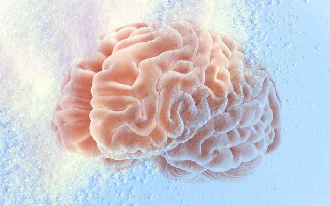
Do you often feel low brain endurance for focus and concentration? Do you often crave sugar and sweets in the afternoon? Or do you feel energized after meals? Glucose, or sugar, is the main source of energy in the human body. And, because the human brain has so many nerve cells or neurons, it is one of the most energy-demanding organs, which utilizes about one-half of all the energy from glucose in the human body. Sugar is important but too much of it can also have its downsides. �
Brain functions, such as memory, thinking, and learning, are relatively associated with glucose levels and how efficiently the brain utilizes this essential energy fuel source. If there isn�t enough glucose, or sugar, in the brain, by way of instance, neurotransmitters, or the human brain�s chemical messengers, don’t develop properly and the communications between neurons can ultimately break down. Additionally, dysglycemia, a common health issue caused by abnormal blood glucose levels, can cause loss of energy for brain function and has also been associated with poor attention and cognitive function. �
�The human brain is dependent on sugar or glucose as its main energy fuel source,� stated Vera Novak, MD, Ph.D., an HMS associate professor of medicine at Beth Israel Deaconess Medical Center. �It just simply cannot be without it.� �
What is Dysglycemia?
As previously mentioned, brain structure and function, such as cognition, can be affected by dysglycemia, or blood glucose abnormalities, in older adults. Researchers conducted a cross-sectional and longitudinal cohort research study, analyzing the association of dysglycemia with brain health. The researchers found that dysglycemia is associated with an increased number of brain infarcts, white matter hyperintensities volume, and decreased total white matter, gray matter, and hippocampus volume cross-sectionally. According to the research study, there was also a decrease in gray matter volume longitudinally. Dysglycemia was ultimately associated with reduced language performance, speed, and visuospatial function. �
�Our results suggest that dysglycemia affects brain health in elderly survivors, evidenced by higher cerebrovascular disease, lower white, and gray matter volume as well as language, visuospatial function, and cognitive speed,� stated the authors. �
Dysglycemia can cause changes in blood glucose levels which may cause a variety of health issues. Dysglycemia is also not necessarily defined by specific blood sugar levels. Instead, having an abnormally low, high, or unstable blood glucose levels suggests an underlying health issue that requires further investigation. Moreover, while type 1 and type 2 diabetes are the most common causes of dysglycemia, other examples of blood sugar level abnormalities can include gestational diabetes and pre-diabetic conditions as well as drug-related and genetically related abnormalities of the blood sugar levels. �
Furthermore, dysglycemia can be a result of hereditary or environmental factors, or it can even be a combination of both. Genes can predispose a person to ultimately develop dysglycemia over time, just as much as several lifestyle habits can, too. A poor diet high in unhealthy fats, sugars, and processed foods can commonly cause a person to develop dysglycemia. Lacking certain vitamins and minerals that enhance the human body�s sensitivity to insulin can also cause dysglycemia. �
Dysglycemia and Brain Health
Although the brain needs glucose or sugar, too much of this energy fuel source can also have several side-effects. A 2012 research study on animals conducted by researchers at the University of California at Los Angeles demonstrated a positive relationship between the consumption of fructose, another form of sugar, and the aging of cells. A 2009 research study, also utilizing animal models and conducted by a team of scientists at the University of Montreal and Boston College, connected excess glucose consumption to memory and cognitive deficiencies. Further research studies are still required. �
The effects of glucose and other forms of sugar on the human brain may be the most profound in diabetes, a group of health issues in which high blood glucose levels persist over a prolonged period of time. Type 1 diabetes is a health issue in which the immune system destroys the cells in the pancreas that produce insulin, a hormone utilized by the human body to maintain and regulate blood glucose levels. Type 2 diabetes, caused by dietary and other environmental factors, is a health issue in which cells become overwhelmed by insulin and fail to properly respond and they ultimately become insulin resistant. �
Long-term diabetes, either type 1 or type 2, can have many consequences for the brain cells, or neurons, as well as the brain. High blood glucose levels can affect the brain�s functional connectivity which connects brain regions that share functional properties and brain matter. It can also cause the brain to atrophy or shrink and it can lead to small-vessel disease, which restricts blood flow in the brain, causing cognitive difficulties and it can cause the development of vascular dementia. �
In her laboratory, Novak evaluated several ways to prevent these effects in people with type 2 diabetes. One of these ways involves a nasal spray known as intranasal insulin (INI). When used, INI enters the brain and binds to receptors in its memory networks, including the hippocampus, hypothalamus, and insular cortex. As signaling within these memory networks become more efficient, cognitive functions in these areas, such as learning and visual perceptions of spatial relationships, improve. �
�Type 2 diabetes accelerates brain aging,� says Novak, �which, in turn, accelerates the progression of functional decline. With intranasal insulin, we�re hoping to find a new avenue for treatment to slow down these effects or prevent them altogether.� �
In a pilot research study, Novak and her colleagues found that a single dose of INI had a positive effect on memory, verbal learning, and spatial orientation. She is now planning the first clinical trial of INI in older adults with type 2 diabetes. The results of the clinical trial are especially relevant because of the high prevalence of dementia and significant cognitive decline among older adults with diabetes. Sugar, or glucose, is fundamental, however, it must be controlled for overall brain health. �
Glucose, or sugar, is an important source of energy fuel for every cell in the human body, especially the brain. However, excess amounts of blood glucose, or sugar, levels can be more harmful than beneficial and it can ultimately cause a variety of brain health issues, including neurological diseases like dementia and Alzheimer’s disease. Dysglycemia, or abnormal blood glucose, or sugar, levels, is a common condition in diabetes. Managing and regulating glucose, or sugar, in patients with diabetes is essential to promote overall brain health and wellness, according to research studies. – Dr. Alex Jimenez D.C., C.C.S.T. Insight
Neurotransmitter Assessment Form
The following Neurotransmitter Assessment Form can be filled out and presented to Dr. Alex Jimenez. Symptoms listed on this form are not intended to be utilized as a diagnosis of any type of disease, condition, or any other type of health issue. �
Do you often feel low brain endurance for focus and concentration? Do you often crave sugar and sweets in the afternoon? Or do you feel energized after meals? Glucose, or sugar, is the main source of energy in the human body. And, because the human brain has so many nerve cells or neurons, it is one of the most energy-demanding organs, which utilizes about one-half of all the energy from glucose in the human body. Sugar is important but too much of it can also have its downsides. �
Brain functions, such as memory, thinking, and learning, are relatively associated with glucose levels and how efficiently the brain utilizes this essential energy fuel source. If there isn�t enough glucose, or sugar, in the brain, by way of instance, neurotransmitters, or the human brain�s chemical messengers, don’t develop properly and the communications between neurons can ultimately break down. Additionally, dysglycemia, a common health issue caused by abnormal blood glucose levels, can cause loss of energy for brain function and has also been associated with poor attention and cognitive function. �
The scope of our information is limited to chiropractic, musculoskeletal, and nervous health issues or functional medicine articles, topics, and discussions. We use functional health protocols to treat injuries or disorders of the musculoskeletal system. Our office has made a reasonable attempt to provide supportive citations and has identified the relevant research study or studies supporting our posts. We also make copies of supporting research studies available to the board and or the public upon request. To further discuss the subject matter above, please feel free to ask Dr. Alex Jimenez or contact us at 915-850-0900.�
Curated by Dr. Alex Jimenez �
References:
Marchione, Victor. �Cognition and Brain Structure Affected by Dysglycemia in Older Adults: Study.� Bel Marra Health – Breaking Health News and Health Information, Bel Marra Health, 10 Jan. 2017, www.belmarrahealth.com/cognition-brain-structure-affected-dysglycemia-older-adults-study/.
Edwards, Scott. �Sugar and the Brain.� Sugar and the Brain | Department of Neurobiology, neuro.hms.harvard.edu/harvard-mahoney-neuroscience-institute/brain-newsletter/and-brain-series/sugar-and-brain.
Additional Topic Discussion: Chronic Pain
Sudden pain is a natural response of the nervous system which helps to demonstrate possible injury. By way of instance, pain signals travel from an injured region through the nerves and spinal cord to the brain. Pain is generally less severe as the injury heals, however, chronic pain is different than the average type of pain. With chronic pain, the human body will continue sending pain signals to the brain, regardless if the injury has healed. Chronic pain can last for several weeks to even several years. Chronic pain can tremendously affect a patient’s mobility and it can reduce flexibility, strength, and endurance.
Neural Zoomer Plus for Neurological Disease
Dr. Alex Jimenez utilizes a series of tests to help evaluate neurological diseases. The Neural ZoomerTM Plus is an array of neurological autoantibodies which offers specific antibody-to-antigen recognition. The Vibrant Neural ZoomerTM Plus is designed to assess an individual�s reactivity to 48 neurological antigens with connections to a variety of neurologically related diseases. The Vibrant Neural ZoomerTM Plus aims to reduce neurological conditions by empowering patients and physicians with a vital resource for early risk detection and an enhanced focus on personalized primary prevention. �
Food Sensitivity for the IgG & IgA Immune Response
Dr. Alex Jimenez utilizes a series of tests to help evaluate health issues associated with food sensitivities. The Food Sensitivity ZoomerTM is an array of 180 commonly consumed food antigens that offers very specific antibody-to-antigen recognition. This panel measures an individual�s IgG and IgA sensitivity to food antigens. Being able to test IgA antibodies provides additional information to foods that may be causing mucosal damage. Additionally, this test is ideal for patients who might be suffering from delayed reactions to certain foods. Utilizing an antibody-based food sensitivity test can help prioritize the necessary foods to eliminate and create a customized diet plan around the patient�s specific needs. �
Formulas for Methylation Support
XYMOGEN�s Exclusive Professional Formulas are available through select licensed health care professionals. The internet sale and discounting of XYMOGEN formulas are strictly prohibited.
Proudly,�Dr. Alexander Jimenez makes XYMOGEN formulas available only to patients under our care.
Please call our office in order for us to assign a doctor consultation for immediate access.
If you are a patient of Injury Medical & Chiropractic�Clinic, you may inquire about XYMOGEN by calling 915-850-0900.
�
For your convenience and review of the XYMOGEN products please review the following link. *XYMOGEN-Catalog-Download �
* All of the above XYMOGEN policies remain strictly in force.
Do you often feel energy level drops in the afternoon? Do you often crave sugar and sweets in the afternoon? Do you often have difficulty concentrating before eating? Various medical conditions can affect the overall health of our body and mind. However, research studies have found that anemia caused by iron deficiency can tremendously affect our brain health. �
Iron deficiency is considered to be one of the most prevalent nutritional health issues, affecting approximately 2.5 billion people worldwide. In developing countries, about 40 percent of children and 50 percent of pregnant women have an iron deficiency. Iron is an essential mineral found in approximately 5 percent of the earth�s crust, however, inefficiency in absorption, low iron levels in staple grain foods, and a variety of medical conditions can make iron deficiency a common problem among humans. In first world countries, iron deficiency is still one of the most common nutrient deficiencies. �
What Causes Iron Deficiency and Anemia?
Poor iron intake and increased iron loss, generally through bleeding or breastfeeding, are several of the main causes of iron deficiency. Pregnant women, breastfeeding women, women with heavy periods, children or picky eaters, vegetarians and vegans, as well as people with digestion health issues which cause decreased iron absorption like celiac disease or post gastric bypass, and people with increased bleeding, such as cancer, ulcers, gastritis, or parasites, are generally at higher risk for iron deficiency. High calcium intake, by way of instance, children who drink a lot of milk, can also affect iron absorption, together with drugs and/or medications, such as antacids and proton-pump inhibitors for gastroesophageal reflux disease. �
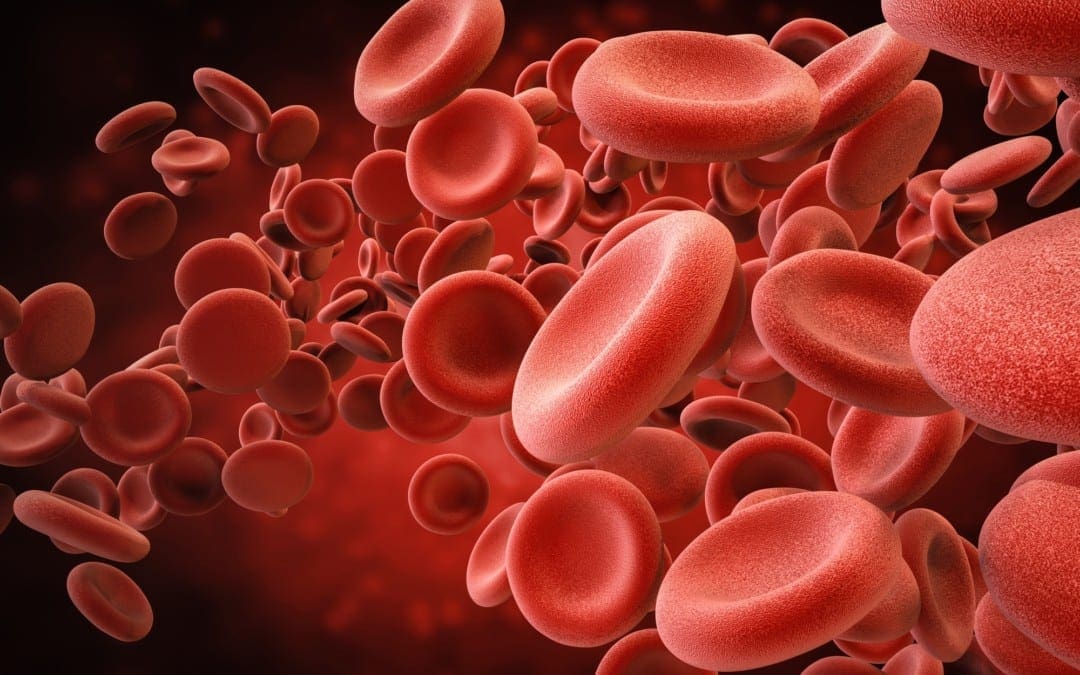
Although low iron levels are well-known for causing anemia because red blood cells need iron as a part of hemoglobin, iron is also needed for the brain and nerves. Severe iron deficiency in younger children can ultimately cause irreversible damage to cognition and result in lower IQ and developmental delays, especially during the most fundamental stages of human development and up to 16 months of age. Even in adults. the most common symptoms associated with iron deficiency are generally neurological symptoms, including fatigue, brain fog, and restless legs that cause insomnia, among other symptoms. �
Pica, the abnormal behavioral compulsion to eat non-nutritional foods like dirt or clay, is tremendously prevalent in regions of the world where iron deficiency is common. In the developed world, pica is a rare health issue, however, it still frequently occurs in children, pregnant women, and among other groups of people that are at higher risk for iron deficiency, including people who have had gastric bypass. Non-neurological symptoms associated with iron deficiency ultimately include pallor, generalized weakness, and higher than usual heart rate along with shortness of breath, especially with exertion. �
What are the Symptoms of Iron Deficiency?
As previously mentioned above, iron deficiency can cause problems associated with cognition and neurological health issues, such as restless legs and insomnia. The exact mechanisms of why this happens are unknown, however, without enough iron in the brain and nerves, there are problems with neurotransmitter signaling, the development of nerve insulation known as myelin, and brain energy metabolism. Reduced central neuron processing is considered to be one of the most critical problems associated with iron deficiency, which can be a cause of psychiatric symptoms and ongoing psychiatric problems. �
Occasionally, iron deficiency may also cause anxiety, depression, irritability, and even poor concentration and restlessness. By way of instance, iron deficiency has a much higher prevalence in children with ADHD but the symptoms can improve with iron supplements. People with iron deficiency have higher risks of developing psychiatric disorders, especially ADHD, and developmental disorders. Evidence has demonstrated that iron deficiency can cause a variety of other health issues. �
Iron enters the brain through the blood-brain barrier via transferrin receptors. Iron uptake into the brain is highly regulated but it also does highly depend on the iron status of the human body. Therefore, people with low iron levels will have much less iron going into the brain and people with high iron levels will have much more iron going into the brain. Several regions of the brain also appear to gather iron and have higher levels than others. Moreover, neurological symptoms can manifest before developing iron deficiency anemia. Thus, healthcare professionals can’t rule out iron deficiency anemia from the most commonly utilized basic screening test, a complete blood count. A better general screen involves ferritin levels, where less than 15 ng/ml presents the diagnosis for iron deficiency but less than 40 ng/ml presents with fatigue, brain fog, restless legs, and other neurological symptoms. Ferritin on its own can be misleading in populations of people with chronic inflammation, including people on dialysis, where ferritin can be high even if the person is diagnosed with iron deficiency. Furthermore, a full iron workup includes hemoglobin, MCV, ferritin, total iron-binding capacity, serum iron, and transferrin saturation. �
What is the Treatment for Iron Deficiency Anemia?
Treating iron deficiency is considerably simple through the utilization of iron supplements or in mild or moderate cases by encouraging the consumption of foods that are high in iron. Occasionally, people with severe absorption health issues will need iron transfusions intravenously. Meat and seafood are the best sources of easily absorbable heme iron, however, non-heme iron is naturally found in leafy greens, beans, and nuts. Make sure to talk to your doctor if you have iron deficiency. �
It is ultimately essential to make sure if you have iron deficiency before treating it with increased amounts of iron supplements. With the exception of blood loss, the only way to reduce excess iron is through the process of skin cells flaking off. Therefore, adult men who take a lot of iron supplements and people with a genetic tendency to absorb more iron from foods are at a higher risk of developing a medical condition, known as hemochromatosis or severe iron overload. �
Excess iron is stored in the liver and can lead to scarring of the liver, known as cirrhosis. Iron overload can also lead to joint and hormonal problems and it can also cause a bronze-ish skin color. Symptoms of hemochromatosis include joint pain, fatigue, and low sex drive as well as a higher risk of developing diabetes. High serum iron is associated with health issues like high blood pressure. People who aren�t iron deficient can consider regular blood donations to prevent accidental iron overload. Iron is one of those types of minerals that should neither be too high or too low but rather, just right. More research studies, especially clinical trials analyzing common medical conditions, such as restless legs, insomnia, and ADHD, are fundamental to help healthcare professionals understand the relationship between iron deficiency anemia and brain health. �
Recent research studies have demonstrated that iron deficiency anemia may be associated with brain health issues. Because the brain and nerves need iron for many functions, iron deficiency can cause a variety of symptoms and medical conditions, including brain fog, fatigue, restless legs with insomnia, anxiety, depression, and cognitive problems, besides anemia or lack of healthy red blood cells. Treatment for iron deficiency anemia may utilize iron supplements, however, it’s important to make sure to talk to a qualified healthcare professional in order to avoid risks and side effects through iron supplementation.� – Dr. Alex Jimenez D.C., C.C.S.T. Insight
Neurotransmitter Assessment Form
The following Neurotransmitter Assessment Form can be filled out and presented to Dr. Alex Jimenez. Symptoms listed on this form are not intended to be utilized as a diagnosis of any type of disease, condition, or any other type of health issue. �
Do you often feel energy level drops in the afternoon? Do you often crave sugar and sweets in the afternoon? Do you often have difficulty concentrating before eating? Various medical conditions can affect the overall health of our body and mind. However, research studies have found that anemia caused by iron deficiency can tremendously affect our brain health. � Iron deficiency is considered to be one of the most prevalent nutritional health issues, affecting approximately 2.5 billion people worldwide. In developing countries, about 40 percent of children and 50 percent of pregnant women have an iron deficiency. Iron is an essential mineral found in approximately 5 percent of the earth�s crust, however, inefficiency in absorption, low iron levels in staple grain foods, and a variety of medical conditions can make iron deficiency a common problem among humans. In first world countries, iron is still considered to be the most common nutrient deficiency. �
The scope of our information is limited to chiropractic, musculoskeletal, and nervous health issues or functional medicine articles, topics, and discussions. We use functional health protocols to treat injuries or disorders of the musculoskeletal system. Our office has made a reasonable attempt to provide supportive citations and has identified the relevant research study or studies supporting our posts. We also make copies of supporting research studies available to the board and or the public upon request. To further discuss the subject matter above, please feel free to ask Dr. Alex Jimenez or contact us at 915-850-0900.�
Curated by Dr. Alex Jimenez �
References:
Deans, Emily. �Heavy Metal: Iron and the Brain.� Psychology Today, Sussex Publishers, 29 Nov. 2015, www.psychologytoday.com/us/blog/evolutionary-psychiatry/201511/heavy-metal-iron-and-the-brain.
Additional Topic Discussion: Chronic Pain
Sudden pain is a natural response of the nervous system which helps to demonstrate possible injury. By way of instance, pain signals travel from an injured region through the nerves and spinal cord to the brain. Pain is generally less severe as the injury heals, however, chronic pain is different than the average type of pain. With chronic pain, the human body will continue sending pain signals to the brain, regardless if the injury has healed. Chronic pain can last for several weeks to even several years. Chronic pain can tremendously affect a patient’s mobility and it can reduce flexibility, strength, and endurance.
Neural Zoomer Plus for Neurological Disease
Dr. Alex Jimenez utilizes a series of tests to help evaluate neurological diseases. The Neural ZoomerTM Plus is an array of neurological autoantibodies which offers specific antibody-to-antigen recognition. The Vibrant Neural ZoomerTM Plus is designed to assess an individual�s reactivity to 48 neurological antigens with connections to a variety of neurologically related diseases. The Vibrant Neural ZoomerTM Plus aims to reduce neurological conditions by empowering patients and physicians with a vital resource for early risk detection and an enhanced focus on personalized primary prevention. �
Food Sensitivity for the IgG & IgA Immune Response
Dr. Alex Jimenez utilizes a series of tests to help evaluate health issues associated with food sensitivities. The Food Sensitivity ZoomerTM is an array of 180 commonly consumed food antigens that offers very specific antibody-to-antigen recognition. This panel measures an individual�s IgG and IgA sensitivity to food antigens. Being able to test IgA antibodies provides additional information to foods that may be causing mucosal damage. Additionally, this test is ideal for patients who might be suffering from delayed reactions to certain foods. Utilizing an antibody-based food sensitivity test can help prioritize the necessary foods to eliminate and create a customized diet plan around the patient�s specific needs. �
Formulas for Methylation Support
XYMOGEN�s Exclusive Professional Formulas are available through select licensed health care professionals. The internet sale and discounting of XYMOGEN formulas are strictly prohibited.
Proudly,�Dr. Alexander Jimenez makes XYMOGEN formulas available only to patients under our care.
Please call our office in order for us to assign a doctor consultation for immediate access.
If you are a patient of Injury Medical & Chiropractic�Clinic, you may inquire about XYMOGEN by calling 915-850-0900.
�
For your convenience and review of the XYMOGEN products please review the following link. *XYMOGEN-Catalog-Download �
* All of the above XYMOGEN policies remain strictly in force.
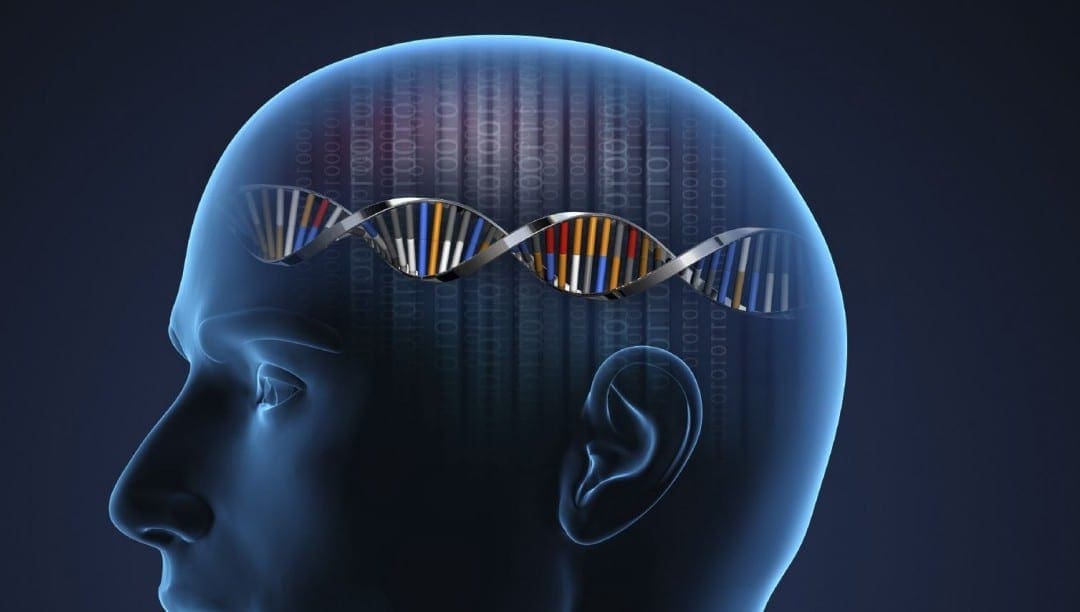
Biomarkers are molecules that can help diagnose a health issue. These have become important for verifying investigations, choosing the best remedies, and monitoring disease progression. One exception, however, includes biomarkers for neurological diseases. Neurological biomarkers are found in the cerebral spinal fluid (CSF) or, in undetectable amounts, in the blood vessels. The human brain is closely guarded by the blood-brain barrier which protects it from damaging compounds circulating throughout the blood vessels. The blood-brain barrier has made it inaccessible to use these biomarkers. �
Biomarkers may be analyzed using the CSF but this also needs an invasive lumbar puncture process. Biomarker signatures, or recent improvements in discovery, in addition to the ability of clusters of biomarkers, are currently helping to make neurological disorders more treatable and more reachable. Treating and preventing neurological disorders, such as chronic traumatic encephalopathy (CTE), Alzheimer’s disease, Parkinson’s disease, autism, and major depressive disorder, is very likely to become less difficult to diagnose with the recent arrival of neurological biomarkers found in the blood. �
Biomarker for Brain Health Issues
Biomarker signatures, found with panels of high-quality antibodies, are yet another safe and effective tool for evaluating neurological disorders and diseases. Assistant professor of neurology and immunobiology at the University of Arizona College of Medicine at Tucson, Kristian Doyle, utilizes biomarkers to examine how the immune system deals with dead brain tissue. The system eliminates brain tissue with a procedure called liquefactive necrosis following a stroke but the pathophysiology of the procedure is unknown. This information is essential because liquefactive necrosis may be neurotoxic. �
“We utilize multiplex immunoassays to describe inflammation within chronic stroke infarcts in the point of liquefactive necrosis, and to describe changes by ordinary stroke comorbidities,” says Doyle. Because over 10 million individuals survive a stroke every year, Doyle expects that biomarkers may help them monitor the development of liquefactive necrosis and start to tailor remedies that mitigate the secondary harm due to this procedure,” he states. Another connection between inflammation and neurotoxicity is analyzed by Alysson Muotri, professor of molecular and cellular medicine and director of the Stem Cell Program at the University of California, San Diego School of Medicine. The Muotri laboratory uses induced pluripotent stem cells (iPSCs) from individuals with schizophrenia and autism to search for biomarkers of those ailments. �
Muotri’s laboratory started analyzing the cytokine interleukin-6 (IL-6) as a biomarker because evidence indicates that chronic exposure to elevated cytokines might be neurotoxic together with elevated levels associated with depression, autism, and schizophrenia. “The gap of one of the many brain disorders could function as cytokines act on particular types or subtypes of nerves, or within a particular brain area,” states Muotri. His laboratory differentiates iPSCs to cells, which they suspect might be releasing cytokines from patients. Also, because IL-6 can also be involved in immune-inflammatory pathways, Muotri supposes a connection between autism and in utero exposure to infection, such as the Zika virus. �
“Our forecast is that the inflammation caused by Zika vulnerability is sufficient to make a neurotoxic environment which could rewire how the human brain is shaped,” he states. “We see that in mice, therefore, we believe some Zika-exposed children are going to develop autism or have intellectual disabilities” Larger biomarker signatures are available with technologies from CDI Laboratories, which provides microarrays of practical human proteins, including over 20,000 to a single variety, to check the antibodies within human liquid biopsy samples, including blood, serum, plasma, CSF, or tissue lysates. The consequent “autoantibody profile” is a helpful tool for study and for diagnoses or prognoses of individuals. �
“We have worked in the area of biomarker discovery for various neurodegenerative diseases like multiple sclerosis, neuropsychiatric lupus, Alzheimer’s disease, and Parkinson’s disease,” states George Dorfman, director of business development in CDI Laboratories, a spin-off firm located in Baltimore, Maryland, and Mayaguez, Puerto Rico which was created from research in the High Throughput Biology Center at Johns Hopkins University. CDI’s stage is particularly beneficial in building panels for biomarker discovery because researchers can start by utilizing patient samples or banked trials to evaluate resistant profiles of cohorts that reveal specific symptoms or no indications in the event of control trials. �
“This provides us with an inherent candidate biomarker panel that offers advice on the following clinical outcome or curative efficacy, which is confirmed to yield the last panel, then interpreted into the state an ELISA-based kit or any other immunodiagnostic format at the clinical setting,” says Dorfman. “In the event of multiple sclerosis, as a patient grows through measures of this disease, their entire body creates novel antibodies or greater present antibody titers against specific proteins, such as myelination proteins. Our panels may discover these, to provide a notion about exactly what patients’ disease development might seem like, and supply a signature which may be interpreted into another evaluation or an FDA-approved diagnostic” CDI’s technologies have also been utilized to create an autoantibody profile for neuropsychiatric lupus, a beneficial diagnostic tool to ultimately help diagnose a neurological disorder that typically lacks obvious clinical signs. �
Understanding Biomarkers for Brain Health
The amount and types of biomarkers, as well as the quantity of information which researchers have to arrange, can help provide better remedies and prevention methods and techniques. “It ought to be no surprise that researchers spend around 80 percent of the time handling and not assessing statistics,” states Scott Marshall, managing director of translational informatics and diagnostic sciences in Precision for Medicine in Frederick, Maryland. The biomarker data management system, PATH, was made to incorporate any sort of biomarker information for further neurological disease diagnosis. �
“The true power of biomarkers comes if you connect this data to clinical information,” states Marshall. Their biomarker information management system supports translational research and biomarker-guided medicine development and puts no limitation on the number of biomarkers that may be tracked. “It can manage multiple biomarker technologies concurrently, such as complicated flow cytometry, next-generation sequencing, immuno-sequencing, epigenetic profiling, and other varieties of assays measuring biological variant too,” states Marshall. Their kind of “translational informatics” instrument is much more efficient than generating reams of information” with no strategy to acquire actionable insights out of these.” �
Research teams utilize Precision to Medicine’s platform for neuro-related programs that vary from illness pathogenesis to creating complicated signatures that are predictive of treatment response. By way of instance, the system was utilized in a research study including the evaluation of transcriptomic and genomic data in the treatment of major depressive disorder. The outcome is a genomically defined subset of individuals utilizing a probability of improvement. “This type of signature can now be evaluated by means of an assay, which may subsequently be developed to accompany diagnostic or free diagnostic to successfully target the correct individual group,” states Marshall. Biomarker data management systems become more fundamental as distinct kinds of biomarkers are examined collectively, ultimately including proteins and miRNAs. �
Combining kinds of biomarkers will very likely boost their usefulness. “Diagnostics is becoming increasingly more important as we know that the interplay between microRNAs, proteins, DNA, and messenger RNA is necessary,” states Pregibon. Clinical decision-making may profit particularly in which the human brain has been blocked by the blood-brain barrier until lately. “The chance to leverage biomarker-driven targeted treatments means that the sufferers that are more inclined to react to treatments are getting them quicker,” states Marshall. “For researchers, that’s the energy of biomarkers.” �
�
The recent ability to be able to detect neurological biomarkers in the blood, despite the blood-brain barries, is largely due in part to new technological advances in diagnosis and detection. Several of these technologies can ultimately increase sensitivity, however, increased sensitivity can help improve earlier detection or diagnosis of biomarkers for neurological diseases and disorders. Researchers and healthcare professionals currently believe that the presence of these biomarkers may be present earlier than we currently understand, which can help improve health issue diagnosis and treatment. – Dr. Alex Jimenez D.C., C.C.S.T. Insight
Neurotransmitter Assessment Form
The following Neurotransmitter Assessment Form can be filled out and presented to Dr. Alex Jimenez. Symptoms listed on this form are not intended to be utilized as a diagnosis of any type of disease, condition, or any other type of health issue.
Biomarkers are molecules that can help diagnose a health issue. These have become important for verifying investigations, choosing the best remedies, and monitoring disease progression. One exception, however, includes biomarkers for neurological diseases. Neurological biomarkers are found in the cerebral spinal fluid (CSF) or, in undetectable amounts, in the blood vessels. The human brain is closely guarded by the blood-brain barrier which protects it from damaging compounds circulating throughout the blood vessels. The blood-brain barrier has made it inaccessible to use these biomarkers. �
Biomarkers may be analyzed using the CSF but this also needs an invasive lumbar puncture process. Biomarker signatures, or recent improvements in discovery, in addition to the ability of clusters of biomarkers, are currently helping to make neurological disorders more treatable and more reachable. Treating and preventing neurological disorders, such as chronic traumatic encephalopathy (CTE), Alzheimer’s disease, Parkinson’s disease, autism, and major depressive disorder, is very likely to become less difficult to diagnose with the recent arrival of neurological biomarkers found in the blood. �
The scope of our information is limited to chiropractic, musculoskeletal, and nervous health issues or functional medicine articles, topics, and discussions. We use functional health protocols to treat injuries or disorders of the musculoskeletal system. Our office has made a reasonable attempt to provide supportive citations and has identified the relevant research study or studies supporting our posts. We also make copies of supporting research studies available to the board and or the public upon request. To further discuss the subject matter above, please feel free to ask Dr. Alex Jimenez or contact us at 915-850-0900.�
Curated by Dr. Alex Jimenez �
References:
Smith, Caitlin. �Biomarkers on the Brain: Putting Biomarkers Together for a Better Understanding of the Nervous System.� Science, 15 Mar. 2018, www.sciencemag.org/features/2017/12/biomarkers-brain-putting-biomarkers-together-better-understanding-nervous-system.
Additional Topic Discussion: Chronic Pain
Sudden pain is a natural response of the nervous system which helps to demonstrate possible injury. By way of instance, pain signals travel from an injured region through the nerves and spinal cord to the brain. Pain is generally less severe as the injury heals, however, chronic pain is different than the average type of pain. With chronic pain, the human body will continue sending pain signals to the brain, regardless if the injury has healed. Chronic pain can last for several weeks to even several years. Chronic pain can tremendously affect a patient’s mobility and it can reduce flexibility, strength, and endurance.
Neural Zoomer Plus for Neurological Disease
�
Dr. Alex Jimenez utilizes a series of tests to help evaluate neurological diseases. The Neural ZoomerTM Plus is an array of neurological autoantibodies which offers specific antibody-to-antigen recognition. The Vibrant Neural ZoomerTM Plus is designed to assess an individual�s reactivity to 48 neurological antigens with connections to a variety of neurologically related diseases. The Vibrant Neural ZoomerTM Plus aims to reduce neurological conditions by empowering patients and physicians with a vital resource for early risk detection and an enhanced focus on personalized primary prevention. �
Food Sensitivity for the IgG & IgA Immune Response
�
Dr. Alex Jimenez utilizes a series of tests to help evaluate health issues associated with food sensitivities. The Food Sensitivity ZoomerTM is an array of 180 commonly consumed food antigens that offers very specific antibody-to-antigen recognition. This panel measures an individual�s IgG and IgA sensitivity to food antigens. Being able to test IgA antibodies provides additional information to foods that may be causing mucosal damage. Additionally, this test is ideal for patients who might be suffering from delayed reactions to certain foods. Utilizing an antibody-based food sensitivity test can help prioritize the necessary foods to eliminate and create a customized diet plan around the patient�s specific needs. �
Formulas for Methylation Support
XYMOGEN�s Exclusive Professional Formulas are available through select licensed health care professionals. The internet sale and discounting of XYMOGEN formulas are strictly prohibited.
Proudly,�Dr. Alexander Jimenez makes XYMOGEN formulas available only to patients under our care.
Please call our office in order for us to assign a doctor consultation for immediate access.
If you are a patient of Injury Medical & Chiropractic�Clinic, you may inquire about XYMOGEN by calling 915-850-0900.
�
For your convenience and review of the XYMOGEN products please review the following link. *XYMOGEN-Catalog-Download �
* All of the above XYMOGEN policies remain strictly in force.
Do you often feel that your energy levels drop in the afternoons? Do you often feel brain fog or have unclear thoughts and poor concentration? Do you often experience brain fatigue with chronic pain and inflammation? Diet and supplements are essential for overall well-being. However, fish oil omega-3s are a common supplement with a variety of health benefits. �
Fish oil is a well-known supplement that comes from fatty fish, such as sardines, anchovies, mackerel, and salmon. Fish oil has two types of omega-3 fatty acids, known as eicosapentaenoic acid (EPA) and docosahexaenoic acid (DHA), both of which are commonly used to support skin and heart health. Fish oil supplements can also promote brain health, especially when it comes to improving memory and mood problems like depression, as well as a variety of other health issues. The purpose of the following article is to discuss how the omega-3 fatty acids in fish oil may support and promote brain health. �
What Are Fish Oil Omega-3s?
Omega-3 fatty acids, best known as omega-3s, are polyunsaturated fats that offer most of the brain health benefits of fish oil. Fish oil has two main types of omega-3 fatty acids, EPA and DHA. These two omega-3s are found in cell membranes and these also have anti-inflammatory effects in the human body. They are also well-known for their fundamental roles in human development and heart health. EPA and DHA are found in fatty fish and fish oil. Because the majority of people do not eat the recommended amounts of fish, many people probably aren’t getting enough EPA and DHA in their regular diets. �
The human body can make EPA and DHA from another type of omega-3 fatty acid, known as alpha-linolenic acid (ALA). ALA is commonly found in a variety of foods, including walnuts, flaxseeds, chia seeds, canola oil, soybeans, and soybean oil. However, the human body can’t properly turn ALA into EPA and DHA, where healthcare professionals and researchers estimate that approximately less than 10 percent of the amount of ALA you eat is properly turned into EPA or DHA. Fish oil is a good option for people who don�t eat a lot of fish but are still looking to gain the health benefits of omega-3 fatty acids. �
How Do Omega-3s Improve Brain Health?
The omega-3 fatty acids EPA and DHA are essential for normal brain function during all stages of life. EPA and DHA play a fundamental role in the development of a baby�s brain. Several research studies have associated pregnant women�s fish intake or fish oil use with higher scores for their children on brain function and intelligence tests in early childhood. �
These omega-3s are also essential for the regulation of normal brain function throughout life. They are commonly found in the cell membranes of brain cells, preserving cell membrane health and facilitating communication between brain cells. �
When animals are fed diets without omega-3 fatty acids, the amount of DHA in their brains decreases, and they often demonstrate deficits in learning and memory. In older adults, decreased levels of DHA in the blood have ultimately been associated with smaller brain size, a prevalent symptom of accelerated brain aging. Clearly, it is essential to make sure that you get enough omega-3 fatty acids in order to avoid these detrimental effects on brain function and development. �
Fish Oil for Memory
The omega-3s found in fish oil play important roles in brain function and development. There are also claims that fish oil can improve brain function in people with memory problems, such as dementia and Alzheimer�s disease. Alzheimer�s disease affects brain function and quality of life in millions of elderly adults. That’s why finding a supplement or natural remedy that can help improve brain function in this population of individuals would be a major, if not a life-changing discovery. �
Unfortunately, a review of the research study found no compelling evidence that omega-3 supplements like fish oil can improve brain function in people with dementia and Alzheimer�s disease. However, several research studies suggest that fish oil supplements may improve brain function in people with more mild types of brain health issues, including mild cognitive impairment (MCI) or age-related cognitive decline. Although these types of brain conditions aren�t as severe as dementia and Alzheimer�s disease, they can still result in memory loss and sometimes other types of impaired brain function. �
One research study gave 485 older adults with age-related cognitive decline either 900 mg of DHA or a placebo every day. After 24 weeks, those taking DHA performed better on memory and learning tests. Similarly, another research study investigated the effects of taking 1.8 grams of omega-3 fatty acids from fish oil supplements daily for 24 weeks. The researchers found improvements in brain function in people with MCI but there were no benefits for those with dementia and Alzheimer�s disease. According to the research study, fish oil supplements may be most beneficial when people start taking them in the early stages of brain function decline. If you wait too long, fish oil may be of little benefit to brain health. �
Fish Oil for Depression
Finding treatments for depression and other mental health issues is still a public health priority. A recent review of several research studies concluded that taking fish oil supplements improved symptoms in people with depression. However, the greatest improvements in depressive symptoms seemed to occur in people who were also taking antidepressants. In addition, people seemed to experience greater effects when the fish oil supplement also had higher doses of EPA. �
It is still unclear how EPA and omega-3s help improve depressive symptoms. Researchers suggest that it may be associated with their effects on serotonin and serotonin receptors in the brain. Others have proposed that omega-3s from fish oil can improve depressive symptoms through their anti-inflammatory effects. Additional evidence suggests that fish oil may also help improve other mental health issues like borderline personality disorder and bipolar disorder. However, more high-quality research studies are required before the medical community can make definitive recommendations. �
Should You Take Fish Oil for Your Brain Health?
According to the evidence gathered from a variety of research studies, you may want to consider taking fish oil omega-3 fatty acids if you are experiencing mild memory loss, depression, or if you want to improve your overall brain health.�There are no official recommendations regarding how much omega-3s from fish oil you need to take to see benefits in brain function and mental health. The amounts of fish oil omega-3 fatty acids used in the research studies varied from each clinical trial. �
The US Food and Drug Administration has set a safe and effective limit for the intake of omega-3 fatty acid supplements to be at 3,000 mg per day. The European Food Safety Authority has set their recommendation a little higher, at no more than 5,000 mg per day. Taking 1,000 to 2,000 mg of omega-3 fatty acids from fish oil daily is likely a good starting point, which is under the recommended limit. People with depression should choose fish oil supplements with higher amounts of EPA. �
It is essential to read labels carefully when evaluating fish oil supplements. A 1,000-mg capsule of fish oil may have less than 500 mg of actual omega-3 fatty acids but this can vary for each brand. In general, fish oil supplements are considered to be safe and effective at dosages under those that were previously mentioned. Make sure you talk with a healthcare professional before taking fish oil omega-3 fatty acid supplements. Because research studies have reviewed their potential effects on blood clotting, this is especially important if you are taking blood-thinning medications or have an upcoming surgery. �
EPA and DHA are omega-3 fatty acids in fish oil that are fundamental for normal brain function and development. People with mild decline in memory or brain function and mood changes like depression may consider taking omega-3s from fish oil because these can improve symptoms and overall brain health. Although fish oil is typically praised for its benefits for heart health, it also has incredible effects on brain and mental health that are worth mentioning.�- Dr. Alex Jimenez D.C., C.C.S.T. Insight
Neurotransmitter Assessment Form
The following Neurotransmitter Assessment Form can be filled out and presented to Dr. Alex Jimenez. Symptoms listed on this form are not intended to be utilized as a diagnosis of any type of disease, condition, or any other type of health issue. �
Do you often feel that your energy levels drop in the afternoons? Do you often feel brain fog or have unclear thoughts and poor concentration? Do you often experience brain fatigue with chronic pain and inflammation? Diet and supplements are essential for overall well-being. However, fish oil omega-3s are a common supplement with a variety of health benefits. �
Fish oil is a well-known supplement that comes from fatty fish, such as sardines, anchovies, mackerel, and salmon. Fish oil has two types of omega-3 fatty acids, known as eicosapentaenoic acid (EPA) and docosahexaenoic acid (DHA), both of which are commonly used to support skin and heart health. Fish oil supplements can also promote brain health, especially when it comes to improving memory, mood problems like depression, as well as neurological diseases and other health issues. The purpose of the article above was to discuss how the omega-3 fatty acids in fish oil support and promote brain health. �
The scope of our information is limited to chiropractic, musculoskeletal, and nervous health issues or functional medicine articles, topics, and discussions. We use functional health protocols to treat injuries or disorders of the musculoskeletal system. Our office has made a reasonable attempt to provide supportive citations and has identified the relevant research study or studies supporting our posts. We also make copies of supporting research studies available to the board and or the public upon request. To further discuss the subject matter above, please feel free to ask Dr. Alex Jimenez or contact us at 915-850-0900.�
Sudden pain is a natural response of the nervous system which helps to demonstrate possible injury. By way of instance, pain signals travel from an injured region through the nerves and spinal cord to the brain. Pain is generally less severe as the injury heals, however, chronic pain is different than the average type of pain. With chronic pain, the human body will continue sending pain signals to the brain, regardless if the injury has healed. Chronic pain can last for several weeks to even several years. Chronic pain can tremendously affect a patient’s mobility and it can reduce flexibility, strength, and endurance.
Neural Zoomer Plus for Neurological Disease
Dr. Alex Jimenez utilizes a series of tests to help evaluate neurological diseases. The Neural ZoomerTM Plus is an array of neurological autoantibodies which offers specific antibody-to-antigen recognition. The Vibrant Neural ZoomerTM Plus is designed to assess an individual�s reactivity to 48 neurological antigens with connections to a variety of neurologically related diseases. The Vibrant Neural ZoomerTM Plus aims to reduce neurological conditions by empowering patients and physicians with a vital resource for early risk detection and an enhanced focus on personalized primary prevention. �
Food Sensitivity for the IgG & IgA Immune Response
Dr. Alex Jimenez utilizes a series of tests to help evaluate health issues associated with food sensitivities. The Food Sensitivity ZoomerTM is an array of 180 commonly consumed food antigens that offers very specific antibody-to-antigen recognition. This panel measures an individual�s IgG and IgA sensitivity to food antigens. Being able to test IgA antibodies provides additional information to foods that may be causing mucosal damage. Additionally, this test is ideal for patients who might be suffering from delayed reactions to certain foods. Utilizing an antibody-based food sensitivity test can help prioritize the necessary foods to eliminate and create a customized diet plan around the patient�s specific needs. �
Formulas for Methylation Support
XYMOGEN�s Exclusive Professional Formulas are available through select licensed health care professionals. The internet sale and discounting of XYMOGEN formulas are strictly prohibited.
Proudly,�Dr. Alexander Jimenez makes XYMOGEN formulas available only to patients under our care.
Please call our office in order for us to assign a doctor consultation for immediate access.
If you are a patient of Injury Medical & Chiropractic�Clinic, you may inquire about XYMOGEN by calling 915-850-0900.
�
For your convenience and review of the XYMOGEN products please review the following link. *XYMOGEN-Catalog-Download �
* All of the above XYMOGEN policies remain strictly in force.
Neurological diseases, including well-known neurodegenerative disorders like Alzheimer’s disease (AD) and Parkinson’s disease (PD) as well as other rare health issues, such as Huntington’s disease (HD) and amyotrophic lateral sclerosis (ALS), affect millions of people worldwide. Unfortunately, these are estimated to increase due to the aging population. Currently, there is no treatment available for any neurodegenerative disorder. Treatments for symptoms are available for several neurological diseases, such as PD and HD, but the therapeutic benefits are limited. Although the causes and symptoms for these health issues are different for each neurodegenerative disorders, their molecular pathogeneses share common underlying factors and characteristics, including excessive levels of reactive oxygen species (ROS), mainly due to mitochondrial impairment, neuroinflammation, and disturbances in protein homeostasis (proteostasis). This increases the possibility of developing a universal treatment that will focus on targeting the common triggers of neurological diseases. �
Nrf2 Activation Pathways and the Human Brain
The transcription factor, Nrf2, regulates the main endogenous defense mechanism against oxidative and xenobiotic stress and inflammation. Nrf2 also plays a fundamental role in the management of mitochondrial function and cellular proteostasis, which suggests the possible benefits of Nrf2 to control neurodegenerative disorders. When under stress, Nrf2 activates the transcriptional upregulation of a large network of cytoprotective genes that allow adaptation and survival. The levels and activity of Nrf2 are controlled through ubiquitination and proteasomal degradation mediated by several ubiquitin ligase systems, including Keap1-Cul3/Rbx1, ?-TrCP-Cul1, and Hrd1. Keap1 is the most well understood key regulator of Nrf2. �
Keap1 functions as a primary sensor for electrophiles and oxidants, which chemically change certain cysteines in Keap1, leading to conformational changes that protect Nrf2 from Keap1-associated degradation. Subsequently, Nrf2 will accumulate and then translocate to the nucleus where it binds as a heterodimer with a small Maf transcription factor to antioxidant response elements in the promoter of its target genes to activate the expression of a large network of detoxification, antioxidant, and anti-inflammatory genes as well as other genes involved in the clearance of damaged proteins. Of particular interest is also the upregulation of genes responsible in the biosynthesis and the regeneration of glutathione (GSH), a major intracellular antioxidant. Moreover, Nrf2 also suppresses proinflammatory responses through transcriptional repression and it is involved in the regulation of mitochondrial function. Keap1 and p62/SQSTM1 are Nrf2-associated proteins and main regulators of negative and positive feedback loop mechanisms. In addition, p62 targets Keap1 for selective degradation through autophagy, therefore contributing to the sustained Nrf2 activation response. �
Aging has been associated with the increase in ROS and chronic inflammation, which suggests a loss of adaptability and/or impairment of Nrf2 signaling, which are particularly pronounced in age-dependent neurodegenerative disorders. Surprisingly, rare mutations in SQSTM1 can cause susceptibility to the human neurodegenerative disorder, ALS as well as frontotemporal lobar degeneration, and are associated with muted Nrf2 activation responses in clinical trials. Research studies suggest a reciprocal correlation and show negative effects of mutant disease-related proteins on Nrf2 signaling, thus suggesting the inhibition of the Nrf2 pathway as a possible mechanism underlying neurodegeneration and health issues. �
Nrf2 Activation for Neurodegenerative Disorders
ALS, an adult-onset neurodegenerative disorder caused by the selective death of motor neurons in the brain and spinal cord, is commonly characterized by progressive muscle weakness and atrophy which is generally considered to be fatal, typically within 5 years of diagnosis. ALS has a predominant sporadic ALS form with no apparent genetic component, however, approximately 5 to 10 percent of cases show an autosomal dominant inheritance pattern or familial form of the disease, known as fALS, with is known to cause gene mutations. The symptoms of sporadic ALS and familial ALS are similar, which suggests the involvement of common pathogenic mechanisms, including oxidative stress and neuroinflammation. �
Research studies show that oxidative stress and neuroinflammation should be the key therapeutic targets of Nrf2 signaling in ALS. Genetic research studies in ALS mouse models have shown a considerable therapeutic effect of increased Nrf2 levels in astrocytes, the main GSH producers for neurons. Furthermore, Nrf2 signaling is fundamental for controlling neuroinflammation in ALS through the management of the effects of activated microglial cells on overall neuronal survival. Consistent with the therapeutic potential of Nrf2 signaling, treatment with small molecule activators, including the extremely potent cyanoenone triterpenoids, has ultimately shown efficacy in research study mouse ALS models. �
The neuroprotective potential of Nrf2 activation has been evaluated in experiments utilizing genetic mouse HD models. HD is an autosomal dominant and highly penetrant neurodegenerative disorder, which results from the pathological expansion of a trinucleotide CAG repeats encoding polyglutamines in HTT protein. Brains from patients with HD usually show marked striatal and cortical atrophy at the time of diagnosis. Once motor or other symptoms become apparent, generally throughout midlife, the affected individuals become increasingly disabled over the course of 15 to 25 years before eventually succumbing to the effects of severe physical and mental deterioration, according to evidence from research studies. �
Complex pathogenic mechanisms have been shown in HD, however, excessive oxidative stress has been recognized as a fundamental driver of pathology. The harmful role of oxidative stress has been described in both HD patients and in experimental clinical trial models and it is potentially due to the neuronal sensitivity of an excess in ROS. The levels of several Nrf2-dependent antioxidant proteins, including glutathione peroxidases, catalase, and superoxide dismutase 1, are increased in human HD brains as compared with non-disease controls, suggesting a partial activation of Nrf2 defense signaling yet insufficient enough to block progressive neurodegeneration. Pharmacological activation of Nrf2 results in the broad antioxidant effects of HD in mouse brains and ameliorates the neurological phenotype. Increased expression of several key inflammatory mediators has been shown in the blood, the striatum, the cortex, and the cerebellum from postmortem patient HD tissues, however, neuroinflammation in HD patients seems to be less pronounced than in ALS or PD patients. �
Neurological Disease Treatment with Nrf2 Activation
Finally, the neurological phenotype of the most common neurological disease and neurodegenerative disorder, PD, can be challenged by Nrf2 activation. PD is characterized by the progressive loss of dopaminergic neurons in the substantia nigra and the profound reduction of dopamine in the striatum. Currently available dopaminergic treatments offer relief from several symptoms but these only address the motor manifestations. Multiple genetic and environmental factors have been suggested in the etiology of PD, however, like ALS, the majority of the clinical cases are sporadic. The discovery that the environmental neurotoxin 1-methyl-4-phenyl-1,2,3,6-tetrahydropyridine (MPTP) causes Parkinson’s disease in humans, led to the development of the MPTP mouse model of disease, which to date, remains one of the most highly utilized animal models of sporadic PD, including the evaluation of drug efficiency. Nrf2 activators showed neuroprotective effects in MPTP mice, which are associated with a reduction of oxidative damage and neuroinflammation. The identification of causative mutations in SNCA, the gene encoding ?-synuclein (aSyn), developed genetic mouse PD models, in which daily oral delivery of the Nrf2 activator dimethyl fumarate (DMF) protected nigral dopaminergic neurons against aSyn toxicity. �
Although oxidative stress and neuroinflammation are pathological hallmarks of AD, a therapeutic role of Nrf2 signaling has developed more slowly, perhaps due to the complexity of disease pathogenesis and readouts of efficiency. Nonetheless, there is a number of recent research studies that demonstrate the efficiency of Nrf2 activators in AD mouse models. � DMF, a U.S. FDA-approved drug (Tecfidera, Biogen-Idec) for the treatment of relapsing multiple sclerosis, activates Nrf2 through the regulation of the Keap1 sensor. DMF is of seemingly low potency and specificity, which prevents neurodegeneration. Drug-like molecules with a similar mechanism of action or with the ability of direct interference with the Keap1/Nrf2 interaction are emerging. The data demonstrate the feasibility to develop Nrf2 activators for treatment. �
In summary, although the causes and symptoms of neurological diseases are different, neurodegenerative disorders share similar molecular mechanisms, which could be regulated and managed with Nrf2 activators. Moreover, targeting Nrf2 signaling may offer a safe and effective treatment approach for a variety of these health issues. Because pharmacological Nrf2 activation targets the broad mechanisms of these health issues, all neurodegenerative conditions would be eligible for treatment. Furthermore, the main goal is to develop noninvasive oral treatment(s) for patients under the supervision of healthcare professionals, which targets both sporadic and familial patients. – Dr. Alex Jimenez D.C., C.C.S.T. Insight
Neurotransmitter Assessment Form
The following Neurotransmitter Assessment Form can be filled out and presented to Dr. Alex Jimenez. Symptoms listed on this form are not intended to be utilized as a diagnosis of any type of disease, condition, or any other type of health issue. �
Neurological diseases, including well-known neurodegenerative disorders like Alzheimer’s disease (AD) and Parkinson’s disease (PD) as well as other rare health issues, such as Huntington’s disease (HD) and amyotrophic lateral sclerosis (ALS), affect millions of people worldwide. Unfortunately, these are estimated to increase due to the aging population. Currently, there is no treatment available for any neurodegenerative disorder. Treatments for symptoms are available for several neurological diseases, such as PD and HD, but the therapeutic benefits are limited. Although the causes and symptoms for these health issues are different for each neurodegenerative disorders, their molecular pathogeneses share common underlying factors and characteristics, including excessive levels of reactive oxygen species (ROS), mainly due to mitochondrial impairment, neuroinflammation, and disturbances in protein homeostasis (proteostasis). This increases the possibility of developing a universal treatment that will focus on targeting the common triggers of neurological diseases. �
The scope of our information is limited to chiropractic, musculoskeletal and nervous health issues or functional medicine articles, topics, and discussions. We use functional health protocols to treat injuries or disorders of the musculoskeletal system. To further discuss the subject matter above, please feel free to ask Dr. Alex Jimenez or contact us at 915-850-0900 . �
Curated by Dr. Alex Jimenez �
References: �
Dinkova-Kostova, Albena T, et al. �Activation of Nrf2 Signaling as a Common Treatment of Neurodegenerative Diseases.� Activation of Nrf2 Signaling as a Common Treatment of Neurodegenerative Diseases | Neurodegenerative Disease Management, 23 May 2017, www.futuremedicine.com/doi/full/10.2217/nmt-2017-0011#.
Additional Topic Discussion: Chronic Pain
Sudden pain is a natural response of the nervous system which helps to demonstrate possible injury. By way of instance, pain signals travel from an injured region through the nerves and spinal cord to the brain. Pain is generally less severe as the injury heals, however, chronic pain is different than the average type of pain. With chronic pain, the human body will continue sending pain signals to the brain, regardless if the injury has healed. Chronic pain can last for several weeks to even several years. Chronic pain can tremendously affect a patient’s mobility and it can reduce flexibility, strength, and endurance.
Neural Zoomer Plus for Neurological Disease
�
Dr. Alex Jimenez utilizes a series of tests to help evaluate neurological diseases. The Neural ZoomerTM Plus is an array of neurological autoantibodies which offers specific antibody-to-antigen recognition. The Vibrant Neural ZoomerTM Plus is designed to assess an individual�s reactivity to 48 neurological antigens with connections to a variety of neurologically related diseases. The Vibrant Neural ZoomerTM Plus aims to reduce neurological conditions by empowering patients and physicians with a vital resource for early risk detection and an enhanced focus on personalized primary prevention. �
Food Sensitivity for the IgG & IgA Immune Response
�
Dr. Alex Jimenez utilizes a series of tests to help evaluate health issues associated with food sensitivities. The Food Sensitivity ZoomerTM is an array of 180 commonly consumed food antigens that offers very specific antibody-to-antigen recognition. This panel measures an individual�s IgG and IgA sensitivity to food antigens. Being able to test IgA antibodies provides additional information to foods that may be causing mucosal damage. Additionally, this test is ideal for patients who might be suffering from delayed reactions to certain foods. Utilizing an antibody-based food sensitivity test can help prioritize the necessary foods to eliminate and create a customized diet plan around the patient�s specific needs. �
Formulas for Methylation Support
XYMOGEN�s Exclusive Professional Formulas are available through select licensed health care professionals. The internet sale and discounting of XYMOGEN formulas are strictly prohibited.
Proudly,�Dr. Alexander Jimenez makes XYMOGEN formulas available only to patients under our care.
Please call our office in order for us to assign a doctor consultation for immediate access.
If you are a patient of Injury Medical & Chiropractic�Clinic, you may inquire about XYMOGEN by calling 915-850-0900.
�
For your convenience and review of the XYMOGEN products please review the following link. *XYMOGEN-Catalog-Download �
* All of the above XYMOGEN policies remain strictly in force.
How often do you feel unclear thoughts or concentration and brain fog? How often do you experience pain, discomfort, and/or inflammation? How often do you feel mental and physical fatigue after eating meals? Inflammation is an essential immune response, however, chronic inflammation, especially in the brain and the joints, can cause a variety of health issues. Fortunately, there are many natural remedies that can help reduce inflammation, including turmeric or curcumin. �
Turmeric, sometimes known as Indian saffron or the golden spice, is a tall plant that grows in Central America and Asia. The turmeric that we commonly see on shelves and in spice cabinets is made of the ground roots of the plant. Moreover, the bright yellow color of processed turmeric has inspired many cultures to use it as a dye. Ground turmeric is also the main ingredient in curry powder. Capsules, powders, teas, and extracts are several ways that turmeric is available commercially. �
Curcumin is the active ingredient found in turmeric and it has many powerful biological properties. Ayurvedic medicine, a traditional Indian system of treatment, recommends utilizing turmeric for a variety of health issues, including chronic pain and inflammation. Western medicine has started to evaluate the use of turmeric as a pain reliever. �In the following article, we will discuss the health benefits and risks of turmeric as well as how these can affect overall health and wellness. �
Health Benefits of Turmeric
According to the Arthritis Foundation, research studies in which turmeric has been utilized helped reduce chronic inflammation in the clinical trial participants. The anti-inflammatory properties of turmeric, or curcumin, may help reduce arthritis joint pain and discomfort. Together with other supplements, such as an Nrf2 activator, research studies have shown that these can reduce inflammatory cytokines by dampening NF-?B activity and down-regulating COX, LOX, iNOS, matrix metalloproteiases, INF-a, and interleukines-1, 2, 6, 8, 12 as well as chemokines. The Arthritis Foundation suggests taking capsules of 400 to 600 milligrams (mg) of turmeric or curcumin up to three times per day for chronic pain and inflammation. �
Many researchers and healthcare professionals believe that turmeric may not only be efficient at reducing inflammation but it may also be safe and effective as a chronic pain reliever. As previously mentioned, the spice is reputed to help relieve arthritis as well as fibromyalgia symptoms. Research tudies seem to support the utilization of turmeric for pain relief, with one source noting that it seemed to work as well as ibuprofen (Advil) in people with arthritis. Although dosing recommendations seem to vary, those who participated in the research study took 800 mg of turmeric in capsules, each day. �
Turmeric has also recently been getting a lot of attention because of its antioxidant abilities. The antioxidant effects of turmeric appear to be so powerful that it may even help stop or prevent your liver from being damaged by toxins. This could be good news for people who take strong medicines for diabetes or any other health issues that may damage their liver through long-term utilization. Curcumin has also shown great promise as a type of cancer treatment. Research studies suggest that curcumin can have protective effects against pancreatic cancer, prostate cancer, and multiple myeloma. �
Turmeric is utilized in curry powder is because it helps add a delicious element to foods. However, turmeric can also play a fundamental role in the digestive process of that food. Because of its antioxidant and anti-inflammatory properties, turmeric can help promote healthy digestion. It’s utilized in ayurvedic medicine as a digestive system healing agent. Today, Western medicine has started to evaluate how turmeric, or curcumin, can help with gut inflammation as well as gut permeability, two measures of your digestive efficiency. Turmeric is even being explored as a possible treatment approach for irritable bowel syndrome (IBS). Turmeric can have many health benefits but curcumin supplements may also have several health risks. �
Health Risks of Turmeric
The same healing agents in turmeric that support healthy digestion can unfortunately also cause irritation when consumed in large amounts. Several research study participants looking at the utilization of turmeric as a type of cancer treatment had to drop out of the clinical trial because their digestive system was so negatively affected by the spice. Turmeric stimulates the stomach to produce more gastric acid. Although this can help with digestion, it can negatively affect digestion for others. �
Turmeric’s purifying properties may also make you bleed more easily. It’s not completely understood why this happens. However, people who take blood-thinning medicines, such as warfarin (Coumadin) should avoid consuming large doses of turmeric or curcumin.�
Researchers and healthcare professionals hypothesize that turmeric’s other benefits, including its capability to lower blood pressure and cholesterol, may have something to do with how turmeric affects your blood. Furthermore, researchers and healthcare professionals believe that the way curcumin is extracted from turmeric can be the cause of this health issue. Approximately 95 percent of curcumin in the U.S. is extracted by carcinogenic solvents. �
You may have also heard that eating foods seasoned with curry can stimulate labor. Although there’s little evidence to back up this claim, research studies suggest that turmeric can help ease symptoms of PMS. But perhaps, there may be some truth behind the old wives’ tale. Because of its blood-thinning effects alone, however, pregnant women are recommended to avoid taking turmeric supplements. Adding small amounts of turmeric as a spice to food or meals shouldn’t be a problem. �
The Takeaway of Turmeric
It appears that there are many health benefits to including turmeric as a part of your regular diet. The golden spice supports a healthy immune system, helps relieve chronic pain, and can even help promote healthy digestion, among other health benefits. However, because of some of its health risks and side effects, turmeric or curcumin may not be worth taking for several people. It’s essential to utilize caution when deciding whether turmeric is something you may want to try. As with any alternative treatment option, talk with your doctor before you utilize turmeric to treat any health issue. If you want to buy a turmeric or curcumin supplement, then there is an excellent selection online with thousands of great customer reviews. �
Turmeric, or curcumin, is a powerful, natural remedy which has been demonstrated to have many health benefits, especially for chronic pain and inflammation, among others. However, too much of the powerful, golden spice can also have several health risks and side effects. It’s fundamental to talk to a healthcare professional to discuss if turmeric or curcumin is a good treatment option. Turmeric, or curcumin, can be utilized for a variety of health issues, including arthritis and fibromyalgia. Make sure to talk to a doctor before using turmeric or curcumin supplements. – Dr. Alex Jimenez D.C., C.C.S.T. Insight
Neurotransmitter Assessment Form
The following Neurotransmitter Assessment Form can be filled out and presented to Dr. Alex Jimenez. Symptoms listed on this form are not intended to be utilized as a diagnosis of any type of disease, condition, or any other type of health issue. �
How often do you feel unclear thoughts or concentration and brain fog? How often do you experience pain, discomfort, and/or inflammation? How often do you feel mental and physical fatigue after eating meals? Inflammation is an essential immune response, however, chronic inflammation, especially in the brain and the joints, can cause a variety of health issues. Fortunately, there are many natural remedies that can help reduce inflammation, including turmeric or curcumin. �
The scope of our information is limited to chiropractic, musculoskeletal and nervous health issues or functional medicine articles, topics, and discussions. We use functional health protocols to treat injuries or disorders of the musculoskeletal system. To further discuss the subject matter above, please feel free to ask Dr. Alex Jimenez or contact us at 915-850-0900 . �
Curated by Dr. Alex Jimenez �
Additional Topic Discussion: Chronic Pain
Sudden pain is a natural response of the nervous system which helps to demonstrate possible injury. By way of instance, pain signals travel from an injured region through the nerves and spinal cord to the brain. Pain is generally less severe as the injury heals, however, chronic pain is different than the average type of pain. With chronic pain, the human body will continue sending pain signals to the brain, regardless if the injury has healed. Chronic pain can last for several weeks to even several years. Chronic pain can tremendously affect a patient’s mobility and it can reduce flexibility, strength, and endurance.
Neural Zoomer Plus for Neurological Disease
Dr. Alex Jimenez utilizes a series of tests to help evaluate neurological diseases. The Neural ZoomerTM Plus is an array of neurological autoantibodies which offers specific antibody-to-antigen recognition. The Vibrant Neural ZoomerTM Plus is designed to assess an individual�s reactivity to 48 neurological antigens with connections to a variety of neurologically related diseases. The Vibrant Neural ZoomerTM Plus aims to reduce neurological conditions by empowering patients and physicians with a vital resource for early risk detection and an enhanced focus on personalized primary prevention. �
Food Sensitivity for the IgG & IgA Immune Response
Dr. Alex Jimenez utilizes a series of tests to help evaluate health issues associated with food sensitivities. The Food Sensitivity ZoomerTM is an array of 180 commonly consumed food antigens that offers very specific antibody-to-antigen recognition. This panel measures an individual�s IgG and IgA sensitivity to food antigens. Being able to test IgA antibodies provides additional information to foods that may be causing mucosal damage. Additionally, this test is ideal for patients who might be suffering from delayed reactions to certain foods. Utilizing an antibody-based food sensitivity test can help prioritize the necessary foods to eliminate and create a customized diet plan around the patient�s specific needs. �
Formulas for Methylation Support
XYMOGEN�s Exclusive Professional Formulas are available through select licensed health care professionals. The internet sale and discounting of XYMOGEN formulas are strictly prohibited.
Proudly,�Dr. Alexander Jimenez makes XYMOGEN formulas available only to patients under our care.
Please call our office in order for us to assign a doctor consultation for immediate access.
If you are a patient of Injury Medical & Chiropractic�Clinic, you may inquire about XYMOGEN by calling 915-850-0900.
�
For your convenience and review of the XYMOGEN products please review the following link. *XYMOGEN-Catalog-Download �
* All of the above XYMOGEN policies remain strictly in force.
How often do you feel more susceptible to pain, discomfort, and inflammation? Rheumatoid arthritis (RA) is an autoimmune disease that affects about 0.5 to 1.0 percent of the adult population throughout the world. Evidence suggests that the autonomic nervous system (ANS) plays a fundamental role in the management of an abnormal immune response and inhibition of inflammation. The ANS regulates cytokine production through the cholinergic anti-inflammatory pathway, including the efferent vagus nerve, the neurotransmitter ACh, and its receptors (?7 nicotinic ACh receptor, ?7 nAChR). �
Turmeric, or curcumin, has historically been utilized as a spice and a medicinal herb in India and China. Evidence suggests that curcumin affects diverse bioactivities. In recent years, a variety of research studies have shown that consuming curcumin considerably ameliorated collagen-induced arthritis (CIA). A clinical trial has shown that curcumin is a safe and effective natural remedy for RA patients. However, pharmacokinetic research studies have shown that its bioavailability can be very poor which raises the question, how does curcumin, or turmeric, produce an anti-inflammatory effect? �
Curcumin, the Gut-Brain Axis, and Inflammation
Researchers believe that they have found how curcumin, or the natural phenol compounds found in turmeric, can ultimately help reduce inflammation. Curcumin-containing turmeric is commonly given to horses by their owners due to its perceived anti-inflammatory properties. As a matter of fact, researchers have shown the ability of curcuminoids to reduce inflammation in aging horses. In one research study, researchers writing in the Journal of Neuroinflammation described their findings with rats that had collagen-induced arthritis to learn more about the performance of curcumin. The research team stated that several research studies had shown that taking curcumin by mouth reduced collagen-induced arthritis. �
During another clinical trial, the findings had shown that curcumin was safe and effective for patients with rheumatoid arthritis. However, the research study had also shown that its bioavailability was poor, which raised questions about how curcumin produced its anti-inflammatory effect. They then evaluated whether the gut-brain axis was involved in its therapeutic action. The researchers found that curcumin reduced collagen-induced arthritis through the gut-brain axis by controlling the function of the cholinergic system. The cholinergic system consists of nerve cells that utilize the neurotransmitter acetylcholine. It is associated with cognition, memory, selective attention, and emotional processing. �
The research study team believes that targeting the gut cholinergic anti-inflammatory pathway could be a promising treatment approach for rheumatoid arthritis patients as well as with other inflammatory disorders, such as irritable bowel syndrome, characterized by an imbalanced autonomic nervous system. Previous research studies also suggested that curcumin activated its effects in a gut-dependent way. The gut is a sensory organ, which sends signals from the lumen to the central nervous system, which in turn sends signals to peripheral tissues or organs. Surprisingly, recent research studies ultimately show that curcumin has regulatory effects on the gut microbiota in several circumstances and that the gut microbiota is involved in the development of arthritis. �It is possible that curcumin affects the cholinergic anti-inflammatory pathway through the gut-brain axis via modulation of the gut microbiota�, stated the research study team. �
How Turmeric Changes the Gut Microbiome
Further research studies are still required to continue to explain the turmeric paradox: efficacy despite poor bioavailability. One possible hypothesis as to how curcumin and turmeric change the gut microbiome, according to the first human clinical research study evaluating its effects, is that the gastrointestinal effects of the spice may have wider-reaching systemic effects. Previous research studies show that curcumin can considerably alter gut microbiota where these alterations of the composition and/ or metabolic activity of gut bacteria may in part explain the therapeutic benefits of curcumin. �
To demonstrate how curcumin alters gut bacteria in humans, a research study involved a group of people taking turmeric (3000 mg turmeric root plus 3.75 mg black pepper�derived extract of piperine alkaloid [BioPerine]), curcumin (3000 mg of curcumin [Curcumin C3 Complex] plus 3.75 mg BioPerine) or placebo, twice daily for 8-weeks. Utilizing gut microbial DNA sequencing it was found that both turmeric and curcumin changed the gut microbiota in a highly similar manner, with the research study team suggesting that curcumin may drive the majority of changes observed in turmeric-treated subjects. �
Overall, turmeric and curcumin increased bacterial species richness. Surprisingly, although the gut microbiota response was highly individual, there were several concordances in response to turmeric/ curcumin. �Responsive� subjects had a similar microbial signature involving uniform increases in species of polysaccharide-degrading and hydrogen-consuming bacteria. �
�This research study in healthy subjects has potentially raised more intriguing questions than it has fully answered and emphasizes the complexity of human intervention research studies intending to evaluate the effects of these potentially powerful herbal medicines,� stated the research study investigators. One hypothesis they suggest is that individual variations in turmeric absorption may result in the observed variations in prebiotic-like effects. �Future research studies that include a larger human cohort will clarify whether the responsive microbiota we identified here is representative and whether less prevalent response signatures in our data may be clearly defined with additional participants,� they concluded. �
If there is definitely a fundamental role of the gut microbiome in regulating individual responses to turmeric and/ or curcumin more research studies in this field could help support the understanding of its health benefits in the context of personalized nutrition, especially for rheumatoid arthritis (RA) patients, among patients with inflammatory health issues. �
Turmeric, or curcumin, is a powerful, natural remedy which has been demonstrated to have many health benefits, especially for brain health. According to many research studies, turmeric or curcumin can help reduce inflammation associated with rheumatoid arthritis (RA), among that of other inflammatory diseases. Although further research studies are still required to establish this as a fact, the current findings suggest that curcumin, or turmeric, is a safe and effective treatment for chronic inflammation. – Dr. Alex Jimenez D.C., C.C.S.T. Insight
Neurotransmitter Assessment Form
The following Neurotransmitter Assessment Form can be filled out and presented to Dr. Alex Jimenez. Symptoms listed on this form are not intended to be utilized as a diagnosis of any type of disease, condition, or any other type of health issue. �
In honor of Governor Abbott’s proclamation, October is Chiropractic Health Month. Learn more about the proposal. �
How often do you feel more susceptible to pain, discomfort, and inflammation? Rheumatoid arthritis (RA) is an autoimmune disease that affects about 0.5 to 1.0 percent of the adult population throughout the world. Evidence suggests that the autonomic nervous system (ANS) plays a fundamental role in the management of an abnormal immune response and inhibition of inflammation. The ANS regulates cytokine production through the cholinergic anti-inflammatory pathway, including the efferent vagus nerve, the neurotransmitter ACh, and its receptors (?7 nicotinic ACh receptor, ?7 nAChR). �
Turmeric, or curcumin, has historically been utilized as a spice and a medicinal herb in India and China. Evidence suggests that curcumin affects diverse bioactivities. In recent years, a variety of research studies have shown that consuming curcumin considerably ameliorated collagen-induced arthritis (CIA). A clinical trial has shown that curcumin is a safe and effective natural remedy for RA patients. While research studies show that its bioavailability can be very poor,� curcumin or turmeric produces an anti-inflammatory effect by ultimately affecting the gut-brain axis and changing the gut microbiome. �
The scope of our information is limited to chiropractic, musculoskeletal and nervous health issues or functional medicine articles, topics, and discussions. We use functional health protocols to treat injuries or disorders of the musculoskeletal system. To further discuss the subject matter above, please feel free to ask Dr. Alex Jimenez or contact us at 915-850-0900 . �
Curated by Dr. Alex Jimenez �
Additional Topic Discussion: Chronic Pain
Sudden pain is a natural response of the nervous system which helps to demonstrate possible injury. By way of instance, pain signals travel from an injured region through the nerves and spinal cord to the brain. Pain is generally less severe as the injury heals, however, chronic pain is different than the average type of pain. With chronic pain, the human body will continue sending pain signals to the brain, regardless if the injury has healed. Chronic pain can last for several weeks to even several years. Chronic pain can tremendously affect a patient’s mobility and it can reduce flexibility, strength, and endurance.
Neural Zoomer Plus for Neurological Disease
�
Dr. Alex Jimenez utilizes a series of tests to help evaluate neurological diseases. The Neural ZoomerTM Plus is an array of neurological autoantibodies which offers specific antibody-to-antigen recognition. The Vibrant Neural ZoomerTM Plus is designed to assess an individual�s reactivity to 48 neurological antigens with connections to a variety of neurologically related diseases. The Vibrant Neural ZoomerTM Plus aims to reduce neurological conditions by empowering patients and physicians with a vital resource for early risk detection and an enhanced focus on personalized primary prevention. �
Formulas for Methylation Support
XYMOGEN�s Exclusive Professional Formulas are available through select licensed health care professionals. The internet sale and discounting of XYMOGEN formulas are strictly prohibited.
Proudly,�Dr. Alexander Jimenez makes XYMOGEN formulas available only to patients under our care.
Please call our office in order for us to assign a doctor consultation for immediate access.
If you are a patient of Injury Medical & Chiropractic�Clinic, you may inquire about XYMOGEN by calling 915-850-0900.
�
For your convenience and review of the XYMOGEN products please review the following link. *XYMOGEN-Catalog-Download �
* All of the above XYMOGEN policies remain strictly in force.
IFM's Find A Practitioner tool is the largest referral network in Functional Medicine, created to help patients locate Functional Medicine practitioners anywhere in the world. IFM Certified Practitioners are listed first in the search results, given their extensive education in Functional Medicine