Claudication is muscle pain that presents when the body is active and stops when the body is at rest, also known as intermittent claudication. Individuals typically report dull aching, cramping, tingling, and/or numbness. Vascular claudication is caused by circulatory problems like poor blood circulation and peripheral artery disease. Still, spinal conditions can also cause neurogenic claudication caused by problems with the spine and nervous system.
Neurogenic Claudication
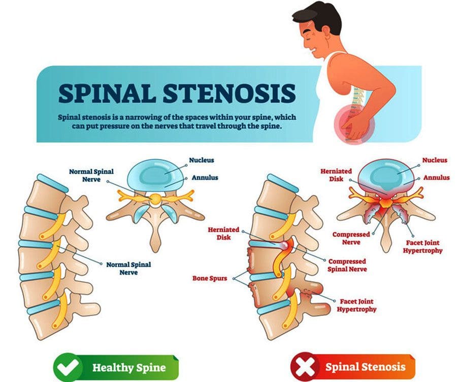
Sciatica is the usual suspect when thigh, hip, buttock, calf, or total leg pain or other sensations are present; however, it could be spinal stenosis with neurogenic claudication. Spinal stenosis is sometimes called pseudo claudication, a narrowing of the space around the low back, which can put pressure on the spinal cord directly and compress the blood vessels around the spine, cutting off oxygen-carrying blood. Pain can start in the lower back and circulate down the legs and cause weakness, tingling, or numbness in the legs and feet. The most common areas of spinal compression include:
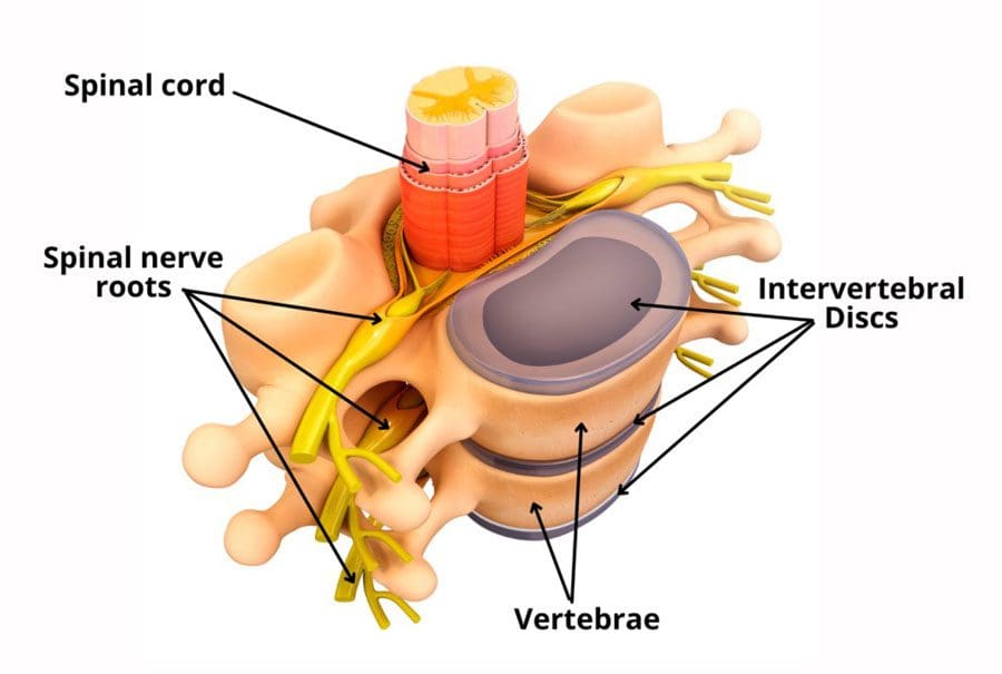
The foramen are the openings on the sides of the spine where nerves exit and connect to the peripheral nervous system.
The narrowing can occur in any of these areas, with the most common cause being lumbar spinal stenosis brought on by lumbar degenerative disease.
Symptoms
The most common symptoms of neurogenic claudication include:
Pain in the lower extremities, including the buttocks, thighs, and calf, only manifests with activities like walking or standing around.
Pain that shows up equally on both sides.
There is no pain when sitting or not walking around.
Radiculopathy or nerve pain that radiates down an affected limb. Sciatica is a typical example.
However, the symptoms of claudication and radiculopathy are different.
Claudication will be felt all along the length of the nerve.
Radiculopathy pain is more localized to the buttock, thighs, and calves and can get worse with activity and is generally present even when at rest.
Treatment
Non-surgical treatment of neurogenic claudication includes medication to help control pain, chiropractic manual therapy, non-surgical spinal decompression, physical rehabilitation therapy, and steroid shots to reduce inflammation. A doctor will recommend stretching, strengthening exercises, and types of activities to help improve the body’s ability to support itself. This could include swimming, walking, and stationary cycling. However, conservative treatment might not be an option for individuals with more severe cases. If conservative treatment options don’t work, surgery could be recommended. A healthcare provider can help explain treatment options. Successful outcomes have been seen in cases that are diagnosed and treated early.
Non-Surgical Spinal Decompression Chiropractor
References
Colak, Ahmet, et al. “A less invasive surgical approach in the lumbar lateral recess stenosis: a direct approach to the medial wall of the pedicle.” The European spine journal: official publication of the European Spine Society, the European Spinal Deformity Society, and the European Section of the Cervical Spine Research Society vol. 17,12 (2008): 1745-51. doi:10.1007/s00586-008-0801-z
Munakomi S, Foris LA, Varacallo M. Spinal Stenosis And Neurogenic Claudication. [Updated 2022 Feb 12]. In: StatPearls [Internet]. Treasure Island (FL): StatPearls Publishing; 2022 Jan-. Available from: https://www.ncbi.nlm.nih.gov/books/NBK430872/
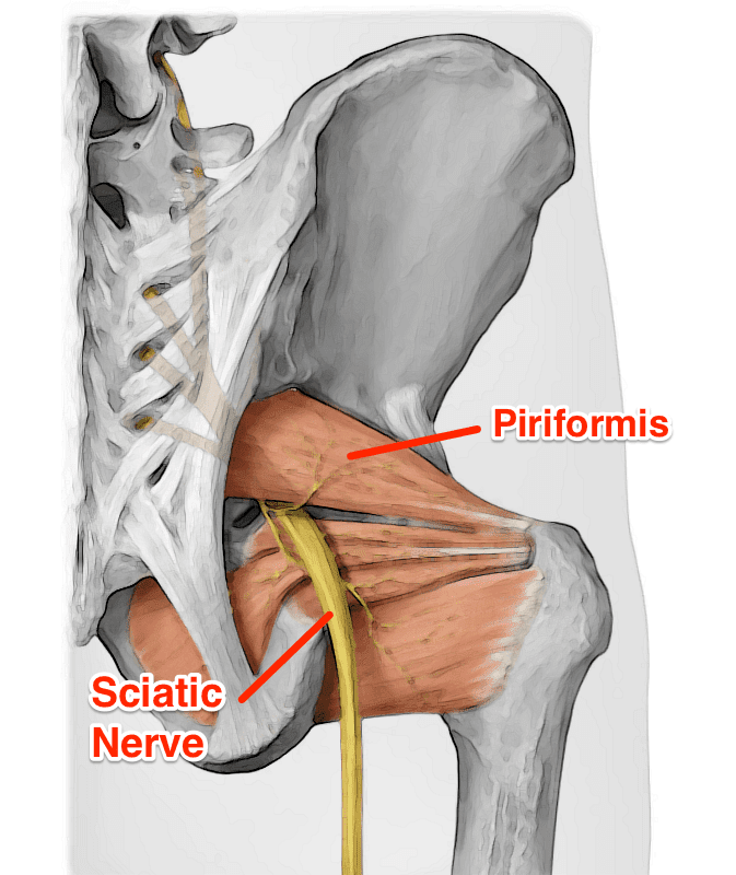
The sciatic nerve is considered the largest in the lower half of the body that helps control sensory and motor functions of the legs. As part of the nervous system, the sciatic nerve resides in the lumbar region of the spine, traveling down to the legs and feet while succumbing to injuries and unwanted factors. When there are injuries or unwanted symptoms that start to affect the lumbar regions of the spine like herniation or a slipped disc, it can press on the sciatic nerve causing sharp, searing pain that can radiate down to the legs and feet. This type of pain can lead to sciatica and dampen a person’s mood if not treated right away. Luckily, there are treatments available for reducing sciatic nerve pain and other issues that affect the body’s lower extremities. Today’s article focuses on a condition that can cause sciatica known as piriformis syndrome, its symptoms, and how decompression therapy can help many people alleviate the sciatic nerve from piriformis syndrome. Referring patients to qualified and skilled providers who specialize in spinal decompression therapy. We guide our patients by referring to our associated medical providers based on their examination when it’s appropriate. We find that education is essential for asking insightful questions to our providers. Dr. Alex Jimenez DC provides this information as an educational service only. Disclaimer
Can my insurance cover it? Yes, it may. If you are uncertain, here is the link to all the insurance providers we cover. If you have any questions or concerns, please call Dr. Jimenez at 915-850-0900.
What Is Piriformis Syndrome?
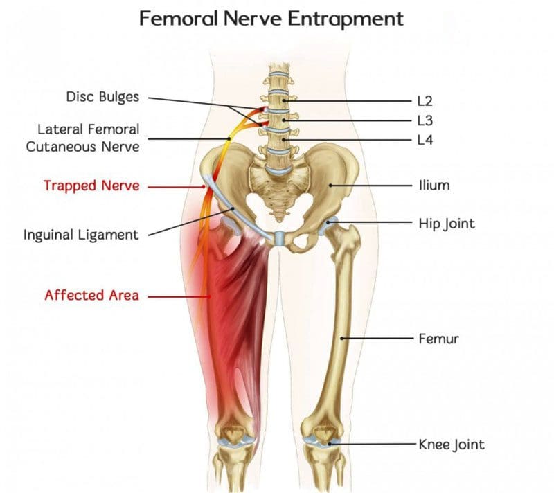
Do you feel muscle spasms occur in your lower back or buttock? How about radiating pain that is traveling down the legs? Do the muscles in the lower body regions feel tender and weak to the touch? Experiencing these symptoms mean that you are suffering from piriformis syndrome. Research studies have defined piriformis syndrome as a condition in which the piriformis muscles in the buttocks region irritate the nearby sciatic nerve, causing it to be trapped. As the sciatic nerve becomes trapped in the piriformis muscle, it can cause sciatica pain-like symptoms that run down the leg. Additional research studies mentioned that since sciatica is a musculoskeletal pain disorder associated with piriformis syndrome, the compressed, irritated sciatic nerve root causes the individual to suffer from painful symptoms that are causing the piriformis muscle to tense up. Piriformis syndrome can affect the sciatic nerve root with or without spinal disorders like herniation, stenosis, or slipped discs.
The Symptoms
When the piriformis muscle aggravates the sciatic nerve, many symptoms can pop up over time, causing painful issues that collide with sciatica and piriformis syndrome. Research studies have shown that piriformis syndrome is a deliberate condition caused by traumatic events, inflammation in the lower back, and spinal degeneration. Most of the causes do hinder a person’s quality of life. Since the sciatic nerve is trapped in the piriformis muscle, it can cause excruciating, burning pain that affects the lower back down to the leg muscles. Other studies have found that other symptoms that are caused by piriformis syndrome are:
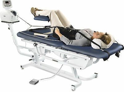
Feeling a limited range of motion on your hips? How about radiating, burning pain that travels down to your feet? Does it hurt to walk up the stairs? Piriformis syndrome can cause sciatica pain-like symptoms that can hinder your ability to walk and function. Decompression therapy can be the solution you are looking for. The video above explains and introduces the DOC decompression table and how it is used to alleviate sciatica pain-like symptoms that are causing pain to the individual. Decompression therapy can help with low back and leg pain by gently pulling the spine to allow the necessary supplements for the spine and to take the pressure off the sciatic nerve roots. Decompression therapy can benefit many individuals suffering from leg pain and who want to get back on their wellness journey. Incorporating spinal decompression as part of your wellness treatment is beneficial. This link will explainhow decompression offers optimal comfort for many people who suffer from piriformis syndrome and get them back to their health and wellness journey.
How Decompression Therapy Can Alleviate Piriformis Syndrome
Since the sciatic nerve is trapped in the piriformis muscle and causes leg pain, some treatments handle piriformis syndrome by decompressing the sciatic nerve. Research studies have found that endoscope decompression surgery can help alleviate piriformis syndrome by relaxing the sciatic nerve to ease the pain from affecting the buttock and leg muscles. For non-surgical decompression therapy, additional research has found that decompression therapy helps widen the spinal disc space in the spine while creating negative pressure in the affected areas. This negative pressure allows the sciatic nerve to relax and reposition the intervertebral disc back in the spine. Decompression treatments combined with physical therapy can even reduce the chances of piriformis syndrome coming back and affecting the sciatic nerve again.
Conclusion
Overall, muscle spasms around the lower body regions can cause piriformis syndrome to develop and cause havoc on the sciatic nerve. Since the piriformis muscle is close to the sciatic nerve, it can trap and aggravate it constantly by sending sciatica pain-like symptoms to the legs. This condition causes muscle weakness and mobility dysfunction in the legs, making a simple walk on the stairs complicated. Treatments like decompression therapy provided in surgical and non-surgical forms can be beneficial for those suffering from piriformis syndrome and sciatica. Decompression therapy allows the negative pressure to release the trapped, irritated sciatic nerve from causing more pain to the legs and helps loosen up the tight muscles in the lower regions of the body. Utilizing decompression as part of your treatment will allow you to continue pain-free your wellness journey.
References
Amjad, Fareeha, et al. “Effects of Non-Surgical Decompression Therapy in Addition to Routine Physical Therapy on Pain, Range of Motion, Endurance, Functional Disability and Quality of Life versus Routine Physical Therapy Alone in Patients with Lumbar Radiculopathy; a Randomized Controlled Trial.” BMC Musculoskeletal Disorders, BioMed Central, 16 Mar. 2022, https://www.ncbi.nlm.nih.gov/pmc/articles/PMC8924735/.
Hicks, Brandon L, et al. “Piriformis Syndrome.” In: StatPearls [Internet]. Treasure Island (FL), StatPearls Publishing, 12 Feb. 2022, https://www.ncbi.nlm.nih.gov/books/NBK448172/.
Hopayian, Kevork, et al. “The Clinical Features of the Piriformis Syndrome: A Systematic Review.” European Spine Journal: Official Publication of the European Spine Society, the European Spinal Deformity Society, and the European Section of the Cervical Spine Research Society, Springer-Verlag, Dec. 2010, https://www.ncbi.nlm.nih.gov/pmc/articles/PMC2997212/.
Revord, John. “Symptoms and Diagnosis of Piriformis Syndrome.” Spine, Spine-Health, 14 Sept. 2012, https://www.spine-health.com/conditions/sciatica/symptoms-and-diagnosis-piriformis-syndrome.
Ro, Tae Hoon, and Lance Edmonds. “Diagnosis and Management of Piriformis Syndrome: A Rare Anatomic Variant Analyzed by Magnetic Resonance Imaging.” Journal of Clinical Imaging Science, Medknow Publications & Media Pvt Ltd, 21 Feb. 2018, https://www.ncbi.nlm.nih.gov/pmc/articles/PMC5843966/.
Vij, Neeraj, et al. “Surgical and Non-Surgical Treatment Options for Piriformis Syndrome: A Literature Review.” Anesthesiology and Pain Medicine, Kowsar, 2 Feb. 2021, https://www.ncbi.nlm.nih.gov/pmc/articles/PMC8241586/.
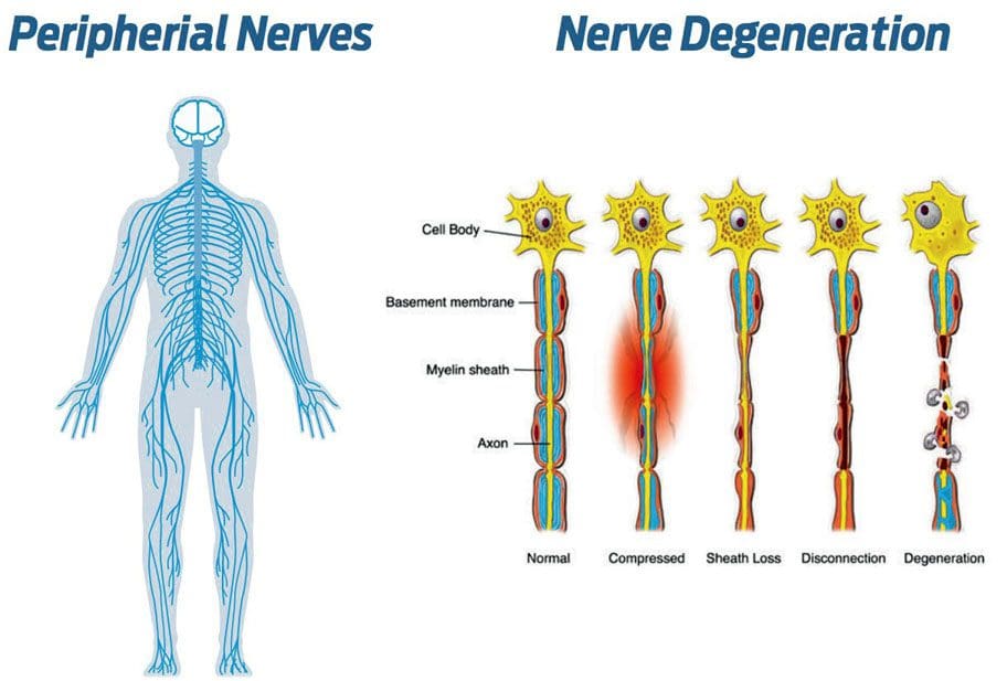
Spinal stress can affect nerve health. Neuropathy happens when disease or damage is sustained in the nerves that transmit messages from the brain through the spinal cord to the whole body. The source of the damage can be inside the spine, where a herniated disc could be squeezing the nerves, impeding or completely blocking blood circulation until deterioration begins to disease or damage nerve receptors. Removing the pressure from the spine and reversing the stress on the nerves can be done through manual or motorized spinal decompression.
Spinal Stress and the Nerves
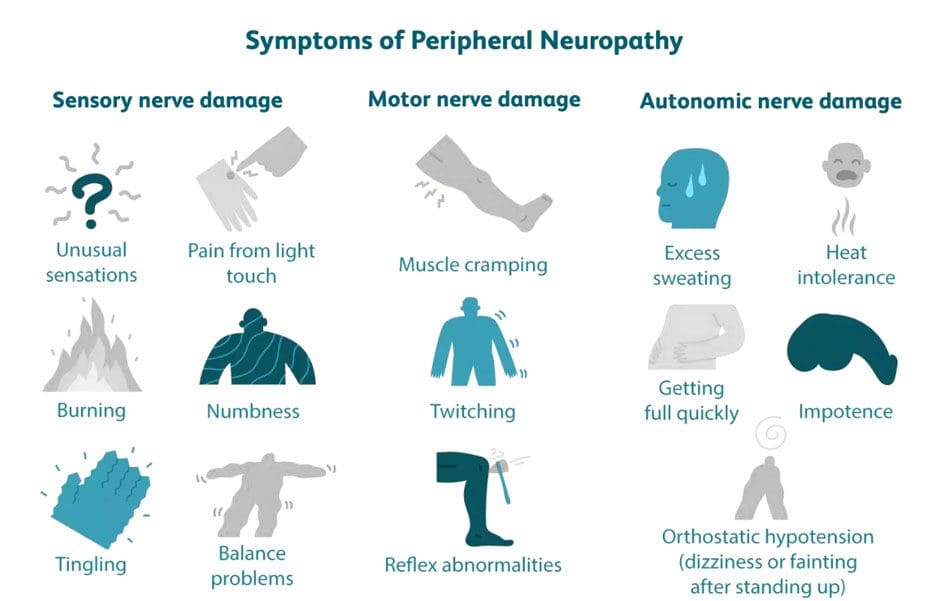
The peripheral nervous system is comprised of three types of nerves that are directly influenced by the central nervous system, each with a distinct function which is why there is a wide range of symptoms associated with neuropathy. The types of nerves include:
Sensory nerves receive sensations from the skin like heat, cold, pleasure, and pain.
Spinal nerves contain sensory and motor fibers giving them sensory and motor functions. The spinal nerves receive sensory messages from the skin, internal organs, and bones. Any disruption from a bent, crushed, or entangled nerve group will not allow proper blood circulation and message transmission, causing delayed responses, tingling, numbness, and pain. If left untreated, it could cause permanent damage that can lead to chronic pain. Decompression therapy accelerates healing as it floods the spine with blood, oxygen, and nutrients.
Peripheral nerves originate from the spinal cord and extend a network of lines throughout the body called dermatomes. Injury to one dermatome can radiate/spread out to other dermatomes and the peripheral areas like the hands and feet. Once communication with the brain is compromised, results can lead to sensations like numbness and severe pain. Several factors can result in peripheral neuropathy, including:
Gordon, Tessa. “Peripheral Nerve Regeneration and Muscle Reinnervation.” International journal of molecular sciences vol. 21,22 8652. 17 Nov. 2020, doi:10.3390/ijms21228652
Menorca, Ron M G et al. “Nerve physiology: mechanisms of injury and recovery.” Hand clinics vol. 29,3 (2013): 317-30. doi:10.1016/j.hcl.2013.04.002
Wang, Mark L et al. “Peripheral nerve injury, scarring, and recovery.” Connective tissue research vol. 60,1 (2019): 3-9. doi:10.1080/03008207.2018.1489381
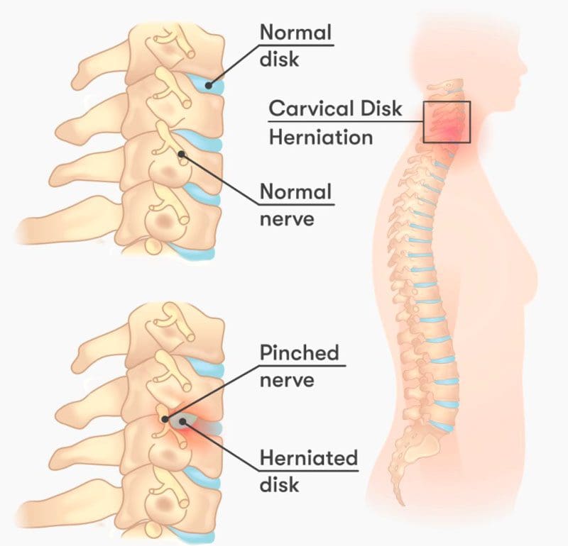
A pinched nerve may not feel like it is healing. This is because of the soreness, aches, discomfort, and tingling feelings/sensations around the affected area. This could be the neck, shoulder, arm, hands, back, legs, and feet. However, when the achiness and tingling move around and shift, it is a sign of the pinched nerve healing.
Amount of Time For Pinched Nerve Healing
Waiting for the nerve to heal is not a recommended treatment option, as most pinched nerves do not fully recover on their own. A pinched nerve usually takes around six weeks to heal with proper treatment. The longer the nerve stays pinched, the more likely there will be permanent damage. To keep the pinched nerve from returning and getting worse, individuals are recommended to incorporate a pre-habilitation plan that involves continuing rehabilitation exercises to strengthen and keep the muscles, ligaments, and nerves loose, and adjusting posture, work, exercise, and diet habits to prevent re-injuring the nerve or cause new injury/s.
Common Nerve Sites
Nerves run throughout the body, so it’s possible to experience a pinched nerve anywhere. The most common pinched nerve sites occur at joints where there is constant movement. These areas include:
Neck
Shoulders
Lower Back
Arms
Hands
Feet
Healing Signs
Individuals often believe that their pinched nerve is getting worse because of soreness, aches and pains, and weird sensations. When the pain stays in one area, that could be a sign that the nerve has not been fully stretched/released and/or that there is still compression taking place. Treatment and healing include feeling the symptoms but in a different way. The symptoms will move up, down, or around depending on where the pinched nerve is. Treatment takes the nerve/s and stretches/elongates them, but the pinch created a nerve crimp, crease, fold that wants to return to the pinched position. This is why continued treatment and stretching are recommended, as a spasm, trauma, or some awkward movement can cause the nerve to re-fold to the pinched position or cause a whole new pinch.
Chiropractic Release
Chiropractic treats pinched/compressed nerves with several therapeutic modalities. These include:
Body Adjustments
Flexion-distraction
Therapeutic massage
Traction
Inversion
Laser therapy
Ultrasound
Combined, these methods can help heal pinched nerves and keep them from recurring.
Body Composition
Skeletal Muscle
Skeletal muscle is a major muscle group. These muscles are attached to the bone by the tendons. Skeletal muscles incorporate nerves, blood vessels, and connective tissue to operate as a unit. Each skeletal muscle consists of cells that come together that form bundles of skeletal muscle fibers.
Strength training stimulates the muscle fibers. When combined with proper nutrition causes hypertrophy/muscle growth.
Muscles contract and shorten to pull bones and joints, allowing body movement.
The nervous system signals the nerves in the muscle/s and triggers these contractions.
Skeletal muscle helps the body:
Maintain posture
Generate body heat
Stability to the bones and joints
References
Bowley, Michael P, and Christopher T Doughty. “Entrapment Neuropathies of the Lower Extremity.” The Medical clinics of North America vol. 103,2 (2019): 371-382. doi:10.1016/j.mcna.2018.10.013
Campbell, W. “Diagnosis and management of common compression and entrapment neuropathies.” Neurologic clinics vol. 15,3 (1997): 549-67. doi:10.1016/s0733-8619(05)70333-9
England, J D. “Entrapment neuropathies.” Current opinion in neurology vol. 12,5 (1999): 597-602. doi:10.1097/00019052-199910000-00014
Kane, Patrick M et al. “Double Crush Syndrome.” The Journal of the American Academy of Orthopaedic Surgeons vol. 23,9 (2015): 558-62. doi:10.5435/JAAOS-D-14-00176
Neuroregenerationcould become an option for spinal cord injury treatments in the future. A spinal cord injury or SCI is when there is damage to the bundle of nerves and cells that send and receive signals from the brain and body. A spinal cord injury can be caused by direct trauma/injury to the cord or damage to the tissue and vertebrae. The damage can result in temporary or permanent changes in:
Sensation
Movement
Strength
Body function/s below the injury site.
There are incomplete and complete injuries. Injuries that cause limited or no cell death can achieve a full recovery. Injuries that are more serious and/or are higher on the spinal cord can cause permanent damage and/or paralysis. Automobile crashes, accidents, and serious falls are the most common causes of spinal cord injuries.
An incomplete injury means the cord can still transmit messages, but there is interference/disturbance.
A complete injury means communication and motor function/voluntary body movement is not transmitting.
Symptoms
Symptoms of a spinal cord injury include:
Unnatural or awkward positioning of the spine or head.
Pain or pressure in the head, neck, or back.
Numbness
Tingling
Loss of or changes in sensation in the hands and feet.
Problems with walking.
Weakness or inability to move parts of the body.
Loss of movement.
Paralysis can occur immediately or develop over time as swelling and bleeding affect the cord.
Loss of bladder and bowel control.
Changes in sexual function.
Difficulty breathing.
SCI Damage Control
A spinal cord injury affects the central nervous system, the body’s central headquarters. Damage can cause complications through what’s called the secondary injury cascade, which is a series of chemical reactions the body activates to help the situation. However, if the chemical response does not stop and stays active, it can worsen the injury. The body recognizes that an emergency has occurred and tries to go into a shut-down mode that kills off some of the cells in the central nervous system. When a spinal injury happens, treatment focuses on stopping the damage as quickly as possible to stop the injury cascade and prevent as much cell death as possible. This act is called neuropreservation, meaning that the team is trying to preserve and save as many nerve cells as possible.
Injury Neuroregeneration Treatment Studies
While current treatment primarily focuses on stopping as much damage as possible then going through physical therapies to maintain spinal alignment and rehabilitate the body, the future of injury treatment is looking towards regrowing and repairing the damaged nerve cells through a process known as neuroregeneration. Repairing nerves that have been damaged could change life for many. Neuroregeneration Treatments being studied include:
Surgery
A study in The Lancet Neurology presents how getting surgery as soon as possible after an injury can provide significant benefits.
The findings could change all of the guidelines for spinal cord injury.
Medication
A study on Riluzole, a medication that has shown promise to slow down nerve cell damage.
A team completed a randomized controlled trial for the medication; soon, the final results will be available.
Scientists are studying ways to grow new nerve cells from an individual’s stem cells without the need for embryonic stem cells.
Specialized stem cells could also be used to help other nerve cells regenerate.
Electrical stimulation
Another approach is using electrical stimulation to restore function in the spinal cord.
Therapy that could help a paralyzed individual walk again.
The Future of Neuroregeneration
Aside from early surgery intervention, most neuroregenerative treatments are not ready or accessible yet. There’s still much more research before it can become a mainstream treatment option. Treatment that involves regenerating nerve cells will take longer than a treatment designed to protect nerve cells. However, more clinical trials are expected to be done in the next few years, with stem cell therapies taking the longest. Some of these therapies could be ready to be used on actual patients in 5-10 years.
Body Composition
The Importance of Measuring Body Composition
Most diet and fitness programs focus on weight loss or gain. However, they tend to overlook that individuals have completely different body compositions. Body composition describes the amount of:
Fat
Bone
Water
Muscle
In the body.
Measuring body composition can tell a body’s unique makeup and help identify areas to work on to improve overall health and wellness. Body composition analysis provides a snapshot of an individual’s health/fitness levels to help achieve health goals from the inside out.
References
Aguilar, Juan et al. “Spinal cord injury immediately changes the state of the brain.” The Journal of neuroscience: the Official Journal of the Society for Neuroscience vol. 30,22 (2010): 7528-37. doi:10.1523/JNEUROSCI.0379-10.2010
Badhiwala, Jetan H; Wilson, Jefferson R; Witiw, Christopher D; et al. (February 2021). The Lancet Neurology Vol. 20, No. 2, P. 117. The Influence of Timing of Surgical Decompression for Acute Spinal Cord Injury: A Pooled Analysis of Individual Patient Data. DOI: 10.1016/S1474-4422(20)30406-3
Chari, Aswin et al. “Surgical Neurostimulation for Spinal Cord Injury.” Brain sciences vol. 7,2 18. 10 Feb. 2017, doi:10.3390/brainsci7020018
A nerve injury is often caused by a sudden traumatic event, like a slip and fall, personal or work injury, an automobile accident, or a sports injury. Overall stresses of the body from poor posture and being overweight can also lead to nerve pain over time, known as cumulative trauma. Where ligaments and bones are not aligned correctly, nerve pain and damage can occur. When nerve pain presents, there is pressure being placed on that nerve/s. Nerve pain symptoms include burning, tingling, or numbness-type sensations in the tissues controlled by that nerve. Orthopedic and neurologic testing will determine what specific nerve is affected. Chiropractic adjustments realign the spine and relieve the pressure on the nerve, thus eliminating the pain and correcting the problem.
Nerve Injury
Too much pressure from surrounding tissues compresses and irritates the nerve and interrupts its ability to function correctly. Pinched nerves are most vulnerable at points in the body where they pass through narrow spaces and have little to no soft tissue protection. Symptoms include:
Pins and Needles Sensation
Numbness
Pain
Weakness
A pinched nerve can decrease the range of motion and cause muscle spasms. If left untreated, a nerve injury can leave an individual with chronic pain and lead to permanent nerve damage.
Tingling and Numbness
Tingling and numbness are unusual or unpleasant physical sensations, most commonly experienced in the arms, hands, fingers, legs, feet, and toes. Tingling and numbness come in two forms:
To determine the appropriate course of treatment, a doctor of chiropractic must diagnose the cause of the nerve injury. Depending on the nature or severity of the sensation, the examination will include:
Muscle tests
Range-of-motion tests
Neurological tests
Orthopedic tests
The chiropractor will palpate the effective areas and order imaging tests like X-rays if necessary. If further testing is needed to diagnose the source of the nerve injury, the doctor may order an MRI or CT scan. Once the underlying condition is diagnosed, a chiropractor will develop a treatment plan to eliminate irritation, correct misalignments causing pressure, and restore proper nerve function. Treatment plans vary from case to case but can include:
Therapeutic Massage
Body adjustments
Spinal manipulation
Heat and Ice
The objective is to relieve/release the pressure on the nerves. Chiropractic adjustments help reposition the muscles and nerves. Deep-tissue massage helps to release tension and eliminate toxins that worsen the sensations. Treatment improves circulation and relieves pressure on the neural pathways necessary to restore normal neural signaling between the body and the brain.
Body Composition
Why The Brain Needs Sugar
The brain needs half of all the body’s energy supply because of its complex nerve cell system. The brain requires glucose for brain cell energy. Because neurons can’t store energy, they need a continuous fuel supply to function correctly from the bloodstream. The ability to think, learn and recall information is closely associated with glucose levels. When blood glucose levels are low, the ability to think is inhibited as the production of chemical messengers/neurotransmitters, are reduced, disrupting communication between the neurons. Natural sugar can boost brain health because it requires glucose for functioning. Sugar is released slowly into the bloodstream when taken naturally from sources like apples and bananas, keeping the energy levels steady, without craving more sugar.
References
Ameh, Victor, and Steve Crane. “Nerve injury following shoulder dislocation: the emergency physician’s perspective.” European journal of emergency medicine: official journal of the European Society for Emergency Medicine vol. 13,4 (2006): 233-5. doi:10.1097/01.mej.0000206190.62201.ad
Nichols, J S, and K O Lillehei. “Nerve injury associated with acute vascular trauma.” The Surgical clinics of North America vol. 68,4 (1988): 837-52. doi:10.1016/s0039-6109(16)44589-5
Ruggiero, S L. “Trigeminal nerve injury and repair.” The New York state dental journal vol. 62,8 (1996): 36-40.
Welch, J A. “Peripheral nerve injury.” Seminars in veterinary medicine and surgery (small animal) vol. 11,4 (1996): 273-84. doi:10.1016/s1096-2867(96)80020-x
WOODHALL, B. “Peripheral nerve injury.” The Surgical clinics of North America (1954): 1147-65. doi:10.1016/s0039-6109(16)34299-2
Neuropathy is a painful condition that causes tingling, numbness, burning sensations in the hands and feet, and other symptoms throughout the body. Neuropathy can make life difficult. There is no cure for neuropathy, but symptoms can be managed with medications, antidepressants, anticonvulsants, and pain relievers. Another treatment option to help relieve neuropathy symptoms is chiropractic.
Symptoms
Symptoms vary from individual to individual depending on their health condition and how the nerves have been impacted. Common symptoms include:
Pain
Numbness
Tingling
Pins-and-needles feeling when touching something hot or cold.
Some individuals lose the sense of feeling like clothing on their body, even though it’s rubbing against the skin but feel as if it is not there.
Other changes can be familiar objects looking different than usual.
Lessened or heightened sense of smell.
Negative impact on mood.
Protective Sheathing Of The Nerves
Neuropathic pain is caused by damage and degeneration to the nerves or the protective covering/sheathing of the nerves. Various causes include:
Diabetes.
Injury.
Infections.
Medication side effects.
Exposure to toxins.
Stages
The symptoms of neuropathy depend on the location and severity of the nerve damage. The stages include:
Numbness and Pain
Stage one consists of numbness and pain.
Some individuals describe a tingling or numbing sensation.
What feels like pinpricks in the hands and/or feet.
This stage can last for months, but most individuals recover within a year.
Constant Pain
Stage two is characterized by continuous pain.
Some individuals may experience shooting pains that come and go.
Intense burning sensations around the waistline.
Numbness on one side of the body with stabbing pain.
This stage can last for a year or more and worsen until the individual is incapacitated.
Loss of motor skills like walking and falling over.
Doctors treat the symptoms so they don’t get worse.
Loss Of Sensation
The final stage is the loss of sensation.
This occurs when the nerve endings are destroyed and can no longer send messages to the brain.
Treatments To Help Relieve Symptoms
Treatments usually involve:
Antidepressants.
Pain medications.
Anti-seizure medications.
Pain-relieving creams.
All can help manage pain and inflammation.
Chiropractic Can Also Help Relieve Symptoms
Chiropractors use hands-on methods to adjust and realign joints, muscles, spinal discs, and ligaments to function more efficiently and bring relief from pressure on the nerves. Neuropathies are often caused by nerve compression in body areas that have been altered by injury or disease that affects ligaments, discs, spinal muscles, sacroiliac joint dysfunction, hip adhesions, leg length discrepancies, etc. These can contribute to pain and numbness in the peripheral nerves that supply the legs, feet, arms, hands, and neck. While a chiropractor cannot cure neuropathy, they can help relieve symptoms, make it much more manageable, and improve quality of life.
Body Composition
Common Cold
The common cold, also known as upper respiratory tract inflammation, is the most common infectious respiratory disease because of its effect on the nose and throat. The average adult will catch 2–3 colds a year, according to the CDC. A virus that causes a cold can enter the respiratory tract directly when inhaling droplets expelled from an infected person or by direct skin contact, like touching the face with a hand that came in contact with the virus. Cold symptoms vary but usually include:
Runny or stuffy nose
Sneezing
Coughing
Headaches
Body aches
The duration of a cold differs; however, most individuals with a healthy immune system recover in 7–10 days. However, individuals with a compromised immune system, asthma, or COPD have an increased risk of developing more serious illnesses like bronchitis or pneumonia. Hundreds of viruses can cause colds. Human Rhinoviruses are common culprits and are constantly mutating, which is why there is no cure. Several medications or natural treatments help alleviate cold symptoms; it is recommended to combat the illness effectively through a healthy immune system response. Doctors recommend proper rest, eating a nutrient-rich diet, and maintaining proper H2O hydration to boost the immune system.
References
D’Angelo, Kevin et al. “The effectiveness of passive physical modalities for the management of soft tissue injuries and neuropathies of the wrist and hand: a systematic review by the Ontario Protocol for Traffic Injury Management (OPTIMa) collaboration.” Journal of manipulative and physiological therapeutics vol. 38,7 (2015): 493-506. doi:10.1016/j.jmpt.2015.06.006
Kissel, Jaclyn A, and Cristina Leonardelli. “Isolated musculocutaneous neuropathy: a case report.” The Journal of the Canadian Chiropractic Association vol. 63,3 (2019): 162-170.
Passioti, Maria et al. “The common cold: potential for future prevention or cure.” Current Allergy and asthma reports vol. 14,2 (2014): 413. doi:10.1007/s11882-013-0413-5
T Francio, Vinicius. “Chiropractic care for foot drop due to peroneal nerve neuropathy.” Journal of bodywork and movement therapies vol. 18,2 (2014): 200-3. doi:10.1016/j.jbmt.2013.08.004
IFM's Find A Practitioner tool is the largest referral network in Functional Medicine, created to help patients locate Functional Medicine practitioners anywhere in the world. IFM Certified Practitioners are listed first in the search results, given their extensive education in Functional Medicine