Dietitians at Integrative and Functional Medicine (DIFM) is a specialty clinic group of nutrition professionals whose center philosophy centers around a holistic, personalized approach to healing and health. Members incorporate many different nutrition remedies such as brain/body modalities in clinical practice supplements and whole foods.
How is nutrition a part of integrative and functional medicine?
Integrative medicine is the practice of medicine that reaffirms the value of the connection between professional and patient, focuses on the entire individual, is informed by evidence, and makes use of all appropriate therapeutic approaches, healthcare professionals, and areas to achieve optimal health and healing.
Functional medicine addresses the underlying causes of illness utilizing a systems-oriented approach and engaging both practitioner and patient at a healing partnership. Functional medicine involves understanding complex, chronic disease’s origins, prevention, and therapy. Hallmarks of a functional medicine approach include:
Patient-centered care. The focus of functional medicine is on care boosting health beyond the absence of illness; as a positive energy. By listening to the patient and studying his or her narrative, the practitioner tailors treatments that address the unique needs of the individual and brings the individual relief.
An integrative, science-based health care strategy. Functional medicine professionals seem “upstream” to think about the intricate web of interactions in the individual’s history, physiology, and lifestyle that can cause disease. The distinctive genetic makeup of each individual is considered, combined with both internal (brain, body, and soul) and external (physical and social environment) variables that affect overall functioning.
Integrating best medical clinics. Functional medicine integrates traditional Western medical practices with what is sometimes regarded as “choice” or “integrative” medicine, creating a focus on prevention through nutrition, diet, and exercise; use of the latest diagnostic techniques; and prescribed combinations of drugs and/or botanical medicines, supplements, therapeutic diets, detoxification programs, or stress-management practices.
Functional Medicine and Nutrition
Functional medication has, for years, been the promoters and teachers of using food as medicine. According to the Institute for Functional Medicine, “functional nutrition highlights the value of top quality meals and phytonutrient diversity to deal with clinical imbalances and move people toward the maximum expression of health. Advanced nutrition evaluation and a comprehensive functional medicine based history leads to a personalized therapeutic intervention made to promote optimal health and protect against diet- and lifestyle-related disease.”
Integrative and functional medicine nutritionists have been uniquely trained and have many years of experience incorporating the art and science of integrative and functional nutrition treatment. They understand that giving you a standard diet plan based on a diagnosis is just not enough to foster optimum recovery. After all, one size does not fit all and they strive to provide the maximum quality of individualized nutrition care to all of our patients.
Practice-Based Evidence for Nutrition
Practice-based evidence promotes the worth of their wisdom and evidence gained by the professional’s clinical observations and experiences. The Dietitians in the Integrative and Functional Medicine (DIFM) practice group of the Academy of Nutrition and Dietetics has developed Standards of Practice (SOP) and Standards of Professional Performance (SOPP) in Integrative and Functional Medicine. The SOP addresses the Nutrition Care Process (NCP) and actions related to person-centered care. The SOPP are statements that describe a level of behaviour in the role.
The Integrative and Functional Medicine Nutrition Therapy (IFMNT) Radial was established within an integrated conceptual framework to assist in IFMNT practice. The structure of this IFMNT Radial allows for the analysis of interrelationships and interactions. The Radial depicts that food is still a source of information that affects, and is influenced by, the five areas also as a factor in disease and health.
The five key areas include: lifestyle, systems (signs and symptoms), heart imbalances, metabolic pathways, and biomarkers. Surrounding the Radial are currently precipitating. The SOP, at the June 2011 issue of the Journal of the American Dietetic Association, is along with the IFMNT Radial.
The scope of our information is limited to chiropractic and spinal injuries and conditions. To discuss options on the subject matter, please feel free to ask Dr. Jimenez or contact us at 915-850-0900 .
By Dr. Alex Jimenez
Additional Topics: Wellness
Overall health and wellness are essential towards maintaining the proper mental and physical balance in the body. From eating a balanced nutrition as well as exercising and participating in physical activities, to sleeping a healthy amount of time on a regular basis, following the best health and wellness tips can ultimately help maintain overall well-being. Eating plenty of fruits and vegetables can go a long way towards helping people become healthy.
Ergonomics in the work place. Back pain is one of the most frequent work-related injuries and is often brought on by ordinary work activities like sitting in an office chair or heavy lifting.� the study of the workplace as it relates to the worker – helps prevent back pain and back injury and help maintain a healthy back.
The objective of an ergonomics program in business is to adapt the workplace to a specific worker, determined by the job description, required tasks, and physical make up of the employee performing these tasks.
Non-accidental injury, where pain occurs as a result of normal activities and needs of the task. This might occur from sitting in an office chair or standing for too long in one position.
Accidental injury results when an unexpected event triggers injury during the task. A load that changes as it is being lifted, and fall and a slip or hitting one’s head on a cabinet door or slips are typical examples. These injuries can jolt other joints, back, and the neck with consequent muscle strain or tearing of soft tissue at the back.
Occupations which are physically demanding and require repetitive lifting (such as in nursing or heavy industry) are at greatest risk for both non-accidental and accidental spine injury. For instance, a number of health workers have problems because patients are of weight and different stature with needs. Often, the patients need help changing position, rising from a chair and walking. Similarly, the physical effort needed to release a trapped individual or save a life is unpredictable. The same problems occur in the building industry where consistencies of tasks are a challenge.
Office Chair Back Injuries
Individuals who sit most of the day, like those working in a computer while sitting in an office chair, are also at high risk for non-accidental spine injury. Office ergonomics, or computer ergonomics, can help minimize the risk such as the dangers associated with prolonged sitting in an office chair, and carpal tunnel syndrome, such as lower back pain, neck strain, and leg pain.
Office Chair: Ergonomics To Reduce Back Pain?
This guide summarize the use of ergonomic concepts, mechanical apparatus and decent body mechanics (biomechanics) that can contribute to reducing back injuries in the work environment for several jobs. Significantly, staying strong, physically fit and flexible improves the probability of preventing back injuries.
There are certain basic ergonomic tips which may help an employee avoid back pain or back injury:
Develop a job description based on the forces within a particular work environment, the time spent performing the task and the biomechanics (which define human moves and seated posture in an office chair) used in the task.
Use body posture as a tool which may be changed to fit the job demands with minimal stress on the muscles, ligaments, bones and joints.
Learn and use proper body mechanics to restrict extra mechanical stress in completing the job.
Maintain fitness and flexibility and create a reserve of strength.
Identifying Poor Posture And Risks
Many potentially harmful situations that lead to back injury can be identified and avoided by following four basic rules of thumb:
Prolonged static posture is your enemy. The healthy body can only tolerate staying in one position for around 20 minutes. That is sitting at a movie theatre, in a desk in an office chair, or on an airplane becomes uncomfortable after a short time. Standing in one area, such as standing on a floor at an assembly line tends to cause back pain. Holding the same position gradually reduces elasticity in the soft tissues (muscles, ligaments, and tendons in the back). Stress builds up and causes discomfort and/or leg discomfort back.
The remedy is simple. Whether you are sitting in an office chair or standing in a line, change positions frequently. Just move. Stand or sit, stretch, have a short walk. After returning to the standing or sitting posture, use an alternate posture for only a couple minutes and some.
Frequent or repetitive stretching to the end range of motion or embarrassing, angled positions can liquefy the joints. Unlike jobs that need seating in an office chair, jobs that require motion can cause discomfort. Such tasks involve lifting overhead lifting from the floor, moving loads, or utilizing force or twisting while managing material and which signal back injuries might be on the way.
Heavy loads offer greater risk. It is important to have the proper tools or get help if the job requires moving objects.
Fatigue�from sitting in an office chair, from work or from insomnia can make people move more awkwardly. If one is overtired or feels fatigued, it is advisable to avoid lifting heavy objects alone or quickly.
These ergonomic rules of thumb will help the worker and their backs. Otherwise the worker is at risk of sustaining or aggravating a back injury.
“The doctor of the future will give no medicine, but will teach his patient in the care of the human frame, in diet and in the cause and prevention of disease”. These wise words were uttered by Thomas Edison more than a hundred years back at the start of the 20th century, the century which could become the conventional system’s expansion we know today regarding alternative treatment options and functional medicine.
What is the future of health care?
The past century has been in many ways an entire departure from Edison’s words. Away from looking at the foods we eat and prevention and toward the Big Pharma, sick care system: diagnose a disease and match it with a corresponding drug. Despite spending more on healthcare than the next 10 top-spending nations combined, the U.S. has the most persistent illness cases and shortest life span of all industrialized nations.
In accordance with a comprehensive study by the Institute of Medicine and the National Research Council, American men rated last in life expectancy, and women were ranked second to last. When it comes to the health of a pregnant mother or her child, the U.S. has the maximum mortality rate among all developed countries. The U.S. also has the third-worst mortality rate from nutritional deficiencies, and also the greatest total age-adjusted mortality rate among all developed nations.
Despite the wonderful progress in life saving surgeries and emergency care, when it comes to chronic health ailments and longevity, we are doing something abysmally wrong. Seeing the shadow surrounding the medical care system, functional medicine has been attempting to supply a solution to this issue.
Yet even today, a lot of people don’t fully comprehend what functional medicine is, or why they need to try it. Well, here are six compelling reasons to give functional medicine a try.
Functional Medicine Interprets Results Differently
On your labs there is typically a reference range that tells you what’s considered “normal.” Anything out of this reference array is typically labeled as “high” or “low” in bold font. A statistical bell curve typical of that specific lab’s populace determines that reference range.
If your laboratory is one number away from being out of the benchmark range, you’re still categorized as “normal.” But disease doesn’t start as soon as you’re out of that diagnostic range. Unfortunately, this means you’re either, trending toward disorder, outside of the benchmark array, or you’re trending toward health.
There are a whole lot of people who go to a healthcare professiona, only to discover why they are struggling with health issues, and their labs return “normal.” They leave without any answers or remedies and’re told there is not anything wrong with them. A medication lab analysis sheds light on unanswered health concerns that fall through the cracks of the standard model of maintenance.
Functional Medicine Provides More Diagnostics
In addition to interpreting the labs that mainstream medical professionals run, functional medicine goes past the label of this disease to check out the full extent of a patient’s physiology. So that it can be paired with a pharmaceutical drug, typically a disease is diagnosed by the labs at the standard model of care. They’re very incomplete from a functional medicine perspective, although the labs your doctor runs are adequate if he is prescribing medications.
Comprehensive labs to examine underlying deficiencies, imbalances, infections and dysfunctions, give excellent insight into these frequently overlooked portions of your health issues.
Functional Medicine Customizes Healthcare
As soon as you’re tagged with a disorder in mainstream medicine, you are given the very same medications everyone else with this disease is given. This cookie cutter strategy works occasionally, but more often than not, it fails miserably. Medication takes into account that we designed a bit differently, so what works for one person may not always be right for you. A detailed health plan that is tailored addresses that you are seeing results.
Functional Medicine and Patients
Mainstream medicine is really bogged down with symptom care. That is why every six months you wait an hour at your physician’s office for a regular visit. This system is failing millions of Americans suffering from chronic health conditions.
In terms of crisis care, emergencies and surgeries, we have among the very best health care systems in the world, but in regards to chronic healthcare, the U.S. is still one of the worst industrialized health care systems in the world.
Functional Medicine and Addressing Dysfunctions
While mainstream medicine is structured to manage symptoms, functional medicine is primarily concerned with addressing the underlying dysfunctions of the body that give rise to symptoms. If someone has high blood sugar, he’s typically medicines that stimulate the pancreas to produce more insulin, which attracts down the blood sugar.
Functional medication asks why a patient has high blood glucose in the first place. Very rarely is somebody sick from a medication deficiency. They may have cellular insulin resistance, brain-adrenal axis dysfunctions causing cortisol along with a gut disease leading to their own blood sugar dis-balance.
In this example there is nothing actually wrong with the pancreas, therefore while the medicine will make their blood sugar numbers seem nicer on a laboratory, it does not address the reasons they’re high in the first place. So functional medicine may be the missing link to getting healthy and getting off drugs and medications.
Remedies Are Essential in Functional Medicine
Functional medicine is not anti-medication, however, practitioners may ask what the individual’s most effective alternative is, and that which causes the fewest side effects. It’s maybe the smartest choice, if a drug fits that criteria. However, it often is not.
Hippocrates, the father of modern medicine, said tens of thousands of years back, “Let food be thy medicine, and medicine thy food,” and practical medication agrees. Food is used as medicine in a state specific manner. Herbal and micronutrient medicines are used to address the dysfunctions found on the labs to encourage healthy function.
It’s important to not forget, though, that even with natural choices and foods, what works for one person might not work for the next. We need to tailor the program for the individual instead of being the alternative model of mainstream medicine, using its magical pills and “one-size-fits-all” approach.
This new era of evidence-based all-natural health care is providing hope for all. As a result of new technologies, webcam consultations with people give the exceptional opportunity to share functional medicine. Edison’s words are coming true. Let the future begin.
The scope of our information is limited to chiropractic and spinal injuries and conditions. To discuss options on the subject matter, please feel free to ask Dr. Jimenez or contact us at 915-850-0900 .
By Dr. Alex Jimenez
Additional Topics: Wellness
Overall health and wellness are essential towards maintaining the proper mental and physical balance in the body. From eating a balanced nutrition as well as exercising and participating in physical activities, to sleeping a healthy amount of time on a regular basis, following the best health and wellness tips can ultimately help maintain overall well-being. Eating plenty of fruits and vegetables can go a long way towards helping people become healthy.
Chronic pain can pose many problems. Traditional medicine delivers a broad selection of treatment options, some more powerful than others. And these choices have a wide spectrum of benefits which may not always be best for everyone.
Should you consider alternative treatments for chronic pain?
There are a variety of traditional medicine treatments and procedures for treating chronic pain. For many pain management specialists, treatment may include steroid injections to decrease inflammation. For others, treatment might suggest procedures, such as narcotics, drugs or medications, to reduce the pain signals.
But, these remedies are only designed to temporarily relieve the symptom of pain. You must return again in a couple of weeks for one more prescription or shot refill. While these remedies can be effective at pain management,they also carry effects that can display as adverse effects to the patient
Common pain control drugs such as oxycodone and hydrocodone are known to have long-term unwanted side effects. Most commonly we think of this extremely addictive nature of the drugs, and our body becomes determined by the drugs to operate without pain. Research has also confirmed the cost that these drugs take in your liver and endocrine system. Levels are disrupted that can start a domino effect that disrupts you mood, metabolism, sleep, and sexual role, among other things. When these types of treatments are not preferred, other treatments should be considered.
How Functional Medicine Differs from Traditional Medicine
Functional medicine has proven tremendously effective at combating chronic pain because of its main focus: Concentrate on the underlying cause, instead of just masking the symptom.
When using medications to dull or numb pain, effectively you are disrupting the signals being transmitted to your brain so that your brain doesn’t feel the pain. But instead of numbing the pain, functional medicine focuses first on determining the source of the individual’s painful symptoms.
Functional Medicine doctors work with patients everyday who deal with chronic pain on a regular basis. Many of these patients believed that they would devote the rest of their lives on tablets and injections and surgical processes to temporarily alleviate the pain. When it is a patient who’s dealing with arthritis, that is truly an autoimmune illness, or somebody who is recovering from an auto accident and has been through numerous surgeries, functional medicine includes a regimen of non-invasive treatments that heal you from the inside out.
At precisely the exact same time, it has been recognized that there’s a time and place for more advanced medical care that may require invasive processes. This should not be your first plan of action.
Allow qualified and experienced healthcare professionals to design a customized program that unites their foundational understanding of chiropractic care together with the quickly-developing research behind functional medicine. Understanding how your system integrates with other body systems is the secret so that you can simply live pain free, breaking free from the cycle of chronic pain and find proper pain management treatments.
The scope of our information is limited to chiropractic and spinal injuries and conditions. To discuss options on the subject matter, please feel free to ask Dr. Jimenez or contact us at 915-850-0900 .
By Dr. Alex Jimenez
Additional Topics: Wellness
Overall health and wellness are essential towards maintaining the proper mental and physical balance in the body. From eating a balanced nutrition as well as exercising and participating in physical activities, to sleeping a healthy amount of time on a regular basis, following the best health and wellness tips can ultimately help maintain overall well-being. Eating plenty of fruits and vegetables can go a long way towards helping people become healthy.
According to the American College of Preventive Medicine, most chronic diseases are preventable and reversible if a comprehensive, individualized approach that addresses genetics, diet, stress, physical activity, and sleep is implemented through integrated functional medicine teams and based on empirical research.
What are the functional medicine treatment approaches?
In this way, health is perceived as more than the absence of illness, just as illness is more than the absence of health.�In order for the body to live up to this principle, it needs to be supplied with the necessary nutrients through a healthy diet, adequate sleep, movement/exercise, and management of stress.
Functional Medicine Approach #1 – A Elimination Diet
Remember that every time someone eats, that changes body chemistry. A functional medicine clinic often guides patients to implement a modified removal diet. Patients are educated to remove certain foods from their diet, such as those containing gluten or dairy, and are encouraged to adjust (increase) the consumption of fruits and vegetables that encompass every color of the rainbow. Patients are advised to remove all added sugars. This practice is often difficult for people; therefore, the FM team must work to encourage their compliance with the elimination diet.
Utilization of an elimination diet requires a patient to remove the most frequent causes of food sensitivity (milk, gluten, high saturated fats, highly processed foods) while tracking clinical symptoms to see if there’s an improvement. In addition, patients are advised to eat protein, healthy fats, nuts and seeds, beans, and beverages to support a more anti-inflammatory way of life. Whenever possible, we urge that individuals select meats that are wild-caught organic, and grass-fed. Basically, patients are directed to consume only “actual” food, not processed.
Patients are advised to follow this diet for 3 months (detoxification period) and log any changes that exist within their physique. Patients are taught to read and understand food labels, to ask questions of restaurants and manufacturers, and to ask their healthcare staff about any food ingredients of concern. At the end of 3 weeks, patients are given the choice to keep with the outlined diet or to go back to their dietary lifestyle.
Functional Medicine Approach #2 – Physical Exercise
The focus then is to review the individual’s improvement on her or his detoxification procedure during the elimination diet. Patients are encouraged to raise questions about any foods that they avoid, or need to have more or less of, add, or refrain from eating. Assessing a patient’s food logs, and directing the steps every patient plans to take with respect to dietary alterations during the week can further help achieve this.
This process is further eased using mindfulness eating techniques. Mindfulness is an exercise in consciousness, or only noticing. We believe that mindfulness is the basis that has been missing for a lot of people, and is the key to helping them conquer food cravings, addictive eating, binge eating, emotional eating, and stress eating, as well as immunity to or limits in their physical activity plan. This technique is also helpful in different aspects such as stress and sleep.
The objective of mindfulness is not to alter anything so much as to allow the mind to go where it wants, and also to be aware if it wanders. Being mindful entails the capacity to detect one’s ideas and sensations (eg, taste, smell, preferences). The aim of mindfulness is to raise patients’ awareness of feelings, their own body functions, and ideas.
The second pillar focuses on physical exercise. Physical exercise is any activity that includes stretching, strengthening, cardiovascular health, or other exercises, and enhances or preserves physical fitness and general wellness and health. In this session, we emphasize the need for strength knowing that aerobic exercises are generally promoted. Strengthening exercises work on muscles to help give equilibrium that is physical and added strength. Cardiovascular (aerobic) exercises may include walking, biking, and swimming, and needs to be carried out regularly for at least 30 minutes each or according to the person’s tolerance levels.
When working with people with chronic pain, it is important to adjust an exercise program to accomodate the patient’s requirements and capacities. While others could be stiff or sore, many chronic pain patients are deconditioned. Some people are prone to pushing though some could be preoccupied with dread of pain which causes an avoidance of the action altogether to complete a job. Often, people wait for a “great day” to finish rigorous activity. A cycle of overactivity can happen on a recurring basis and cause unwanted effects, such as injury or re-injury.
During this particular session, patients receive instruction on time-based actions to help them pace themselves while completing daily tasks. In pacing, time provides the guide for activity participation, instead of the feeling of pain. To put it differently, patients must measure the amount of time that they could engage before sensing pain, instead of waiting to grow to signal them to stop. Pacing helps to keep a consistent action level over time, which can be rehabilitative and involves taking breaks.
Functional Medicine Approach #3 – Sleep Hygiene
In the third session, the supplier starts with a review to assess a patient’s progress toward his or her personal objectives. The focus would be to introduce education about the psychology of proper sleep hygiene and stress control. Many patients that suffer with chronic pain normally have unsatisfactory or poor sleep patterns.
During this particular session, patients are educated about sleeping influencers and are invited to make changes to some element that may be impeding sleep in a negative way. Providers may also suggest stimulation control and provide guidance designed to associate bedtime with all the rapid onset of sleep and also to establish a normal sleep-wake schedule that’s consistent with the person’s circadian sleep cycle.
The psychologist and individual also identify any psychological issues and stressors that may exert a negative impact on sleep. Patients are taught to use relaxation techniques to help reduce anxiety and initiate sleep and are directed through a progressive muscle relaxation (PMR) workout which can be employed at home to promote sound sleep. PMR is a method which will help reduce muscle tension by alternately tensing and relaxing the muscles. PMR entails a physical and mental component. The component involves tensing and relaxing different muscle groups, whereas the mental component focuses on differentiating between feelings of anxiety and relaxation. With exercise, the patient learns how to effectively introduce relaxation to attain a decrease in muscle strain, which reduce stress as well as enhance sleep.
Functional Medicine Approach #4 – Stress Management
The group therapy protocol concludes from the fourth semester with a concise overview of important topics in the previous sessions, with an emphasis on progress made toward human goals, problem-solving against some barriers to treatment recommendations, and encouraging each player to make personal goals for posttreatment.
Patients are challenged to maintain their diet regime going ahead or opt to reintroduce foods back into their diets. Patients who opt to incorporate back foods are encouraged to include select foods, one at a time, each for one day. Patients are taught to integrate the food back into the diet if no detectable symptoms or sensitivity reactions happen.
This consideration is presented to reinforce the notion that incorporating back foods might come in the resurfacing of symptoms that were removed or greatly diminished when certain foods were removed from the diet, allowing the individual to create a decision regarding his or her priorities according to her or his level of commitment. Although this can be a 4-session application, patients are also encouraged to create follow-up appointments for individual consultation visits to explore targeted concerns and requirements. The goal of the program is to educate and support self-care for the length of the program, but also for a lifetime, not only among chronic pain sufferers.
The scope of our information is limited to chiropractic and spinal injuries and conditions. To discuss options on the subject matter, please feel free to ask Dr. Jimenez or contact us at 915-850-0900 .
By Dr. Alex Jimenez
Additional Topics: Wellness
Overall health and wellness are essential towards maintaining the proper mental and physical balance in the body. From eating a balanced nutrition as well as exercising and participating in physical activities, to sleeping a healthy amount of time on a regular basis, following the best health and wellness tips can ultimately help maintain overall well-being. Eating plenty of fruits and vegetables can go a long way towards helping people become healthy.
Functional Medicine can help with according to the Centers for Disease Control and Prevention, or the CDC, chronic diseases and ailments, such as heart disease, stroke, cancer, type two diabetes, obesity, and arthritis, are the most common, expensive, and preventable of all health problems. The prevalence of chronic pain is higher than that of cancer, diabetes, and heart disease combined.
What is the prevalence of chronic pain?
In the USA, 86% of all healthcare spending in 2010 has been directed at people with one or more chronic ailments. Alarming projections indicate future generations may have shorter, less healthy lifestyles, and health care costs are estimated to grow to $4.153 trillion. Behaviours, such as being inadequate nutrition, sedentary, tobacco use, and alcohol intake, lead to much of distress, this illness, and death linked to chronic diseases and ailments.
According to the American College of Preventive Medicine, many chronic diseases are preventable and reversible in the event a comprehensive, individualized strategy that addresses genetics, diet, stress, physical activity, and sleep is executed through integrated functional medicine teams and based on empirical research. Health is perceived as more than just as illness is greater than the lack of health in the person’s body.
What’s Functional Medicine?
Functional medicine (FM) addresses the underlying causes of illness, using a systems-oriented approach and engaging both practitioner and patient at a healing partnership. The practitioner can support the healing process by viewing illness and health as part of a cycle, all components of the biological system interact dynamically with the surroundings by changing the attention of clinic to a patient-centered approach. Functional medicine also takes as its focus, one relationship: the sacred trust between the person and the doctor who chooses to be the patient of the provider. Functional medicine is further directed by 6 core fundamentals:
Recognizing the biochemical individuality of every Individual, based on the theories of genetics and environmental influence
Emphasis on a patient-centered rather than a disease-centered approach to remedy
Trying to find a dynamic equilibrium among the internal and external experiences
Familiarity with the intricate relations of internal physiological things
Identification of health as a positive vitality, not merely the absence of disorder
Promotion of organ preservation because the capacity to enhance the well-being span, not only the lifespan of every individual. The role of professionals would be to spend time listening to their histories and taking a look at the interactions among genetic, environmental, and lifestyle factors that could influence complicated and long-term health disease, such as chronic pain. Experiences can result in the upkeep of chronic pain, exercise, diet, thoughts, feelings, and environmental consequences.
Science has given support to what may be known unconsciously, how we live, the quality of our relationships, how the food that we eat, and how we use our own bodies, have a much bigger effect than genetics ever will. By fixing these poor habits, in other words, pain is treated by functional medicine. This is also a basic principle of health. Functioning correctly, FM helps practitioners treat patients, to prevent, and cure chronic conditions efficiently and at lower cost compared to traditional medical paradigm.
The “I” in disease underlines how disease affects the body or thoughts of the individual, and also the “w” in health leads us to work together to attain a condition of being in great physical and psychological health. Thus, the approach into the management of pain is delivered in a group format. The group therapy protocol includes 4 sessions which are approximately 60 to 75 minutes each in duration. The treatment team consists of a dietitian an osteopath doctor, and a health psychologist. Patients are coached to modify their surroundings and live an anti inflammatory lifestyle through 4 important pillars: 1) diet, 2) exercise, 3) stress control( and 4) sleep hygiene.
The scope of our information is limited to chiropractic and spinal injuries and conditions. To discuss options on the subject matter, please feel free to ask Dr. Jimenez or contact us at 915-850-0900 .
By Dr. Alex Jimenez
Additional Topics: Wellness
Overall health and wellness are essential towards maintaining the proper mental and physical balance in the body. From eating a balanced nutrition as well as exercising and participating in physical activities, to sleeping a healthy amount of time on a regular basis, following the best health and wellness tips can ultimately help maintain overall well-being. Eating plenty of fruits and vegetables can go a long way towards helping people become healthy.
About 30% of people with fibromyalgia experience nervousness, depression, or some form of mood disturbance. Researchers have not yet determined whether fibromyalgia causes these conditions or vice versa, but what has become clear is that when your psychological state succumbs to your physical pain, your pain gets stronger. That’s why your physician may recommend you seek a psychiatrist, psychologist, or a counselor.
How can mental and emotional support help with fibromyalgia?
Fibromyalgia is a complex condition. Its symptoms will often impact your life in a way that transcend pain and are varied. The pain and fatigue alone could be sufficient to negatively alter your lifestyle, thus affecting your mood. To take control of your symptoms, you may have to have a multi-disciplinary strategy, incorporating psychology, physical therapy, and medications, to help provide overall relief from all fibromyalgia symptoms.
The Difference Between Anxiety and Depression
Many individuals frequently group anxiety and depression together. Although it is true that you may be anxious and depressed, the two are not quite the same and it may be important to understand this to find relief.
Intense, chronic sadness characterizes depression. Actual depression is much more significant, although you might say you are depressed after a particularly bad day at work.
People manage depression in their own way. You cry or lash out in anger. You eat in reaction to your annoyance or may spend days in bed. No matter the reason, what is most important is recognizing the shift in your behavior. If you find yourself thinking, “I never used to feel like that. My life used to be greater,” then speak with your physician or therapist.
Anxiety, on the other hand, is known for its consuming feelings of fear, panic, and worry which are excessive. You may feel as though your heart is racing, so much in fact that your nervousness may be confused with a heart issue.
Finding the Proper Mental Health Professional
There are various kinds of professionals, including licensed professional counselors (LPCs), psychologists, and psychiatrists that are trained to diagnose and cure whatever mental or emotional pain you are experiencing. Your health care provider will help you select which one will best help you.
LPCs might diagnose and treat fibromyalgia and they may often be required to have a master’s degree in counseling mental and psychological disorders. Psychologists, a separate set of non-physician mental health professionals, have doctorate degrees and cure psychological issues using therapies (ie, cognitive-behavioral therapy). Psychiatrists are medical doctors who are licensed to prescribe.
Recognizing that your pain is physical can be hard, and visiting a mental health professional may be daunting, but doing so can decrease your fibromyalgia pain. In the event that you don’t need drugs, visiting a health specialist may be an extremely beneficial experience. You may talk about your expertise with fibromyalgia, which is therapeutic in itself.
If you detect a change in your outlook on life and have fibromyalgia, do not be afraid to seek out the help of mental health professional. The ultimate purpose is to allow you to feel better about yourself and recover a life.
The scope of our information is limited to chiropractic and spinal injuries and conditions. To discuss options on the subject matter, please feel free to ask Dr. Jimenez or contact us at 915-850-0900 .
By Dr. Alex Jimenez
Additional Topics: Wellness
Overall health and wellness are essential towards maintaining the proper mental and physical balance in the body. From eating a balanced nutrition as well as exercising and participating in physical activities, to sleeping a healthy amount of time on a regular basis, following the best health and wellness tips can ultimately help maintain overall well-being. Eating plenty of fruits and vegetables can go a long way towards helping people become healthy.
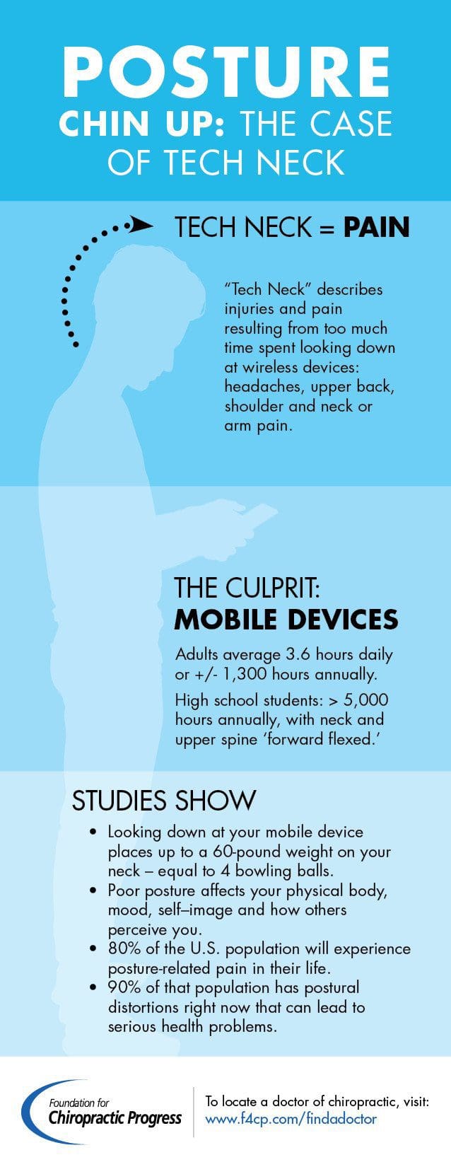
Did you know people send an average of 250 million texts daily? Along with the convenience that technology provides, also comes the need to avoid or minimize injuries. This is particularly true of young people, who are still growing.
With the ever increasing daily use of mobile devices such as smartphones, tablets and handheld games, chiropractors are seeing an increase in corresponding Repetitive Strain Injuries (RSI’s), known by names like text neck and Blackberry thumb. RSIs are injuries of the musculoskeletal and nervous systems that are often caused by repetitive activities, forceful exertions, vibrations, mechanical compression (pressing against hard surfaces), or sustained awkward positions.
What Is Text Neck?
Text neck shows itself as curved shoulders, head hanging forward and down and is caused by poor posture from being�hunched over a mobile device for a long time. This prolonged poor posture is often related to chronic headaches, shoulder, neck pain and can have long term impact.
For every inch of forward head posture, it can increase the weight of the head�on the backbone by an additional 10 pounds.
Physiology Of Joints & Technology
Young men and women are especially at risk as they are heavy users of advancing technology i.e. smartphones and handheld gaming devices.
Text neck and neck strain can cause postural abnormalities and change the growth pattern, especially in the spine.
Technology isn’t going anywhere, so how can we help our children minimize the risks? The trick is to stress the importance of posture and how to attain it, since text neck is a postural abnormality.
Chiropractic And Strong Posture
Recommendations To Avoid Text Neck
There are several things parents and young people can incorporate into their daily activities to alleviate the symptoms of text neck, related RSIs and fortify their posture:
Sit up straight with chest out and shoulders back.
Bring your arms up to eye level so you don’t have to look down to see the screen.
If you must look down, tuck your chin into your neck instead of hanging your head forward.
If you use your mobile device for extensive typing, consider investing in an external keyboard.
Rest your forearms on a pillow while typing to minimize neck tension.
Avoid using mobile devices in bright sunlight. Straining to see the screen often leads forward chin movement which, strain the head muscles.
Try For A Balanced Lifestyle
The best way to minimize the risk of RSIs related to mobile devices is to balance the use of these devices and all around techology.
Balance is critical. Encourage your child to take breaks from devices that are mobile and get regular physical activity to offset the effects of leaning over a smartphone, tablet or computer.
“You want to neutralize the stress,” says Doctor of chiropractic Brian Gushaty. “Strenuous physical activity for the upper body, such as racquet sports, can provide a good counterbalance for the strain caused by poor posture.”
Another key element is to introduce your child to a regular stretching program:
Hand stretches and squeezing a stress ball can help fingers.
Pull shoulder blades down and back to help alleviate neck and shoulder strain.
Stretch the chest by standing up straight with arms down at your side. Turn forearms until thumbs are pointing at the wall behind you.
Posture strengthening programs, like Straighten Up Alberta, is a fun, fast and effortless method to incorporate stretching into your daily routine.
If you are worried your child is suffering from a repetitive strain injury like text neck, speak to a health care provider. A chiropractor is trained to treat RSI’s in all age groups and can provide advice on achieving a balanced healthy lifestyle for your whole family.
More than half of people with fibromyalgia experience proper sleep issues. You may have a hard time falling asleep or wake up several times during the night if you have this painful condition. You don’t spend enough time at the deeper sleep stages. Or you possibly suffer from all three.
How can poor sleep affect fibromyalgia?
It isn’t simply important that there’s a sleep problem in the first place, but more so, that there is a problem causing you to experience sleeping issues. Without proper rest, chronic pain will aggravate and fatigue can increase. Finding a solution will not cure your fibromyalgia, but it is going to lower your pain and tiredness. And since those are the very painful fibromyalgia symptoms, that may be consolation enough.
The Value of Proper Sleep with Fibromyalgia
The worth of sleep goes beyond simply giving you a rest. It has biochemical and psychological significance. A few reasons your body needs a good night’s sleep include:
Proper Sleep allows your body to fix damaged tissues.
Dreaming promotes good physical and psychological wellness.
Some essential hormones, growth hormone, for instance, are secreted during sleep or soon before waking.
You concentrate better and are less fatigued with a good night’s sleep. Deficiency of quality rest can cause what is known as the fibro fog (the inability to focus and concentrate because of fibromyalgia’s extreme fatigue).
Many researchers consider fibromyalgia sufferers don’t get enough deep sleep. Fundamentally, sleep researchers have identified three kinds of sleep, mild sleep (stages 1 and 2), deep sleep (stages 3 and 4), and rapid eye movement (REM) sleep.) If you don’t spend enough time in deep sleep, your body reduces the production of hormones that are important. Pain may be increased by decreased production of these hormones .
Likewise, if you don’t experience sufficient REM sleep, your body may produce less cortisol (although the hormone, which controls blood pressure and blood surgar, may be released at any given time during proper sleep). People with fibromyalgia may have reduced levels of cortisol, which contributes to their fatigue.
7 Tips to Help Achieve Better Sleep
Anti-depressants. Many people find that low doses of tricyclic anti-depressants help attain a more profound sleep. The drugs make people feel exhausted, and then fall asleep. Talk to your doctor about possible side effects.
Don’t watch TV or surf the Internet on your computer instantly before going to bed. These activities boost activity from the brain, which makes it more difficult to fall asleep.
Get more exercise.Your pain and fatigue may save you from exercising, but mild exercise may help you to get a more therapeutic sleep.
Herbal supplements. Valerian, kava kava, and melatonin are alternative medications which have helped some people fall asleep. Valerian helps with insomnia, kava kava also treats insomnia, along with stress and nervousness, and cortisol helps reset the body’s natural rhythm. Always speak to your doctor before taking herbal or other supplements to avoid a potentially significant interaction with drugs you take, especially if they’re prescription or over-the-counter drugs.
Mattress choice. You may be on the market for a new mattress, if you are not sleeping on a bed that promotes a good night’s sleep. You will find a variety of mattresses that can make a big difference in your quality of sleep.
Prescription sleep treatments. There are an assortment of FDA-approved drugs specifically for sleep disorders, such as zolpidem (Ambien) and eszoplicone (Lunesta).
Simulate the breathing of deep sleep. This may “trick” your body into sleeping by taking slow deep breaths which mimic those of the deeper sleep stages. You will feel relaxed and better able to fall asleep.
If you are experiencing proper sleep problems, talk with your physician. You will learn the best treatment choices to give you the excellent sleep you need to help suppress your fibromyalgia symptoms.
The scope of our information is limited to chiropractic and spinal injuries and conditions. To discuss options on the subject matter, please feel free to ask Dr. Jimenez or contact us at 915-850-0900 .
By Dr. Alex Jimenez
Additional Topics: Wellness
Overall health and wellness are essential towards maintaining the proper mental and physical balance in the body. From eating a balanced nutrition as well as exercising and participating in physical activities, to proper sleep for a healthy amount of time on a regular basis, following the best health and wellness tips can ultimately help maintain overall well-being. Eating plenty of fruits and vegetables can go a long way towards helping people become healthy.
Physical therapy often takes a hands-on approach, which might make you cringe if you’re experiencing pain from several hypersensitive tender points. However, in managing your fibromyalgia symptoms, gentle and effective are used by physical therapy, and will most likely play a part in the recovery process.
Can physical therapy help ease fibromyalgia?
There are a variety of physical therapy techniques. Passive treatments include hydrotherapy, heat therapy, deep tissue massage, electrical muscle stimulation, and ultrasound and relax the body. Your physical therapy program will often start with passive treatments. When you feel ready, you will begin active treatments that protect against fibromyalgia pain and strengthen your body. Your physical therapist may work with you to develop a suitable strategy.
Passive Physical Therapy Treatments for Fibromyalgia
Deep Tissue Massage: Unless you’re in an extreme amount of pain, deep tissue massage is an ideal fibromyalgia treatment because it uses a good deal of pressure to ease deep muscle tension and spasms. Spasms prevent muscle motion in the affected level, which is just one reason people with fibromyalgia experience a diminished range of movement. Physical therapy techniques, including deep tissue massage, can assist you to use your muscles more efficiently. This treatment could be combined with cold or heat remedies to improve the benefits.
Heat Therapy: Heat treatment is one of the most preferred methods of reducing chronic aches and pains associated with fibromyalgia. The body’s natural recovery process is triggered by heat by relaxing your muscles and speeding up blood flow. Extra oxygen is delivered by extra blood and nutrients. Blood removes waste byproducts from muscle spasms.
Heat may not fully eliminate the origin of your pain, but it could effectively lower your pain. This treatment is used in a couple of ways–via dry heat (a heating system or a sterile, warm towel) or moist heat (steam heating or some moist, warm fabric).
When utilizing heat treatment on your own after physical treatment ends, never overheat painful areas. If you are using a heating pad, set it. It is not overly hot, when using a towel that is hot, touch it to make sure. Heat potentially cause burns but also may not only exacerbate your fibromyalgia pain.
Hydrotherapy: As the name suggests, hydrotherapy involves water. As a treatment, hydrotherapy may involve sitting at a bath to ease pain, relax muscles, without adding strain, and condition your body.
Electric Muscle Stimulation: It isn’t debilitating, although electric muscle stimulation seems intense. This technique reduces muscle spasms and is generally believed to trigger the release of endorphins, which can be your body’s natural pain killers.
Ultrasound: This treatment utilizes sound waves to create a gentle heat that increases blood circulation to your deep tissues. Ultrasound helps decrease pain, inflammation, stiffness, and muscle spasms and is successful in relieving variety of motion limitations to people with inflammatory problems.
Active Physical Therapy Treatments for Fibromyalgia
Active treatments help address core stability, flexibility, strength, and joint motion. An exercise program may also be prescribed to attain outcomes. This will not curb recurrent pain but will benefit your general health. Your physical therapist will work with you to develop a schedule based on your particular symptoms and wellness.
Active treatments include:
Core stability: Your core (abdominal) muscles have a greater impact on your general health than you could think. In supporting your spine strong core muscles serve as great allies to your back muscles. It’s called the core since it is the powerhouse of your body. Naturally, your body is provided by a core with a stable centre stage.
Muscle flexibility and strengthening: Your variety of motion will probably be restricted if you’re experiencing fibromyalgia pain. Utilizing customized strengthening and stretching exercises, your physical therapist can help you strengthen and lengthen your muscles, and improve joint movement. Pain is better handled by strong muscles.
Hydrotherapy: Water-based exercises may be recommended to present gentle aerobic conditioning.
Your physician will teach you self-care principles so you recognize how to best treat your fibromyalgia symptoms. The ultimate goal is for you to develop the knowledge to help control your symptoms.
It’s essential that you understand the exercises and continue them after the proper therapy ends. You won’t like results if you fail to continue with a fitness regimen. You can reduce additional fibromyalgia pain, by caring for your body on your own.
The scope of our information is limited to chiropractic and spinal injuries and conditions. To discuss options on the subject matter, please feel free to ask Dr. Jimenez or contact us at 915-850-0900 .�
By Dr. Alex Jimenez
Additional Topics: Wellness
Overall health and wellness are essential towards maintaining the proper mental and physical balance in the body. From eating a balanced nutrition as well as exercising and participating in physical activities, to sleeping a healthy amount of time on a regular basis, following the best health and wellness tips can ultimately help maintain overall well-being. Eating plenty of fruits and vegetables can go a long way towards helping people become healthy.
IFM's Find A Practitioner tool is the largest referral network in Functional Medicine, created to help patients locate Functional Medicine practitioners anywhere in the world. IFM Certified Practitioners are listed first in the search results, given their extensive education in Functional Medicine


 Individuals who sit most of the day, like those working in a computer while sitting in an office chair, are also at high risk for non-accidental spine injury. Office ergonomics, or computer ergonomics, can help minimize the risk such as the dangers associated with prolonged sitting in an office chair, and carpal tunnel syndrome, such as lower back pain, neck strain, and leg pain.
 Many potentially harmful situations that lead to back injury can be identified and avoided by following four basic rules of thumb:
 These ergonomic rules of thumb will help the worker and their backs. Otherwise the worker is at risk of sustaining or aggravating a


 For every inch of forward head posture, it can increase the weight of the head�on the backbone by an additional 10 pounds.