Ergonomics involves the study and engineering of improving work tools/products to help employees and improve the physical demands of their jobs.
Contents
How does sciatica fit into this?
People that have to sit or stand for long periods in their jobs can develop back/hip conditions that can lead to:
Back
Buttock
Leg
Foot pain
And this can make working painfully difficult if not impossible contributing to lost workdays.
Check out these tips and apply ergonomic principles to everyday activities like:
Using a sit-to-stand up desk
Adjust sitting posture
Adjusting standing posture
Proper movement
These tips can help get you through the workday and the workweek with less pain.
Have a Seat
Sitting for long periods is not good for the spine or sciatic pain.
Try to stand up every 20 minutes and walk around the workspace.
Choose a well-designed ergonomic chair.
Add low back support with a lumbar pillow or even a rolled-up towel at the base of the chair.
Added tips to reduce sciatica while sitting:
Do not cross your legs
Keep feet flat on the floor
Keep hips and knees bent at a 45-degree angle
If the chair has wheels, roll around and make it an exercise, instead of twisting/turning the body in an awkward position that can exacerbate the pain.� Use the chair to move as a single unit.
Stand Up
Changing up posture is a wise way to exercise the spine on the job.
Mix it up when it comes to sitting and standing.
Sitting all day is connected to a variety of health problems that go beyond back pain. These include:
Obesity
Type 2 diabetes
There are great benefits when you can sit and stand at work. This can help the work routine immensely.
Standing lowers the risk of type 2 diabetes: Studies have shown people that sit for long periods at work had higher levels of fasting blood sugar.
Standing lowers the risk of cardiovascular problems: Research has linked people who spend even just two hours a day sitting have an increased risk for cardiovascular health problems by one-hundred percent.
Standing helps burn extra calories: A study found regular use of sit-stand desks at work can help burn calories and prevent weight gain when combined with proper diet and exercise.
An easy way is to use a sit-stand desk or sit-to-stand desk.
A sit-stand desk allows adjusting desk height to work seamlessly from sitting to standing.
Before purchasing this type of equipment, research the different styles and types to find the right model for you.
Although standing is important, don’t stand in one place or position for an extended time. Move around and stretch out.
If the job requires standing, rest one foot on a box or stool. and alternate every 10 to 15 minutes.
When standing with sciatica:
Take care when getting up from sitting to a standing position.
When getting up try not to bend at the waist, as this can stretch and aggravate the nerve.
Slide to the front of the seat and stand up by straightening the legs.
Keep Work Within Arms Reach
Keep your work close to avoid bending forward, as this also aggravates the nerve.
Keep your shoulders relaxed, and rest the elbows and arms on the desk or chair arms.
Computer Ergonomics
Create a sciatica-friendly workstation.
Position the monitor at eye level
Keep the keyboard and mouse close
Avoid reaching too far
Choose a proper ergonomic chair
Avoid leaning or slumping forward
Muscle Smarts
Don’t move or lift objects that require great muscular force, like pushing a sofa or picking up a table.
Carrying a:
Purse
Briefcase
Groceries
Luggage
It can be challenging, so carry an equal amount of weight to keep the body balanced.
Anything you don’t need, leave at home. You don’t need the extra weight.
Sleeping On The Proper Mattress Matters
After a long day, get off your feet and rest.
However, if the mattress you sleep on does not support your spine, then sciatica can become a permanent condition.
A soft and lumpy mattress does not properly support the spine, which leads to muscle fatigue and restless sleep.
If these measures:
Time
Ice
Heat
Over-the-counter medications
Don’t help reduce the back and leg pain, definitely call a doctor or chiropractor.
They can determine what is causing sciatica and will create a customtreatment plan to get you working at your best in the shortest amount of time possible.
El Paso, TX Best Sciatica Chiropractor Treatment
Sandra Rubio discusses how Dr. Alex Jimenez and his staff can help relieve your sciatica symptoms. Chiropractic care can improve pain and discomfort as well as reduce irritation and inflammation caused by sciatica. In addition, a chiropractor like Dr. Jimenez can also provide nutritional and fitness advice for sciatic nerve pain. Other treatment methods, like deep-tissue massage, can help relieve sciatica symptoms. Dr. Jimenez is the homeopathic, non-surgical choice for sciatic nerve pain and its associated symptoms.
NCBI Resources
You may be suffering from sciatica if you have ever experienced a shooting, nerve-like pain down one of your legs. The sciatic nerve can be impacted by a number of different things, including injury and degenerative diseases, that can lead to sciatica. Fortunately, chiropractic can be extremely effective for the treatment of sciatica.
A car accident can easily damage the spine and soft tissues. An auto accident may cause a misalignment of the spine, a herniated disc, or other injuries that cause symptoms of sciatica.
Many sufferers of sciatica do not realize that their workplace activities � including repetitive motions and sitting in one position for long periods of time � can lead to sciatica.
Do you have low brain endurance when it comes to focus and concentration? Do you often feel like you must drink coffee or exercise to improve brain function? Have you been experiencing noticeable variation in your mental speed? How often do you pick up your smartphone and forget why? Many women commonly struggle to remember everyday tasks throughout their 40’s and 50’s. Research studies have determined one prevalent cause for midlife brain fog in women: hormones. �
Contents
What is Brain Fog?
Brain fog is a health issue associated with feelings of confusion and disorientation. It can make a person feel as if they’re not thinking, understanding, and remembering things as they should. Other terms utilized to describe brain fog include memory fog, forgetfulness, fogginess, and cognitive or cognition issues. People with brain fog frequently forget names they used to know, lack the ability to remember things without writing them down, and they may not feel as sharp as they used to be. �
One research study that began analyzing women at age 35 demonstrated that many women report forgetfulness and concentration problems throughout their menopausal transition. Healthcare professionals can help regulate this process for women who are transitioning into menopause as well as allow them to know how women�s brains can be sensitive to fluctuating levels of estrogen for mood and cognitive ability. Hormones also play an important role in midlife brain fog. �
Brain Fog and Hormones
During an interview with Dr. Marcie Richardson, Women Living Better (WLB) advisor, she states, �there’s a lot of research studies which haven’t proven a definitive connection between brain fog and changing hormones, however, we do know that there are estrogen receptors found in the human brain”. She adds, “but, there is very clear scientific evidence for sleep affecting cognitive function, so if you are dealing with disrupted sleep, it is likely contributing to your cognitive problems�. �
The December 2018 New York Times article by Jane Brody cited a 2010 research study that followed women for 6-years to see whether her anxiety, depression, sleeping problems, and vasomotor symptoms, were the cause of a decline in her cognitive function right before menopause or in late perimenopause. However, it concluded these were not the causes. �
Brain Fog Remedies to Consider
It�s essential to keep in mind that research studies demonstrate that women can begin to experience brain fog early in the menopausal transition which will then resolve on its own. Many women worry that their brain fog may be a sign or symptom of dementia and Alzheimer’s disease, but these have not been associated with each other in research studies to date. While there aren�t any definitive treatments for brain fog, there are several natural remedies or coping techniques available, such as recognizing the symptom, utilizing tools like notes to self, setting alarms for reminders as well as acknowledging and accepting when a name doesn�t come back to you right away, generally when you are no longer feeling stressed out about it. Keep in mind that disrupted sleep, which comes with hormonal changes for many women, can also contribute to brain fog. �
According to Research Studies
A sample of several recent research studies associated with midlife brain fog and cognition problems in adult women has been demonstrated below. As you can see, it�s inconclusive. The research study on midlife brain fog in women suggests that: �
Many types of problems with memory are associated with lower ratings of health and depressed mood. Problems with current memory and remembering past events are associated with higher levels of reported stress, which many women commonly attribute to the increased burden of having to meet multiple role demands, among other demands.
Women in the earliest and middle stages of perimenopause, as well as those who utilize hormones, have more problems associated with memory than the previously mentioned women in late-stage menopause or perimenopause.
About 72 percent of women reported problems remembering names at least some of the time. About 50 percent have a problem remembering where they put things, recent phone numbers, things others told them or they told others, keeping up a correspondence and forgetting what they were doing. None of these are considered a serious problem.
Another research study, from 2009, concluded that �consistent with transitioning women�s perceived memory difficulties, perimenopause was associated with a decrement in cognitive performance, characterized by women not being able to learn as well as they had throughout premenopause. Improvement rebounded to premenopausal levels in postmenopause, suggesting that menopause transition-related cognitive difficulties may be time-limited, according to the research study�. �
A recent meta-analysis set out to �synthesize the existing research studies of the relationship between menopausal stage and neuropsychological performance and depression�. However, it concluded that although �the menopausal transition is a time of increased vulnerability for cognitive decline and increased risk of depressive symptoms and depressive disorders, these results from the research studies looked at can’t necessarily be generalized�. Although it�s reassuring that there was no documentable cognition decline across research studies, the weakness in these research studies is that cognition decline is generally measured as the ability to complete a test in an experimental setting, a setting with one focus and no interruptions. �
The research study doesn�t accurately emulate real-life settings or measure the type of forgetfulness midlife women report. � A fourth research study also found that cognitive function does not change linearly across perimenopause. Decreases in attention/working memory, verbal learning, verbal memory, and fine motor speed may be most evident in the first year after the final menstrual period, according to the research study. Further research studies are required to determine the association between midlife brain fog and hormone changes in perimenopause and menopause in adult women. �
Symptoms for menopause for women in their 40’s or 50’s are different for each woman, and these can commonly include anything from night sweats to weight gain to thinning hair. However, many women have also reported feeling symptoms of brain fog. According to research studies, hormonal changes in women going through menopause or perimenopause can cause brain fog. However, further research studies are required to demonstrate how hormones can ultimately cause brain fog and other mental health issues. – Dr. Alex Jimenez D.C., C.C.S.T. Insight
Neurotransmitter Assessment Form
The following Neurotransmitter Assessment Form can be filled out and presented to Dr. Alex Jimenez. Symptoms listed on this form are not intended to be utilized as a diagnosis of any type of disease, condition, or any other type of health issue. �
In honor of Governor Abbott’s proclamation, October is Chiropractic Health Month. Learn more about the proposal. �
Do you have low brain endurance when it comes to focus and concentration? Do you often feel like you must drink coffee or exercise to improve brain function? Have you been experiencing noticeable variation in your mental speed? How often do you pick up your smartphone and forget why? Many women commonly struggle to remember everyday tasks throughout their 40’s and 50’s. Research studies have determined one prevalent cause for midlife brain fog in women: hormones. �
The scope of our information is limited to chiropractic, musculoskeletal and nervous health issues or functional medicine articles, topics, and discussions. We use functional health protocols to treat injuries or disorders of the musculoskeletal system. To further discuss the subject matter above, please feel free to ask Dr. Alex Jimenez or contact us at 915-850-0900 . �
Curated by Dr. Alex Jimenez �
Additional Topic Discussion: Chronic Pain
Sudden pain is a natural response of the nervous system which helps to demonstrate possible injury. By way of instance, pain signals travel from an injured region through the nerves and spinal cord to the brain. Pain is generally less severe as the injury heals, however, chronic pain is different than the average type of pain. With chronic pain, the human body will continue sending pain signals to the brain, regardless if the injury has healed. Chronic pain can last for several weeks to even several years. Chronic pain can tremendously affect a patient’s mobility and it can reduce flexibility, strength, and endurance.
Neural Zoomer Plus for Neurological Disease
�
Dr. Alex Jimenez utilizes a series of tests to help evaluate neurological diseases. The Neural ZoomerTM Plus is an array of neurological autoantibodies which offers specific antibody-to-antigen recognition. The Vibrant Neural ZoomerTM Plus is designed to assess an individual�s reactivity to 48 neurological antigens with connections to a variety of neurologically related diseases. The Vibrant Neural ZoomerTM Plus aims to reduce neurological conditions by empowering patients and physicians with a vital resource for early risk detection and an enhanced focus on personalized primary prevention. �
Formulas for Methylation Support
XYMOGEN�s Exclusive Professional Formulas are available through select licensed health care professionals. The internet sale and discounting of XYMOGEN formulas are strictly prohibited.
Proudly,�Dr. Alexander Jimenez makes XYMOGEN formulas available only to patients under our care.
Please call our office in order for us to assign a doctor consultation for immediate access.
If you are a patient of Injury Medical & Chiropractic�Clinic, you may inquire about XYMOGEN by calling 915-850-0900.
�
For your convenience and review of the XYMOGEN products please review the following link.*XYMOGEN-Catalog-Download �
* All of the above XYMOGEN policies remain strictly in force.
If you are experiencing any of these situations, then you might be experiencing a low intake of fiber in your diet, causing inflammation.
Throughout several decades, Americans have lost much diversity in their diets, impacting their gut microbiome, and the contribution to the autoimmune disorder epidemic. The vast majority of people have a less than perfect diet that is consists of high in calories, short on nutrients, and low on fiber intake. Research has stated that about only 10 percent of Americans have met their daily fiber requirements.
The diet is a significant environmental trigger in autoimmune diseases. Dietary approaches can provide the most effective means of an individual to returning balance and the dysfunction with the gastrointestinal system. Researchers have found out that the role of dietary fibers can help with rheumatoid arthritis as there is new and developing research on this discovery.
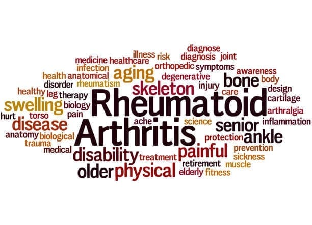
What is Rheumatoid Arthritis?
Rheumatoid arthritis is a long term, progressive, and disabling autoimmune disease. It causes inflammation, swelling, and pain in and around the joints and organs of the body. It affects up to 1 percent of the world’s population and over 1.3 million people in America, according to the Rheumatoid Arthritis Support Network.
Rheumatoid arthritis is also a systemic disease, which means that it affects the whole body, not just the joints. It occurs when an individual’s immune system mistakes their body’s healthy tissues for foreign invaders. As the immune system responds to this, inflammation occurs in the target tissue or organ. Symptoms of rheumatoid arthritis can include:
Pain, swelling, and stiffness in more than one joint
Symmetrical joint involvement
Joint deformity
Unsteadiness when walking
Fever
A general feeling of being unwell
Loss of function and mobility
Weight loss
Weakness
Fiber and Inflammation
Individuals who eat healthily knows that eating fibers in their diet can help reduce the risk of developing various conditions. The AHAEP (American Heart Association Eating Plan) has stated that people should be eating a variety of food fiber sources in their diet. The total dietary fiber intake that a person should be eating is 25 to 30 grams a day from foods, not supplements. Currently, adults in the United States eat about 15 grams a day on their fiber, which is half of the recommended amount.
Eating a high fiber diet can provide many rewards to the body. Eating fruits, vegetables, beans, nuts, and whole grains can provide a boost of vitamins, minerals, protein, and healthy nutrients in the body. Studies have been shown that eating a high fiber diet can help lower the markers of inflammation, which is a critical factor in many forms of arthritis.
The body needs two types of fibers, which are soluble and insoluble. Soluble fibers are mixed with water to form a gel-like consistency, which slows digestion and helps the body absorb nutrients better and helps lower total cholesterol and LDL cholesterol. Insoluble fibers help the digestive system run more efficiently as it adds bulk to stool, which can help prevent constipation.
There have been a few studies that found that people who eat high fiber diets have lower CRP (C-reactive protein) levels in their blood. CRP is a marker for inflammation and is linked to rheumatoid arthritis. When a person eats a high fiber diet, it not only reduces inflammation to their bodies, but it helps lower the body weight as well. High fiber-rich foods feed the beneficial bacteria living in the gut, and then it is releasing substances to the body, promoting lower levels of inflammation.
A study has been shown that patients with rheumatoid arthritis that they consumed either a high fiber bar or cereal for 28 days while continuing with their current medication had decreased levels of inflammation. Researchers noticed that they had an increase of T regulatory cell numbers, a positive Th1/Th17 ratio, a decrease in bone erosion, and a healthy gut microbiome.
Gut Health and Inflammation
The gut plays a crucial role in the immune function as well as digesting and absorbing food in the body. The intestinal barrier provides an effective protective barrier from pathogenic bacteria but also being a healthy environment for beneficial bacteria. With a high fiber diet, it can lead to the production of SCFAs (short-chain fatty acids) in the gastrointestinal tract, thus playing an essential role in T regulatory cell activation, which regulates the intestinal immune system. When inflammation comes to play in the gut, it can disrupt the intestinal permeability barrier and cause a disruption, leading to leaky gut. Probiotics and a high fiber diet can help prevent inflammation and provide a healthy gut function.
Conclusion
Eating a high fiber diet is essential to prevent inflammation, not on the joints, but everywhere in the body. Even though individuals eat half of the recommended amount of fiber in their diets, due to their hectic lifestyle, eating a high fiber diet is beneficial. Incorporating fiber in their diet gradually is ideal as well as drinking water with the fibers to make the process work more effectively in the body. Some products can help aid the body by supporting not only the gastrointestinal function and muscular system but making sure that the skin, hair, nail, and joints are healthy as well.
October is Chiropractic Health Month. To learn more about it, check out Governor Abbott�s declaration on our website to get full details on this historic moment.
The scope of our information is limited to chiropractic, musculoskeletal and nervous health issues as well as functional medicine articles, topics, and discussions. We use functional health protocols to treat injuries or chronic disorders of the musculoskeletal system. To further discuss the subject matter above, please feel free to ask Dr. Alex Jimenez or contact us at 915-850-0900 .
References:
at UCSF Medical Center, Healthcare Specialist. �Increasing Fiber Intake.� UCSF Medical Center, 2018, www.ucsfhealth.org/education/increasing_fiber_intake/.
Brazier, Yvette. �Rheumatoid Arthritis (RA): Symptoms, Causes, and Complications.� Medical News Today, MediLexicon International, 16 Oct. 2018, www.medicalnewstoday.com/articles/323361.php.
Hakansson, Asa, and Goran Molin. �Gut Microbiota and Inflammation.� Nutrients, MDPI, June 2011, www.ncbi.nlm.nih.gov/pmc/articles/PMC3257638/.
Jurgelewicz, Michael. �New Study Demonstrates the Role of Fiber in Rheumatoid Arthritis.� Designs for Health, 11 Oct. 2019, blog.designsforhealth.com/node/1125.
Unknown, Unknown. �More Fiber, Less Inflammation?� Www.arthritis.org, 25 June, 2015, www.arthritis.org/living-with-arthritis/arthritis-diet/anti-inflammatory/fiber-inflammation.php.
Q: My primary healthcare provider recently diagnosed me with a herniated disc in the lumbar spine. They referred me to get chiropractic treatment, but I�m nervous because it’s new to me and I’m afraid of being adjusted wrong, paralyzed, etc. Can I trust chiropractic treatment to work?
A: It�s normal to be nervous about going to a chiropractic clinic.
If you’re not sure whether chiropractic is for you, there is scientific evidence that shows how chiropractic techniques like spinal manipulation/spinal adjustment and forms of manual/mechanical therapy are safe and effective for relieving pain and other musculoskeletal pain, conditions, and symptoms.
I encourage everyone to try chiropractic treatment as a non-surgical treatment option for a herniated disc.
Contents
It Is Your Decision
At the first appointment, a chiropractor will take a medical history and perform a thorough exam to determine the nature of the symptoms and their possible causes, which include a herniated disc.
Sometimes with a herniated disc, there may be no symptoms at all.
But usually a herniated disc causes:
Back pain
Referred pain or pain that is felt in other parts of the body like the legs, feet, etc.
An irritated spinal nerve can cause symptoms in the legs
This can lead to neurological symptoms like:
Tingling
Numbness
Weakness in the legs
Once the chiropractor determines your symptoms, they may use one or several techniques to relieve the back pain and other symptoms.
Techniques used by chiropractors for disc-related problems include:
Specific self-treatment exercises to improve motion & decrease back pain
Cox technique like spinal traction using special tables
Spinal manipulation
Hands-on techniques that relieve pain and restore movement to the spine and body
These techniques have been proven to be very safe. There are other techniques a chiropractor can recommend for various conditions, as each has their own style and method.
A chiropractic treatment plan will also include:
Education
Self-management instructions
This is to teach you how to control/eliminate pain with proper posture and proper body mechanics.
Whichever treatment the chiropractor recommends, he or she will discuss it with you, including the benefits and risks.
Although the treatments listed above will most likely be a part of your treatment plan, your chiropractor will answer your questions and work with you to select a treatment that meets your specific goals and preferences.
Don’t Be Nervous A Chiropractor Monitors Treatment Progress
If symptoms do not improve within a reasonable time frame, then a chiropractor may refer the patient to other treatments to manage disc-related pain, including:
Physical therapy
Acupuncture
Spinal injections
Surgery
Fortunately, self-management and time can be the best treatment. Allowing the body to heal itself is the way to go. But if rest is not enough then chiropractic treatment may be just what is needed to kick in the body’s self-healing function.
If you decide to give chiropractic treatment a try, don’t be nervous, as a chiropractor will monitor progress throughout the treatment.
In any case, chiropractors are qualified to discuss the benefits and risks of other treatments, depending on the condition.
Hopefully, this article has given you the basics of chiropractic medicine and how it works so you can make the best choice for your herniated disc/s.
Low Back Pain Management El Paso, TX Chiropractor
Denise suffered an auto accident injury which resulted in back pain. When she realized she could not sit, walk or sleep for lengthy periods of time without having painful symptoms, Denise found chiropractic care with Dr. Alex Jimenez at El Paso, TX. Once she received therapy for her automobile accident injuries, Denise experienced relief from her symptoms and she was able to execute her regular tasks once again. Thanks to the education and maintenance Dr. Alex Jimenez supplied, Denise regained her initial health and health.
Back pain is more most common, with roughly nine out of ten adults undergoing it at some time in their lifetime, and five functioning adults developing it annually. Some quote around 95 percent of Americans will experience back pain at some time in their lifetime. It is undoubtedly the typical cause of chronic pain since it’s also a substantial contributor to missed work and handicap. In the United States alone, acute cases of lower back pain are the fifth most frequent reason for doctor visits and cause 40% of missed days off work. What’s more, it is the leading cause of disability worldwide.
NCBI Resources
A herniated disc is a common spinal condition that typically affects the cervical spine (neck region) or the lumbar spine (lower back), although it can occur in any part of the spine. Most often, a herniated disc happens at the L4 � L5 and the L5 � S1.� This is because this portion of the spine, the lumbar region, bears the bulk of the body�s weight.
Stomach pain, burning or aching 1-4 hours after eating
Sense of fullness during and after meals
If you are experiencing any of the situations, then you might want to try these six types of food to help boost your immune system.
The Immune System
The immune system is the body�s defense mechanism that provides a robust anatomical barrier.� The gastrointestinal tract is one of the barriers. It has many defense mechanisms such as peristalsis, gastric acid, bile acids, digestive enzymes, flushing, thiocyanate, defensins, and gut flora in the body. The gut flora is the critical focus for many health professionals; however, all the essential defense mechanisms rely heavily on the gastrointestinal tract to function efficiently.
There are ways to benefit the immune system as one of the ways is to plan meals that are filled with necessary nutrients that can fight off infections. Prebiotic and probiotic-rich foods help enhance microbial diversity in the gut, while vitamin C-rich foods can mop up the free radicals that have entered the body. Another benefit is to avoid foods that promote infections like heavily processed foods, added sugars, and sodas. When it is not consumed in the body, it can help boost immunity and enrich the gut microbiome. Here are the six foods to help boost the immune system in the body.
Yellow Bell Peppers
Due to being the most natural vegetable to find at a local grocery store or farmer’s markets around the world, yellow bell peppers contain more vitamin C than oranges. Since oranges contain about 78% of vitamin C, yellow bell peppers contain about 152% of vitamin C and numerous vitamins and minerals. Bell peppers (yellow, red, orange and green) contain the following:
Vitamin B6: Bell peppers contain pyridoxine, which is an essential nutrient for the formation of red blood cells.
Vitamin K1: This vitamin is also known as phylloquinone, which is vital for bone health and blood clotting.
Potassium: This mineral is essential for improving heart health.
Folate: Also known as vitamin B9, this vitamin has a variety of functions to the body and is highly essential to take during pregnancy.
Vitamin E: This is a powerful antioxidant that is essential for healthy nerves and muscles.
Vitamin A: Red bell peppers are high in beta carotene when consumed converts to vitamin A in the body.
Vitamin C helps boost the immune system by influencing the development and function of lymphocytes, and with about half a cup of yellow bell peppers will give the body those lymphocytes.
Guava
Guava is a traditional remedy for a range of health conditions that a person may encounter. These tropical fruits are seasonal throughout the winter. They contain about 140% of vitamin C and rich with lycopene, which is excellent for the immune system as it plays an essential role in the activities of the enzymes. Lycopene is a powerful antioxidant that has been implicated in having a potentially beneficial impact on several chronic diseases, including cancer.
Studies have been shown that the guava fruit and the leaves have been known to have a positive effect on a range of illnesses and symptoms, including:
Type 2 diabetes
Menstrual cramps
Diarrhea
Flu
Blood pressure
Osteoarthritis
Cancer
Broccoli
Broccoli is high in phytonutrients like vitamins A, C, and E while also containing sulforaphane. Sulforaphane is activated when broccoli or any cruciferous vegetables are chewed, cut, or damaged. Raw broccoli or broccoli sprouts contain the highest level of sulforaphane when it is not boiled or cooked. Studies have been shown that consuming broccoli has been associated with reducing many lifestyle-related health conditions like:
Obesity
Diabetes
Improves digestion
Regulate the immune system
Helps support healthy-looking skin
Decrease inflammation
Lowers blood pressure
Turmeric
Turmeric is an excellent immune-boosting food since it supports healthy inflammatory pathways in the body. Inflammation in the body is implicated in the pathophysiology of many health-compromising situations that can lead to chronic illnesses. So consuming pro healthy inflammation foods like turmeric or incorporating turmeric in dishes is an ideal way to boost the immune system.
The active component in turmeric is curcumin and has potent biological properties like anti-oxidative, anti-cytotoxic, and neurorestorative properties, making it an essential staple in an immune-boosting food. Here are some of the benefits that turmeric provides to the body:
Anti-inflammatory properties
Pain relief on the joints
Improves liver function
Reducing the risk of cancer
Preventing gut inflammation
Green Tea
Green tea helps the body relax and contains L-theanine that helps the formation of healthy T-cells. Green tea also contains EGCG (epigallocatechin gallate) and is packed filled with flavonoids to help boost the body’s immune system. Here are some of the health benefits that green tea provides:
Cancer prevention
Lowers the risk of cardiovascular diseases
Lowers cholesterol
Decrease the risk of a stroke
Lowers the risk of type 2 diabetes
Help lose weight
Helps lowers inflammation on the skin
Improves brain function
Helps reduce the risk of Alzheimer�s disease
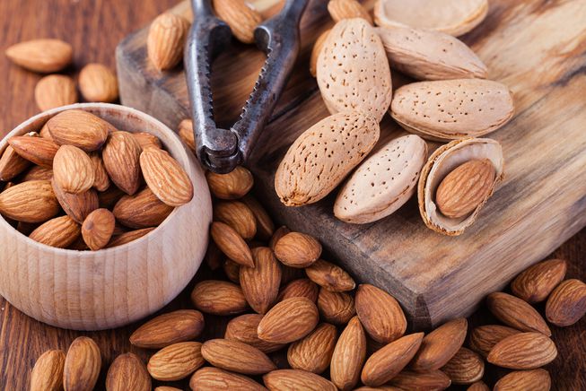
Almonds
Almonds are packed filled with vitamins, minerals, protein, and fibers. It contains vitamin E and helps boost the immune system since it is a free radical scavenging antioxidant. They are easy to find in any grocery store, and the health benefits that almonds can provide are:
Lowering cholesterol
Reduce the risk of cancer
Provide heart health benefits
Reduce type 2 diabetes
Manage weight
Conclusion
Eating these six foods can be beneficial to support a healthy immune system. They are bursting with plant-based nutrition that the body needs to make sure that chronic illnesses like inflammation in the gut. Some products help support the immune system as well as making sure that the gastrointestinal system and the sugar metabolism is supported. Eating a variety of foods that has antioxidants and anti-inflammatory properties is beneficial to the body. With the cold and flu season approaching, it is highly relevant to consume these foods to help fight against the cold and flu and providing assistance to the immune system.
October is Chiropractic Health Month. To learn more about it, check out Governor Abbott�s declaration on our website to get full details on this historic moment.
The scope of our information is limited to chiropractic, musculoskeletal and nervous health issues as well as functional medicine articles, topics, and discussions. We use functional health protocols to treat injuries or chronic disorders of the musculoskeletal system. To further discuss the subject matter above, please feel free to ask Dr. Alex Jimenez or contact us at 915-850-0900 .
Reference:
Ahmed, Touqeer, et al. �Curcuminoids Rescue Long-Term Potentiation Impaired by Amyloid Peptide in Rat Hippocampal Slices. – Semantic Scholar.� Undefined, 1 Jan. 1970, www.semanticscholar.org/paper/Curcuminoids-rescue-long-term-potentiation-impaired-Ahmed-Gilani/c66297f8d0f3b633fac263cbb81f82de1893387a.
Arnarson, Atli. �Bell Peppers 101: Nutrition Facts and Health Benefits.� Healthline, 27 Mar. 2019, www.healthline.com/nutrition/foods/bell-peppers.
Burgess, Lana. �Health Benefits of Guava: How to Use It, Nutrition, and Risks.� Medical News Today, MediLexicon International, 20 Mar. 2019, www.medicalnewstoday.com/articles/324758.php.
Du, Guang-Jian, et al. �Epigallocatechin Gallate (EGCG) Is the Most Effective Cancer Chemopreventive Polyphenol in Green Tea.� Nutrients, MDPI, 8 Nov. 2012, www.ncbi.nlm.nih.gov/pmc/articles/PMC3509513/.
Kim, DS, et al. “Curcuminoids from Curcuma Longa L. (Zingiberaceae) That Protect PC12 Rat Pheochromocytoma and Normal Human Umbilical Vein Endothelial Cells from BetaA(1-42) Insult.� Neuroscience Letters, U.S. National Library of Medicine, 27 Apr. 2001, www.ncbi.nlm.nih.gov/pubmed/11297823.
Luo, Cong, and Xian-Guo Wu. �Lycopene Enhances Antioxidant Enzyme Activities and Immunity Function in N-Methyl-N’-Nitro-N-Nitrosoguanidine-Enduced Gastric Cancer Rats.� International Journal of Molecular Sciences, Molecular Diversity Preservation International (MDPI), 2011, www.ncbi.nlm.nih.gov/pmc/articles/PMC3116194/.
Menon, Venugopal P, and Adluri Ram Sudheer. �Antioxidant and Anti-Inflammatory Properties of Curcumin.� Advances in Experimental Medicine and Biology, U.S. National Library of Medicine, 2007, www.ncbi.nlm.nih.gov/pubmed/17569207.
Nordqvist, Joseph. �Almonds: Health Benefits, Nutrition, and Risks.� Medical News Today, MediLexicon International, 14 Dec. 2017, www.medicalnewstoday.com/articles/269468.php.
Team, Biotics Education. �Key Foods to Boost the Immune System.� Biotics Research Blog, 15 Oct. 2019, blog.bioticsresearch.com/key-foods-to-boost-the-immune-system.
van Gorkom, Gwendolyn N Y, et al. �Influence of Vitamin C on Lymphocytes: An Overview.� Antioxidants (Basel, Switzerland), MDPI, 10 Mar. 2018, www.ncbi.nlm.nih.gov/pubmed/29534432.
Vermeulen, Martijn, et al. �Bioavailability and Kinetics of Sulforaphane in Humans after Consumption of Cooked versus Raw Broccoli.� Journal of Agricultural and Food Chemistry, U.S. National Library of Medicine, 26 Nov. 2008, www.ncbi.nlm.nih.gov/pubmed/18950181.
Ware, Megan. �Broccoli: Health Benefits, Nutritional Information.� Medical News Today, MediLexicon International, 8 Dec. 2017, www.medicalnewstoday.com/articles/266765.php.
Ware, Megan. �Green Tea: Health Benefits, Side Effects, and Research.� Medical News Today, MediLexicon International, 28 Mar. 2017, www.medicalnewstoday.com/articles/269538.php.
Ware, Megan. �Turmeric: Benefits and Nutrition.� Medical News Today, MediLexicon International, 24 May 2018, www.medicalnewstoday.com/articles/306981.php.
Stomach pain, burning or aching 1-4 hours after eating
Feel hungry an hour or two after eating
Digestive problems when lying down or bending forward
If you are experiencing any of these situations, then you should try some micronutrients for your GI tract health.
GI Tract Health
Over two thousand years ago, Hippocrates recognized that the gut plays a significant role in overall health and that modern scientific research has substantiated and solidified this view. With GI (gastrointestinal) health, advanced testing, and intricate healing protocols focused a lot when it comes to the GI tract. Some patients may benefit from the precise analysis of the makeup from their gut flora or the specific food elimination and reintroduction strategies, but not to overlook the fundamentals. Addressing the basics like general micronutrient repletion or supplementation with the foundational nutrients can be targeted for therapeutic purposes and can go a long way for an individual�s healing.
The Micronutrients
These are some of the fundamental micronutrients that the body needs to perform the everyday task. These can mostly be found in foods or in supplements and vitamins that are consumed, and even though high restriction diets can deplete these nutrients, they are still crucial for not only our gut health but for the entire body system as well.
Glutamine
The amino acid glutamine is a trusty workhorse for a healthy gut function in the body. Even though it is technically not an essential amino acid, it serves as an energy source for epithelial cells that makes up the intestinal lining for the intestines. Various circumstances like trauma, burns, or recovery from significant operations or illnesses can increase the body’s demand for glutamine.
Another amino acid is taurine is beneficial for individuals who need help with the digestion of dietary fats. Taurine is unique due to not being used in any structural protein; however, it has other roles in the body. Taurine can be synthesized from cysteine and can be obtained from animal foods specifically, sadly though it is nonexistent in plant food. Bile acids that are bound with taurine are secreted by the liver; the making of this compound is critical for bile acid function and proper fat absorption in the body.
Potassium is the core nutrient that plays a role in a healthy GI function, especially when it comes to intestinal motility. Some disorders like fatigue and cardiac arrhythmias can be the result of potassium deficiency, and inadequate potassium may lead to delayed gastric emptying and intestinal paralysis. If the body is not treated soon, it can lead to chronic illnesses in the GI, causing unpleasant effects like bloating, abdominal pain, and constipation.
All food supplies have an abundance of potassium, but certain medications can reduce potassium levels. Factors like excessive alcohol consumption or strict chronic dieting for weight loss can be the result of inadequate potassium intake and the body status of a person.
B vitamins, especially vitamin B6, are highly essential to the GI tract because they make sure that the brain is also healthy as well. Deficiency of vitamin B6 can cause these symptoms:
Tingling, numbness, and pain in the hands and feet
Anemia
Seizures
Depression
Confusion
Weak immune system
Vitamin B6 is a water-soluble vitamin that produces the neurotransmitters serotonin and norepinephrine and forming myelin for the body. This vitamin can help boost brain function and can improve memory function. Some of the other benefits it can provide to the body are:
Lowers the risk of dementia
Reduce the severity of nausea during pregnancy
Protection from air pollution
Ensures the normal functioning of digestive enzymes
Conclusion
Even though these are the necessary foundational micronutrients and amino acids for their roles in the GI tract, it is crucial for individuals who have these micronutrient deficiencies. Even though the popularity of highly restrictive diets emphasizes on caloric restrictions for weight loss for individuals, it can limit the intake of certain nutrient-dense foods. It can cause disruptions to the gastrointestinal tract. When a person surrounds themselves with an abundance of foods with these micronutrients can live a healthy life. Some products combined with these micronutrient foods can provide support to the gastrointestinal system and help boost the sugar metabolism for the body.
October is Chiropractic Health Month. To learn more about it, check out Governor Abbott�s declaration on our website to get full details on this historic moment.
The scope of our information is limited to chiropractic, musculoskeletal and nervous health issues as well as functional medicine articles, topics, and discussions. We use functional health protocols to treat injuries or chronic disorders of the musculoskeletal system. To further discuss the subject matter above, please feel free to ask Dr. Alex Jimenez or contact us at 915-850-0900 .
References:
Brazier, Yvette. �Vitamin B-6: Benefits, Dosage, Food Sources, and Deficiency Symptoms.� Medical News Today, MediLexicon International, 27 Mar. 2017, www.medicalnewstoday.com/articles/219662.php.
Cadman, Bethany. �L-Glutamine for IBS: Benefits, Side Effects, and Research.� Medical News Today, MediLexicon International, 7 Feb. 2018, www.medicalnewstoday.com/articles/320850.php.
Caporuscio, Jessica. �What Is Taurine? Benefits and Side Effects.� Medical News Today, MediLexicon International, 26 Sept. 2019, www.medicalnewstoday.com/articles/326476.php.
Higdon, Jane. �Potassium.� Linus Pauling Institute, 14 Oct. 2019, lpi.oregonstate.edu/mic/minerals/potassium#deficiency.
Mawer, Rudy. “What Is Taurine? Benefits, Side Effects, and More.” Healthline, 27 Nov. 2018, www.healthline.com/nutrition/what-is-taurine.
Megan Ware, RDN. �Potassium: Health Benefits and Recommended Intake.� Medical News Today, MediLexicon International, 10 Jan. 2018, www.medicalnewstoday.com/articles/287212.php.
Team, DFH. �Micronutrients in GI Health.� Designs for Health, 11 Oct. 2019, blog.designsforhealth.com/node/1123.
Tinsley, Grant. “Glutamine: Benefits, Uses, and Side Effects.” Healthline, 13 Jan. 2018, www.healthline.com/nutrition/glutamine.
FDA recognizes and approves spinal decompression and its ability to eliminate herniated discs.
On the verge of back surgery, a mason discovered the non-surgical solution to work-related chronic back pain.
A new male patient who works in construction came to see me as a last resort to lessen his back pain brought on from damaged/herniated discs.
His primary caregiver recommended back surgery, but that would have put him on disability for months.
Fortunately, before saying yes to the surgery, a co-worker recommended chiropractic care.
Bricklayers/masons have the highest rate of back injuries with non-paid sick leave.
Constant bending over, even with a back brace, takes its toll on the spine, which in this case resulted in two herniated discs.
Pain medications helped in the beginning but with constant use, put him in a constant brain fog state, along with the expense, which took its toll on the family budget.
Contents
Disc Injury & Back Surgery
The doctor did not discuss spinal decompression therapy
�A non-surgical back treatment that slowly and gently stretches the spine.
This stretching lessens the pressure on the compressed nerve root (herniated disc) and results in less and even complete alleviation.
The patient came twice a week with myself and the team working on him over the course of a month, however, every case is different so treatments vary depending on the condition.
With each treatment, the two herniated discs were slowly reverted back to their natural position. This is able to be achieved with less pressure between the discs.
Towards the end of treatment, the patient’s pain was gone by about 90%.
With two weeks of rest, the patient was able to return to work.
The best part was that there was no surgery, pain medications, disability, and hospital bills.
Spine treatment alternative
Chiropractic/Decompression therapy is way less expensive than medication and surgery. It is:
Non-surgical
Recovery time is faster
Completely drug-free
People suffering every day with herniated/injured discs should consider the chiropractic decompression option. You do not have to learn to live with chronic back pain.
If you suffer from:
Herniated discs
Bulging discs
Degenerative disc disease
I encourage you to discuss the condition with an experienced chiropractor. There are many proven alternatives to back surgery and pain meds. People need to be aware of these alternatives for chronic back pain. The right-back pain treatment can definitely improve the quality of life.
Herniated Disc El Paso, TX
Sandra Rubio developed two herniated discs and a bulging disc after suffering from an accident at a young age, which caused her intense pain throughout her youth.
When she became a mother, her symptoms became severe.
After visiting doctors without results, Sandra found chiropractor Dr. Alex Jimenez and found relief from her sciatica and migraines.
The herniated disc treatment she received from Dr. Alex Jimenez was non-surgical.
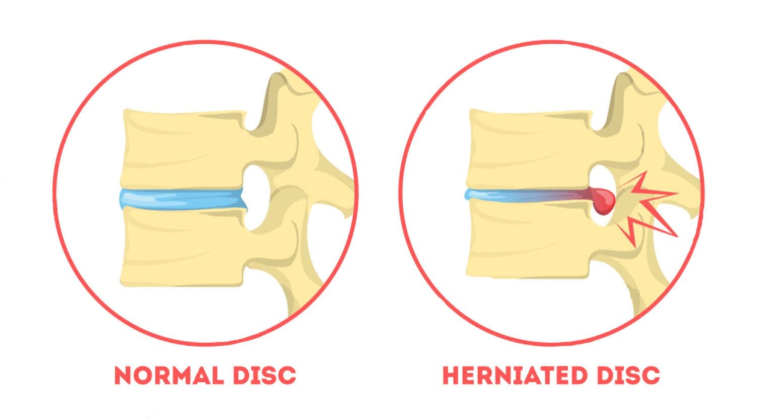
A herniated disc, also known as a slipped disc, is a medical condition in which:
Atear from the outer intervertebral disc allows the soft, central area to bulge out beyond the outer rings.
Disc herniation is usually a result of:
Degeneration (wear/tear)
Trauma (auto accident/sports injury)
Lifting injuries
Straining movement
The tear can release the compounds, which cause inflammation and can cause severe pain even if the nerve root not compressed.
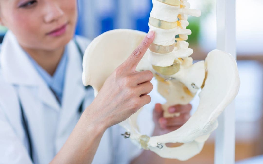
A physical examination is usually the first step in diagnosing a herniated disc. The chiropractor will examine the spine while the patient is standing, and while they’re lying down. Depending on the severity and location of the herniation, they may note a decrease in spine curvature.
Radicular pain will be assessed, when the spine is:
Unmoving
In motion
With pressure applied
Other tests may be administered.
X-rays may also be taken, but an MRI is usually more accurate and shows more detail.
Chiropractic has been very effective in helping patients manage their pain and regain their mobility so they can return to their normal life. Therefore, it should be your first option for treatment before you go down the road with drugs or surgery.
NCBI Resources
It is often referred to as a ruptured disc or slipped disc and occurs when the disc moves or slips out of place. It can also be the result of a disc that has a small tear and is leaking the jelly-like substance that is inside. This can put pressure on the surrounding nerves, causing pain and discomfort.
Increase in sports-related fractures among young and active people
Any type of bone fracture, especially when the spine is involved, comes with the most common and debilitating symptom is severe pain.
Managing pain correctly is vital to the proper healing of a fractured bone.
Unfortunately, the common treatments prescribed to manage fracture pain can cause significant side effects, especially when used beyond the short-term or acute phase of pain.
Bone fractures cannot be always be avoided, but when it comes to osteoporosis, everyone can take steps to help minimize the risk of developing the condition.
Contents
How to Prevent Osteoporosis and Bone Fracture
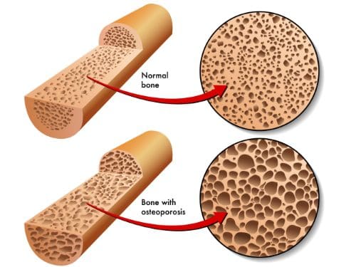
First, understand that osteoporosis is not a normal part of the aging process.
It is an irreversible and degenerative disease that causes bones to become porous over time.
Prevention should begin as early as possible, as this will benefit an individual later in life.
It’s never too late to begin taking steps!
Protecting the bones begins with the most important thing and that is diet.
Most individuals’ diet does not fill the recommended daily values of calcium and Vitamin D.
Both are essential for strong bone health and density.
Diet must be well-balanced with an abundance of:
Green leafy vegetables
Fruit
Dairy sources high in calcium:
Milk
Yogurt
Cheese
However, vitamin D is typically highest in sources of wild-caught fatty fish like salmon and tuna.
Regular exercise is the next important step to help reduce the risk and keep bones strong.
Do exercises that are both:
Weight-bearing (high/low-impact aerobics or walking/jogging)
Muscle-strengthening (weightlifting and exercise bands)
Yoga and Pilates can also help to improve:
Strength
Balance
Flexibility
These are essential in the prevention of bone fractures from falls.
Engage in exercise that you enjoy, this way you will stick with it on a regular basis.
Try for two to three sessions a week if you�re beginning and try to work up to five.
While diet and exercise are extremely important to prevent osteoporosis, there are some areas that should be removed from the lifestyle or limited.
Smoking
Alcohol
These chemicals in bothcigarettes and heavy alcohol consumption are known to be significant contributors to bone loss.
Injury Medical & Chiropractic Clinic offers not only chiropractic treatment, but exercise, and diet programs set up by professional life/health coaches that are customized to each patient. Set up an appointment today, we can help!
Chronic Body Pain Treatment El Paso, TX Chiropractor
Aracely Norte suffered a slip-and-fall accident which tremendously limited her ability to work, affecting her quality of life. Due to the chronic pain she experienced, Aracely had difficulty engaging in her regular, everyday responsibilities. After her lawyer recommended Dr. Alex Jimenez, chiropractor, Aracely found the relief she was looking for.
Chronic pain is a common issue that can occur due to a variety of reasons, including injuries and underlying conditions, however, chiropractic care can help eliminate chronic pain symptoms from the source.
NCBI Resources
As with most conditions, prevention is the most�effective treatment. If you have a family history or fall under any of the risk factors, there are things you can do to minimize the effects or prevent the conditions completely.
Your chiropractor can talk to you about lifestyle changes, exercise, and�diet�as well as supplements that you can take. Chiropractic adjustments can also be effective for many patients with osteopenia and osteoporosis as long as the chosen technique is a low force technique like Activator.
If you are experiencing any of these situations, then try eating flavonoid filled foods to regulate your metabolic health.
Flavonoids
Going to the grocery store is an excellent way to restock on certain food items and getting food that is filled with flavonoids. Nearly all fruits and vegetables are filled with this chemical component and are proven to be beneficial to the body. Flavonoids are a class of plant-derived polyphenolic compounds that represents a larger class of phytochemicals and phytonutrients. They are responsible for protecting plants from threats like insects and animals, while also having many beneficial health effects on metabolic disorders in humans.
With their chemical structure, they are group into six primary subclasses: anthocyanins, flavanols, flavan-3-ols, flavanones, flavones, and isoflavones. They concentrated more on the skins and seeds of plants, and when they are consumed into the body, they have antioxidant and anti-inflammatory properties.
Heart Disease and Cancer
Flavonoid-rich foods have been linked to many health benefits and have been known to protect the body against heart disease and cancer. A recent study stated that individuals who consume a moderate to the high amount of flavonoid-rich foods have the lowest risk of developing cardiovascular diseases and cancer mortality. Individuals who are heavy alcohol users and cigarette smokers have a higher risk of developing these chronic illnesses.
Flavonoid Beneficial Effects on Metabolic Health
Studies provided evidence that flavonoids from citrus fruits possess serval biological activities and have emerged as efficient therapeutics for the treatment of CVD (cardiovascular disease). Citrus flavonoids can scavenge free radicals, improve glucose tolerance and insulin sensitivity, modulate lipid metabolism, and adipocyte differentiation, suppress inflammation and apoptosis, and improve endothelial dysfunction.
A journal review demonstrated how natural-occurring flavonoids could prevent diabetes and its many complications. Flavonoids can target specific molecules that are involved in regulating pathways that support beta-cell proliferation, insulin signaling and secretion, reducing apoptosis, regulating glucose metabolism in the liver, improving carbohydrate digestion, glucose uptake, and deposition in the body. In human nutrition, quercetin (the most abundant dietary flavonoid) was shown to stimulate GLUT4 translocation to the molecular signaling that sets the motion during muscle contractions in the body.
Another study summarized that the role of flavonoid in metabolic diseases was able to elevate the energy system by activating the sympathetic nervous system, increasing epinephrine and thyroid hormone release, stimulating thermogenesis, and induce browning of� WAT (white adipose tissue). Browning WAT and up-regulating BAT (brown adipose tissue) can help increase the energy expenditure and improves lipids and glucose metabolism. When this happens, flavonoids stimulate the AMPK-PGC-1?, Sirt1, and PPAR? signaling pathways. These critical pathways are involved in preventing obesity and metabolic derangement due to their role in energy metabolism in the body.
Flavonoids Prevent Neuroinflammation
Blueberries are an excellent source of flavonoids that may help brain function in older adults. They have protective effects against the development of neurocognitive disorders like Alzheimer’s and other dementia diseases. Studies have shown that anthocyanins have the responsibility for improving cognitive function and working memories on the individual�s brain.
Studies have been shown that flavonoids target astrocytes, and these are star-shaped glial cells of the CNS (central nervous system). When they are healthy, they are crucial for functional control of the CNS since they are the primary cells that are responsible for neurotropic growth, synaptic plasticity, neurogenesis, cell migration, and differentiation. When glial cells are overactivated and dysfunctional, they are associated with the pathogenesis of brain diseases and cancers, hence why flavonoid therapy is a safe treatment of brain pathologies.
Sources of Flavonoids
Flavonoids are easily attainable through eating plant-based foods and beverages. Fruits, vegetables, tea, dark chocolate, and red wine are great examples because they are filled with not only flavonoids, but they also contain anti-inflammatory and antioxidants properties. The phrase “eat the rainbow” takes a whole new meaning for anyone who is trying to eat healthier. Colorful foods with deep reds, purples, oranges, yellows, greens, blues, and black hues are filled with flavonoids. Health professionals often recommend that it is best to avoid white foods that lack nutrients like refined bread, pasta, and sugars. However, white/tan-colored foods like garlic, cauliflower, mushrooms, ginger, onions, and parsnips offer oxidant-fighting properties that are perfect for getting rid of free radicals in the body.
Conclusion
Flavonoids are a class of plant-derived polyphenolic compounds that is in a variety of fruits and vegetables that are easily attainable for anyone to eat. When it is consumed into the body, it has many beneficial health effects on the body. They contained anti-inflammatory and antioxidants that the body needs to fight off free radicals that may have entered the body through environmental factors. Some products can be paired with flavonoid foods that can offer metabolic support as well as supporting the body’s sugar metabolism. So go out and eat the flavonoid food rainbow.
October is Chiropractic Health Month. To learn more about it, check out Governor Abbott�s proclamation on our website to get full details on this historic moment.
The scope of our information is limited to chiropractic, musculoskeletal and nervous health issues as well as functional medicine articles, topics, and discussions. We use functional health protocols to treat injuries or chronic disorders of the musculoskeletal system. To further discuss the subject matter above, please feel free to ask Dr. Alex Jimenez or contact us at 915-850-0900 .
References:
Aron, Patricia M, and James A Kennedy. “Flavan-3-Ols: Nature, Occurrence, and Biological Activity.” Molecular Nutrition & Food Research, U.S. National Library of Medicine, Jan. 2008, www.ncbi.nlm.nih.gov/pubmed/18081206.
Barreca, Davide, et al. �Flavanones: Citrus Phytochemical with Health-Promoting Properties.� BioFactors (Oxford, England), U.S. National Library of Medicine, 8 July 2017, www.ncbi.nlm.nih.gov/pubmed/28497905.
Bondonno, Nicola P, et al. �Flavonoid Intake Is Associated with Lower Mortality in the Danish Diet Cancer and Health Cohort.� Nature Communications, Nature Publishing Group UK, 13 Aug. 2019, www.ncbi.nlm.nih.gov/pmc/articles/PMC6692395/.
Cannon, Barbara, and Jan Nedergaard. �Brown Adipose Tissue: Function and Physiological Significance.� Physiological Reviews, U.S. National Library of Medicine, Jan. 2004, www.ncbi.nlm.nih.gov/pubmed/14715917.
Erdman, John W, et al. �Effects of Cocoa Flavanols on Risk Factors for Cardiovascular Disease.� Asia Pacific Journal of Clinical Nutrition, U.S. National Library of Medicine, 2008, www.ncbi.nlm.nih.gov/pubmed/18296357.
Lila, Mary Ann. �Anthocyanins and Human Health: An In Vitro Investigative Approach.� Journal of Biomedicine & Biotechnology, Hindawi Publishing Corporation, 2004, www.ncbi.nlm.nih.gov/pmc/articles/PMC1082894/.
Mahmoud, Ayman M, et al. �Beneficial Effects of Citrus Flavonoids on Cardiovascular and Metabolic Health.� Oxidative Medicine and Cellular Longevity, Hindawi, 10 Mar. 2019, www.ncbi.nlm.nih.gov/pmc/articles/PMC6431442/.
Matias, Isadora, et al. �Functions of Flavonoids in the Central Nervous System: Astrocytes as Targets for Natural Compounds.� Neurochemistry International, Pergamon, 2 Feb. 2016, www.sciencedirect.com/science/article/pii/S0197018616300092?via%3Dihub.
Panche, A N, et al. �Flavonoids: an Overview.� Journal of Nutritional Science, Cambridge University Press, 29 Dec. 2016, www.ncbi.nlm.nih.gov/pmc/articles/PMC5465813/.
Richter, Erik A, and Mark Hargreaves. �Exercise, GLUT4, and Skeletal Muscle Glucose Uptake.� Physiological Reviews, U.S. National Library of Medicine, July 2013, www.ncbi.nlm.nih.gov/pubmed/23899560.
Team, DFH. �Stock Your Fridge with Flavonoids.� Designs for Health, 1 Oct. 2019, blog.designsforhealth.com/node/1116.
Trayhurn, P, and J H Beattie. �Physiological Role of Adipose Tissue: White Adipose Tissue as an Endocrine and Secretory Organ.� The Proceedings of the Nutrition Society, U.S. National Library of Medicine, Aug. 2001, www.ncbi.nlm.nih.gov/pubmed/11681807.
Yu, Jie, et al. �Isoflavones: Anti-Inflammatory Benefit and Possible Caveats.� Nutrients, MDPI, 10 June 2016, www.ncbi.nlm.nih.gov/pmc/articles/PMC4924202/.
How often do you have a hard time remembering your appointments? Has it become harder for you to learn new things? How often do you feel you have something that must be done? Or even, how often do you feel more susceptible to pain? If it is your very first time experiencing what is commonly referred to as midlife brain fog, which involves a ditsy episode of forgetfulness, it could be frightening, especially knowing that psychological decline is mostly inevitable with age. �
Research studies reveal that the human brain begins to noticeably slow down from the time we hit 40, and around 17 percent of individuals over 65 will wind up with some type of moderate cognitive impairment, like intermittent problems concentrating, locating the proper term, focusing, or even recalling where they have set their car keys, among others. �
Stress is very prevalent in our middle age, also and the reality is that between 6 to 15 percent of people that fulfill the standards for “moderate cognitive impairment” will often go on to develop dementia and Alzheimer’s disease. However, this problem does not need to occur. New research studies indicate that brain fog can ultimately be managed accordingly. �
The brain is based on an intricate variety of compounds to maintain mood in check and also to operate correctly, but should you disturb that equilibrium, you can quickly experience mood changes, not able to sleep, and also struggle to focus properly. Moreover, if you’re eating the incorrect foods, getting inadequate sleep or exercise, overindulging in social networking and TV, stress, and too small downtime, then you will almost surely be destabilizing essential human brain compounds. �
However, you can reverse those trends and take control of your brain health in as little as two weeks if you eliminate the blocks that keep you stuck and give your mind the substances it needs to function efficiently. The purpose of the following article is to show what you can do to prevent and avoid midlife brain fog as well as improve overall health and wellness. �
Contents
BOOST BRAIN FATS
A fantastic source of healthy fats in your daily diet may help you feel better. Enjoy lots of olive oil, which is packed with anti-inflammatory chemicals, found in some research studies to help prevent Alzheimer’s disease and depression, as well as fatty fish and select organic meat. Research studies reveal that half a year of nutritional supplements is sufficient to enhance brain function. Also, make sure to pick extra virgin olive oil for salad dressings and olive oil for cooking, virgin olive oil is not safe at high temperatures. Avoid soybean oil because it is packed with unsaturated omega-6 fats which may not be so beneficial. �
AVOID SWEETENERS
Artificial sweeteners may be saving you a couple of calories but it is impossible that these aren’t giving your brain the nutrients it requires for optimum performance. Your mind requires a source of blood glucose to keep it functioning and it is deprived by artificial sweeteners. Worse, sweeteners are demonstrated to interrupt the degree of good bacteria in the intestine, so disrupting the creation of the happy hormone serotonin, a lot of which is fabricated in the gastrointestinal tract. �
TURN OFF YOUR PHONE
Scaling down on social media usage and electronic equipment will help reduce midlife brain fog. All of those lights, dings, and advertisements scrolling across the display give our brains a very small bit of dopamine, as it would for a compulsive gambler sitting in front of a slot machine. Switch off your phone or its own ringer as frequently as possible and do not leave it charging on your bedroom so that it does not disturb your sleeping, even subconsciously. Aim to have just one day of the weekend free. Dump the Kindle through the night and read novels instead. Cut back multitasking, concentrate on doing one thing at a time and provide that all of your attention. This may be a potent antidote to the onslaught of distractions of networking. �
SWITCH OFF THE TV
Engaging in leisure activities helps stimulate the mind. Research studies demonstrate that studying, playing board games and musical instruments, dance, traveling, knitting, and gardening reduce the risk of cognitive decline and guard you against midlife brain fog. But TV does exactly the contrary. Furthermore, research studies reveal that watching TV raises your risk of cognitive impairment up to 20 percent, whereas studying reduces it by 5 percent, according to the same research study. �
SPICE IT ALL UP
Turmeric includes a plant chemical called curcumin, which has significant anti-inflammatory and antioxidant properties and raises levels of a protein named BDNF (brain-derived neurotrophic factor) that is dubbed the “Miracle-Gro” for the mind. Along with making you feel better, turmeric will make you think better by increasing dopamine within the brain. �
Research studies demonstrate that for combating Alzheimer’s disease, low doses of garlic on lengthy periods of time are somewhat more powerful than very substantial doses. So instead of relying upon an occasional Indian takeaway to the turmeric fix, the goal should be to consume 1 food containing garlic using a grind of fresh black pepper (making the garlic more readily absorbed by your system ) daily. Put in a teaspoon of garlic into stews, soups and salad dressings. �
Saffron, yet another frequent ingredient in curry, may also inhibit Alzheimer’s disease, as well as the carnosic acid from the frequent herb rosemary, which can also boost your brain health (the odor alone can even help enhance memory) while rosemary was demonstrated to boost your ability to recall information. Spicing it all up can ultimately help brain fog. �
GO TO BED EARLY
In addition to fostering learning, disposition, and imagination, sleep serves as the brain “self-cleaning” cycle to stop brain fog and eliminate the plaques involving nerve cells which lead to Alzheimer’s disease. A fantastic night’s sleep may improve alertness and fortify the brain’s links, assisting you to combine the memories that you encoded during daily. Poor sleep leads to elevated levels of stress hormones, like cortisol, and enhances dopamine levels, which makes you unhappy, unmotivated and unfocused. Do anything you can to get up to eight hours of sleep each night and keep it continued throughout the week. �
ENJOY COFFEE
Contemplate drinking coffee (without sugar or milk), a healthy food that may help protect against cognitive decline and protect against depression and dementia. Drink espresso macchiato (black coffee with somewhat foamed milk) or espresso over ice with a dash of soy milk. Both without amounts under 50 calories. You can enjoy up to three cups every day. �
Is inflammation the final trip wire for Alzheimer’s disease?� Neuroinflammation is considered to be the final epigenetic trip wire for the genetic predisposition of Alzheimer’s disease . Brain fog can make thinking, understanding, and remembering basic information challenging. A variety of healthy lifestyle habits and modifications can help prevent, as well as avoid, midlife brain fog and improve overall health and wellness. – Dr. Alex Jimenez D.C., C.C.S.T. Insight
Neurotransmitter Assessment Form
�
The following Neurotransmitter Assessment Form can be filled out and presented to Dr. Alex Jimenez. Symptoms listed on this form are not intended to be utilized as a diagnosis of any type of disease, condition, or any other type of health issue. �
In honor of Governor Abbott’s proclamation, October is Chiropractic Health Month. Learn more about the proposal. �
Have you been experiencing noticeable variations in your mental speed? Do you suffer from pain, discomfort, and inflammation? Have you been experiencing fatigue, especially after meals or exposure to chemicals, scents, or pollutants?�Brain fog can cause a variety of symptoms, including memory and concentration as well as vision problems. In the article above, midlife brain fog can be prevented and avoided by following a variety of lifestyle habits and modifications. �
The scope of our information is limited to chiropractic, musculoskeletal and nervous health issues or functional medicine articles, topics, and discussions. We use functional health protocols to treat injuries or disorders of the musculoskeletal system. To further discuss the subject matter above, please feel free to ask Dr. Alex Jimenez or contact us at 915-850-0900 . �
Curated by Dr. Alex Jimenez �
Additional Topic Discussion: Chronic Pain
Sudden pain is a natural response of the nervous system which helps to demonstrate possible injury. By way of instance, pain signals travel from an injured region through the nerves and spinal cord to the brain. Pain is generally less severe as the injury heals, however, chronic pain is different than the average type of pain. With chronic pain, the human body will continue sending pain signals to the brain, regardless if the injury has healed. Chronic pain can last for several weeks to even several years. Chronic pain can tremendously affect a patient’s mobility and it can reduce flexibility, strength, and endurance.
Neural Zoomer Plus for Neurological Disease
�
Dr. Alex Jimenez utilizes a series of tests to help evaluate neurological diseases. The Neural ZoomerTM Plus is an array of neurological autoantibodies which offers specific antibody-to-antigen recognition. The Vibrant Neural ZoomerTM Plus is designed to assess an individual�s reactivity to 48 neurological antigens with connections to a variety of neurologically related diseases. The Vibrant Neural ZoomerTM Plus aims to reduce neurological conditions by empowering patients and physicians with a vital resource for early risk detection and an enhanced focus on personalized primary prevention. �
Formulas for Methylation Support
XYMOGEN�s Exclusive Professional Formulas are available through select licensed health care professionals. The internet sale and discounting of XYMOGEN formulas are strictly prohibited.
Proudly,�Dr. Alexander Jimenez makes XYMOGEN formulas available only to patients under our care.
Please call our office in order for us to assign a doctor consultation for immediate access.
If you are a patient of Injury Medical & Chiropractic�Clinic, you may inquire about XYMOGEN by calling 915-850-0900.
�
For your convenience and review of the XYMOGEN products please review the following link.*XYMOGEN-Catalog-Download �
* All of the above XYMOGEN policies remain strictly in force.
IFM's Find A Practitioner tool is the largest referral network in Functional Medicine, created to help patients locate Functional Medicine practitioners anywhere in the world. IFM Certified Practitioners are listed first in the search results, given their extensive education in Functional Medicine


 Rheumatoid arthritis is also a systemic disease, which means that it affects the whole body, not just the joints. It occurs when an individual’s immune system mistakes their body’s healthy tissues for foreign invaders. As the immune system responds to this, inflammation occurs in the target tissue or organ. Symptoms of rheumatoid arthritis can include:
 The body needs two types of fibers, which are soluble and insoluble. Soluble fibers are mixed with water to form a gel-like consistency, which slows digestion and helps the body absorb nutrients better and helps lower total cholesterol and LDL cholesterol. Insoluble fibers help the digestive system run more efficiently as it adds bulk to stool, which can help prevent constipation.
There have been a few studies that found that people who eat high fiber diets have lower CRP (C-reactive protein) levels in their blood. CRP is a marker for inflammation and is linked to rheumatoid arthritis. When a person eats a high fiber diet, it not only reduces inflammation to their bodies, but it helps lower the body weight as well. High fiber-rich foods feed the beneficial bacteria living in the gut, and then it is releasing substances to the body, promoting lower levels of inflammation.
The body needs two types of fibers, which are soluble and insoluble. Soluble fibers are mixed with water to form a gel-like consistency, which slows digestion and helps the body absorb nutrients better and helps lower total cholesterol and LDL cholesterol. Insoluble fibers help the digestive system run more efficiently as it adds bulk to stool, which can help prevent constipation.
There have been a few studies that found that people who eat high fiber diets have lower CRP (C-reactive protein) levels in their blood. CRP is a marker for inflammation and is linked to rheumatoid arthritis. When a person eats a high fiber diet, it not only reduces inflammation to their bodies, but it helps lower the body weight as well. High fiber-rich foods feed the beneficial bacteria living in the gut, and then it is releasing substances to the body, promoting lower levels of inflammation.
 The gut plays a crucial role in the immune function as well as digesting and absorbing food in the body. The intestinal barrier provides an effective protective barrier from pathogenic bacteria but also being a healthy environment for beneficial bacteria. With a high fiber diet, it can lead to the production of SCFAs (short-chain fatty acids) in the gastrointestinal tract, thus playing an essential role in T regulatory cell activation, which regulates the intestinal immune system. When