More and more people are choosing chiropractic care for their pack pain and other issues. In fact, they are leaving their traditional medical practices with their pills and surgeries, and opting for the all-natural, non-invasive practice of chiropractic. So why this seemingly sudden exodus over to chiropractic care? It seems that there are several potential reasons.
The American College of Physicians recommends chiropractic for low back pain.
The American College of Physicians published its updated guidelines in 2017, recommending chiropractic and other non-drug treatments, as a first line treatment for chronic and acute lower back pain. Among the treatments listed was spinal manipulation which was �strongly recommended.� It is very effective when combined with exercises like Pilates, yoga, and tai chi.
Studies show chiropractic to be effective for low back pain.
A study published in the April 2017 issue of the Journal of the American Medical Association cited spinal manipulation therapy to be helpful in treating low back pain. The researchers reviewed and analyzed a number of randomized clinical trials that focused on spinal manipulation as a treatment for low back pain. The results were consistent with each, showing that chiropractic treatment, specifically spinal manipulation, was effective in helping low back pain patients with improving function while minimizing pain during treatment courses of up to six weeks.
95% of people who used chiropractic in the past would do it again.
In the 2016 Gallup-Palmer College of Chiropractic Annual Report, 95% of people who have used chiropractic in the past say that it is an effective treatment. What�s more, 97% of people who used a chiropractor in the previous year said that if they had back or neck pain they would seek chiropractic treatment again. More than half of adults who have never seen a chiropractor said they would seek chiropractic treatment if they developed neck or back pain.
In a consumer survey chiropractic was rated higher than all other treatments for back pain.
A Consumer Reports survey published in July 2011 ranked chiropractic higher than Pilates, yoga, and even medications for relieving back pain, osteoarthritis, and neck pain. Many chiropractic patients like the whole-body approach that includes lifestyle recommendations, dietary advice, and supplements for treating pain, immobility, and range of motion issues. Many of the respondents said they opted for the natural treatments like chiropractic in order to avoid the harmful and undesirable side effects of prescription medication.
People are concerned about the opioid epidemic.
With the Centers for Disease Control (CDC) declaring prescription drug (specifically opioid) abuse an epidemic, people are searching for safer, non-drug options. It is startlingly easy for a person to get addicted to pain medication, even while under a doctor�s care. Because of this, natural treatments are becoming more and more popular to people who want to avoid that potential for addiction or who are at a higher risk for becoming addicted.
With spinal fusion surgery on the rise, patients with back pain are seeking less invasive options.
Spinal fusion surgery has been on a steep rise, increasing by 500% in recent years. Many patients are choosing to seek out natural treatments like chiropractic in order to avoid a surgery this drastic. It also helps that chiropractic looks at the whole person and the patient may get advice on lifestyle habits they need to change, dietary adjustments, and exercises that they can do at home.
Alessio Nencioni, Irene Caffa, Salvatore Cortellino and Valter D. Longo
Abstract | The vulnerability of cancer cells to nutrient deprivation and their dependency on specific metabolites are emerging hallmarks of cancer. Fasting or fasting-mimicking diets (FMDs) lead to wide alterations in growth factors and in metabolite levels, generating environments that can reduce the capability of cancer cells to adapt and survive and thus improving the effects of cancer therapies. In addition, fasting or FMDs increase resistance to chemotherapy in normal but not cancer cells and promote regeneration in normal tissues, which could help prevent detrimental and potentially life-threatening side effects of treatments. While fasting is hardly tolerated by patients, both animal and clinical studies show that cycles of low-calorie FMDs are feasible and overall safe. Several clinical trials evaluating the effect of fasting or FMDs on treatment-emergent adverse events and on efficacy outcomes are ongoing. We propose that the combination of FMDs with chemotherapy, immunotherapy or other treatments represents a potentially promising strategy to increase treatment efficacy, prevent resistance acquisition and reduce side effects.
Dietary and lifestyle-related factors are key determinants of the risk of developing cancer, with certain cancers being more dependent on dietary habits than others1�9 . Consistent with this notion, obesity is estimated to account for 14% to 20% of all cancer-related mortality in the United States7 , leading to guidelines on nutrition and physical activity for reducing the risk of developing cancer6 . In addition, given the emerging propensity of cancer cells, but not of normal tissues, to disobey anti-growth signals (owing to oncogenic mutations)10 and their inability to properly adapt to fasting conditions11,12, there is growing interest in the possibility that certain calorie-limited diets could also become an integral part of cancer prevention and, perhaps, of cancer treatment as a means to increase efficacy and tolerability of anticancer agents11�13.
Even though in the past decade we have witnessed unprecedented changes and remarkable advances in cancer treatment14,15, there remains a crucial need for more effective and, possibly, curative approaches for tumours but also, and just as importantly, for strategies to reduce the side effects of cancer treatments15,16. The issue of treatment-emergent adverse events (TEAEs) is one of the key hurdles in medical oncology15,16. In fact, many patients with cancer experience acute and/or longterm side effects of cancer treatments, which may require hospitalization and aggressive treatments (such as antibiotics, haematopoietic growth factors and blood transfusions) and profoundly affect their quality of life (for example, chemotherapyinduced peripheral neuropathy)16. Thus, effective toxicity-mitigating strategies are warranted and anticipated to have major medical, societal and economic impact15,16.
Fasting forces healthy cells to enter a slow division and highly protected mode that protects them against toxic insults derived from anticancer drugs while sensitizing different types of cancer cells to these therapeutics11,12,17. This discovery implies that a single dietary intervention could potentially help address different and equally important aspects of cancer therapy.
In this Opinion article, we discuss the biological rationale for using fasting or fasting-mimicking diets (FMDs) to blunt TEAEs but also to prevent and treat cancer. We also illustrate the caveats of this experimental approach18,19 and the published and ongoing clinical studies in which fasting or FMDs have been applied to patients with cancer.
Systemic & Cellular Fasting Response
Fasting leads to changes in the activity of many metabolic pathways associated with the switch into a mode able to generate energy and metabolites using carbon sources released primarily from adipose tissue and in part from muscle. The changes in the levels of circulating hormones and metabolites translate into a reduction in cell division and metabolic activity of normal cells and ultimately protect them from chemotherapeutic insults11,12. Cancer cells, by disobeying the anti-growth orders dictated by these starvation conditions, can have the opposite response of normal cells and therefore become sensitized to chemotherapy and other cancer therapies.
Systemic Response To Fasting
The response to fasting is orchestrated in part by the circulating levels of glucose, insulin, glucagon, growth hormone (GH), IGF1, glucocorticoids and adrenaline. During an initial post-absorptive phase, which typically lasts 6�24hours, insulin levels start to fall, and glucagon levels rise, promoting the breakdown of liver glycogen stores (which are exhausted after approximately 24hours) and the consequent release of glucose for energy.
Glucagon and low levels of insulin also stimulate the breakdown of triglycerides (which are mostly stored in adipose tissue) into glycerol and free fatty acids. During fasting, most tissues utilize fatty acids for energy, while the brain relies on glucose and on ketone bodies produced by hepatocytes (ketone bodies can be produced from acetyl-CoA generated from fatty acid ?-oxidation or from ketogenic amino acids). In the ketogenic phase of fasting, ketone bodies reach concentrations in the millimolar range, typically starting after 2�3 days from the beginning of the fast. Together with fat-derived glycerol and amino acids, ketone bodies fuel gluconeogenesis, which maintains glucose levels at a concentration of approximately 4mM (70mg per dl), which is mostly utilized by the brain.
Glucocorticoids and adrenaline also contribute to direct the metabolic adaptations to fasting, helping maintain blood sugar levels and stimulating lipolysis20,21. Notably, although fasting can at least temporarily increase GH levels (to increase gluconeogenesis and lipolysis and to decrease peripheral glucose uptake), fasting reduces IGF1 levels. In addition, under fasting conditions, IGF1 biological activity is restrained in part by an increase in the levels of insulin-like growth factor binding protein 1 (IGFBP1), which binds to circulating IGF1 and prevents its interaction with the corresponding cell surface receptor22.
Finally, fasting decreases the levels of circulating leptin, a hormone predominantly made by adipocytes that inhibits hunger, while increasing the levels of adiponectin, which increases fatty acid breakdown23,24. Thus, in conclusion, the hallmarks of the mammalian systemic response to fasting are low levels of glucose and insulin, high levels of glucagon and ketone bodies, low levels of IGF1 and leptin and high levels of adiponectin.
Cellular Response To Fasting
The response of healthy cells to fasting is evolutionarily conserved and confers cell protection, and at least in model organisms, has been shown to increase lifespan and healthspan12,22,25�31. The IGF1 signalling cascade is a key signalling pathway involved in mediating the effects of fasting at the cellular level. Under normal nutrition, protein consumption and increased levels of amino acids increase IGF1 levels and stimulate AKT and mTOR activity, thereby boosting protein synthesis. Vice versa, during fasting, IGF1 levels and downstream signalling decrease, reducing AKT-mediated inhibition of mammalian FOXO transcription factors and allowing these transcription factors to transactivate genes, leading to the activation of enzymes such as haem oxygenase 1 (HO1), superoxide dismutase (SOD) and catalase with antioxidant activities and protective effects32�34. High glucose levels stimulate protein kinase A (PKA) signalling, which negatively regulates the master energy sensor AMP-activated protein kinase (AMPK)35, which, in turn, prevents the expression of the stress resistance transcription factor early growth response protein 1 (EGR1) (Msn2 and/or Msn4 in yeast)26,36.
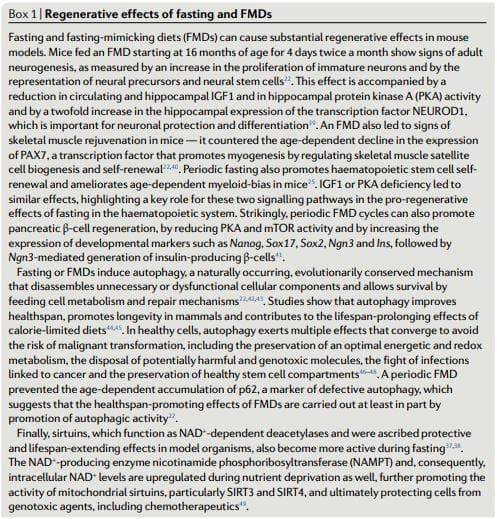
Fasting and the resulting glucose restriction inhibit PKA activity, increase AMPK activity and activate EGR1 and thereby achieve cell-protective effects, including those in the myocardium22,25,26. Lastly, fasting and FMDs (see below for their composition) also have the ability to promote regenerative effects (Box 1) by molecular mechanisms, some of which have been implicated in cancer, such as increased autophagy or induction of sirtuin activity22,37�49.
Dietary Approaches In Cancer FMDs
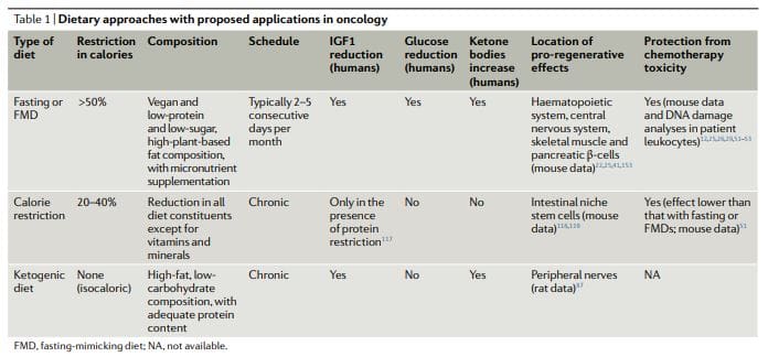
The dietary approaches based on fasting that have been investigated more extensively in oncology, both preclinically and clinically, include water fasting (abstinence from all food and drinks except for water) and FMDs11,12,17,25,26,50�60 (Table 1). Preliminary clinical data indicate that a fast of at least 48hours may be required to achieve clinically meaningful effects in oncology, such as preventing chemotherapy-induced DNA damage to healthy tissues and helping to maintain patient quality of life during chemotherapy52,53,61.
However, most patients refuse or have difficulties completing water fasting, and the potential risks of the extended calorie and micronutrient deficiency associated with it are difficult to justify. FMDs are medically designed dietary regimes very low in calories (that is, typically between 300 and 1,100kcal per day), sugars and proteins that recreate many of the effects of water-only fasting but with better patient compliance and reduced nutritional risk22,61,62. During an FMD, patients typically receive unrestricted amounts of water, small, standardized portions of vegetable broths, soups, juices, nut bars, and herbal teas, as well as supplements of micronutrients. In a clinical study of 3 monthly cycles of a 5-day FMD in generally healthy subjects, the diet was well tolerated and reduced trunk and total body fat, blood pressure and IGF1 levels62. In previous and ongoing oncological clinical trials, fasting or FMDs have typically been administered every 3�4 weeks, for example, in combination with chemotherapy regimens, and their duration has ranged between 1 and 5 days52,53,58,61,63�68. Importantly, no serious adverse events (level G3 or above, according to Common Terminology Criteria for Adverse Events) were reported in this studies52,53,58,61.
Ketogenic Diets
Ketogenic diets (KDs) are dietary regimens that have normal calorie, high-fat and low-carbohydrate content69,70. In a classical KD, the ratio between the weight of fat and the combined weight of carbohydrate and protein is 4:1. Of note, FMDs are also ketogenic because they have high-fat content and have the ability to induce substantial elevations (?0.5mmol per litre) in the levels of circulating ketone bodies. In humans, a KD can also reduce IGF1 and insulin levels (by more than 20% from baseline values), although these effects are affected by the levels and types of carbohydrates and protein in the diet71. KDs can reduce blood glucose levels, but they normally remain within the normal range (that is,>4.4mmol per litre)71.
Notably, KDs may be effective for preventing the increase in glucose and insulin that typically occurs in response to PI3K inhibitors, which was proposed to limit their efficacy72. Traditionally, KDs have been used for treating refractory epilepsy, mainly in children69. In mouse models, KDs induce anticancer effects, particularly in glioblastoma70,72�86. Clinical studies indicate that KDs probably have no substantial therapeutic activity when used as single agents in patients with cancer and suggest that potential benefits of these diets should be sought in combination with other approaches, such as chemotherapy, radiotherapy, antiangiogenic treatments, PI3K inhibitors and FMDs72,73.
KDs were reported to have neuroprotective effects in peripheral nerves and in the hippocampus87,88. However, it remains to be established whether KDs also have proregenerative effects similar to fasting or FMDs (Box 1) and whether KDs also can be used to protect living mammals from the toxicity of chemotherapy. Notably, the regenerative effects of fasting or FMDs appear to be maximized by the switch from the starvation-response mode, which involves the breakdown of cellular components and the death of many cells, and the re-feeding period, in which cells and tissues undergo reconstruction22. Because KDs do not force entry into a starvation mode, do not promote a major breakdown of intracellular components and tissues and do not include a refeeding period, they are unlikely to cause the type of coordinated regeneration observed during the FMD refeeding.
Calorie Restriction
While chronic calorie restriction (CR) and diets deficient in specific amino acids are very different from periodic fasting, they share with fasting and FMDs a more or less selective restriction in nutrients, and they have anticancer effects81,89�112. CR typically involves a chronic 20�30% reduction in energy intake from the standard calorie intake that would allow an individual to maintain a normal weight113,114. It is very effective in reducing cardiovascular risk factors and cancer incidence in model organisms, including primates108,109,114.
However, CR can cause side effects, such as changes in physical appearance, increased cold sensitivity, reduced strength, menstrual irregularities, infertility, loss of libido, osteoporosis, slower wound healing, food obsession, irritability, and depression. In patients with cancer, there are substantial concerns that it may exacerbate malnutrition and that it will unavoidably cause excessive loss of lean body mass18,113�116. CR reduces fasting blood glucose levels, though they remain within the normal range114. In humans, chronic CR does not affect IGF1 levels unless a moderate protein restriction is also implemented117.
Studies show that by reducing mTORC1 signaling in Paneth cells, CR augments their stem cell function and that it also protects reserve intestinal stem cells from DNA damage118,119, but it is unknown whether pro-regenerative effects in other organs are also elicited by CR. Thus, the available data suggest that fasting and FMDs create a metabolic, regenerative and protective profile that is distinct and probably more potent than that elicited by a KD or CR.
Fasting & FMDs In Therapy: Effects on hormone and metabolite levels
Many of the changes in the levels of circulating hormones and metabolites that are typically observed in response to fasting have the capability to exert antitumour effects (that is, reduced levels of glucose, IGF1, insulin and leptin and increased levels of adiponectin)23,120,121 and/or to afford protection of healthy tissues from side effects (that is, reduced levels of IGF1 and glucose). Because ketone bodies can inhibit histone deacetylases (HDACs), the fasting-induced increase of ketone bodies may help slow tumor growth and promote differentiation through epigenetic mechanisms122.
However, the ketone body acetoacetate has been shown to accelerate, instead of reduce, the growth of certain tumors, such as melanomas with mutated BRAF123. Those changes for which there is the strongest evidence for a role in the beneficial effects of fasting and FMDs against cancer are the reductions in the levels of IGF1 and glucose. At the molecular level, fasting or an FMD reduces intracellular signaling cascades including IGF1R�AKT�mTOR�S6K and cAMP�PKA signaling, increases autophagy, helps normal cells withstand stress and promotes anticancer immunity25,29,56,124
Some yeast oncogene orthologues, such as Ras and Sch9 (functional orthologue of mammalian S6K), are able to decrease stress resistance in model organisms27,28. In addition, mutations that activate IGF1R, RAS, PI3KCA or AKT, or that inactivate PTEN, are present in the majority of human cancers10. Together, this led to the hypothesis that starvation would cause opposite effects in cancer versus normal cells in terms of their ability to withstand cell stressors, including chemotherapeutics. In other words, starvation can lead to a differential stress resistance (DSR) between normal and cancer cells.
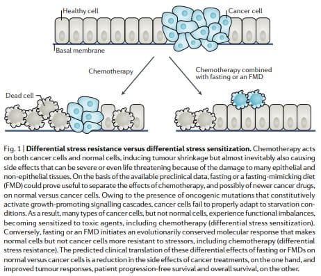
According to the DSR hypothesis, normal cells respond to starvation by downregulating proliferation associated and ribosome biogenesis and/or assembly genes, which forces cells to enter a self-maintenance mode and shields them from the damage caused by chemotherapy, radiotherapy and other toxic agents. By contrast, in cancer cells, this self-maintenance mode is prevented through oncogenic changes, which cause constitutive inhibition of stress response pathways12 (Fig. 1). Consistent with the DSR model, short-term starvation or the deletion of proto-oncogene homologues (that is, Sch9 or both Sch9 and Ras2) increased protection of Saccharomyces cerevisiae against oxidative stress or chemotherapy drugs by up to 100-fold as compared with yeast cells expressing the constitutively active oncogene homologue Ras2val19.
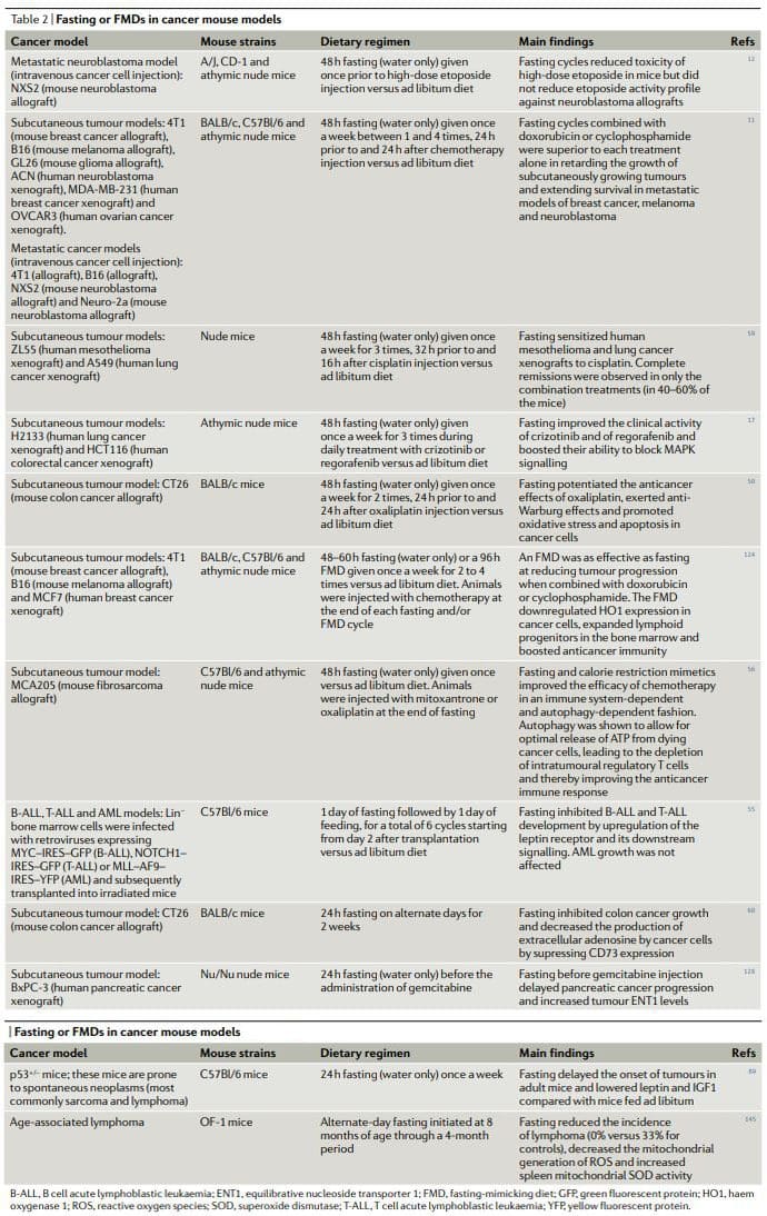
Similar results were obtained in mammalian cells: exposure to low-glucose media protected primary mouse glia cells against toxicity from hydrogen peroxide or cyclophosphamide (a prooxidant chemotherapeutic) but did not protect mouse, rat and human glioma and neuroblastoma cancer cell lines. Consistent with these observations, a 2-day fasting effectively increased the survival of mice treated with high-dose etoposide compared with non-fasted mice and increased the survival of neuroblastoma allograftbearing mice compared with non-fasted tumor-bearing mice12.
Subsequent studies found that reduced IGF1 signaling in response to fasting protects primary glia and neurons, but not glioma and neuroblastoma cells, from cyclophosphamide and from pro-oxidative compounds and protects mouse embryonic fibroblasts from doxorubicin29. Liver IGF1-deficient (LID) mice, transgenic animals with a conditional liver Igf1 gene deletion that exhibit a 70�80% reduction in circulating IGF1 levels (levels similar to those achieved by a 72-hour fast in mice)29,125, were protected against three out of four chemotherapy drugs tested, including doxorubicin.
Histology studies showed signs of doxorubicin-induced cardiac myopathy in only doxorubicin-treated control mice but not in LID mice. In experiments with melanoma-bearing animals treated with doxorubicin, no difference in terms of disease progression between control and LID mice was observed, indicating that cancer cells were not protected from chemotherapy by reduced IGF1 levels. Yet, again, tumour-bearing LID mice exhibited a remarkable survival advantage compared with the control animals owing to their ability to withstand doxorubicin toxicity29. Thus, overall, these results confirmed that IGF1 downregulation is a key mechanism by which fasting increases chemotherapy tolerability.
Both dexamethasone and mTOR inhibitors are widely used in cancer treatment, either because of their efficacy as anti-emetics and anti-allergics (that is, corticosteroids) or for their antitumour properties (that is, corticosteroids and mTOR inhibitors). However, one of their main and frequently dose-limiting side effects is hyperglycaemia. Consistent with the notion that increased glucose�cAMP� PKA signalling reduces resistance to toxicity of chemotherapeutic drugs12,26,126, both dexamethasone and rapamycin increase toxicity of doxorubicin in mouse cardiomyocytes and mice26. Interestingly it was possible to reverse such toxicity by reducing circulating glucose levels through either fasting or insulin injections26.
These interventions reduce PKA activity while increasing AMPK activity and thereby activating EGR1, indicating that cAMP� PKA signalling mediates the fasting-induced DSR via EGR1 (ref. 26). EGR1 also promotes the expression of cardioprotective peptides, such as the atrial natriuretic peptide (ANP) and the B-type natriuretic peptide (BNP) in heart tissue, which contributes to the resistance to doxorubicin. Furthermore, fasting and/or FMD might protect mice from doxorubicin-induced cardiomyopathy by boosting autophagy, which may promote cellular health by reducing reactive oxygen species (ROS) production through the elimination of dysfunctional mitochondria and by removal of toxic aggregates.
In addition to reducing chemotherapyinduced toxicity in cells and increasing survival of chemotherapy-treated mice, cycles of fasting induce bone marrow regeneration and prevent the immunosuppression caused by cyclophosphamide in a PKA-related and IGF1-related manner25. Thus, compelling preclinical results indicate the potential of fasting and FMDs to increase chemotherapy tolerability and to avoid major side effects. Because initial clinical data lend further support to this potential, these preclinical studies build a strong rationale for evaluating FMDs in randomized clinical trials with TEAEs as a primary end point.
Differential Stress Sensitization: Increasing The Death of Cancer Cells
If used alone, most dietary interventions, including fasting and FMDs, have limited effects against cancer progression. According to the differential stress sensitization (DSS) hypothesis, the combination of fasting or FMDs with a second treatment is much more promising11,12. This hypothesis predicts that, while cancer cells are able to adapt to limited oxygen and nutrient concentrations, many types of cancer cells are not able to execute changes that would allow survival in the nutrient-deficient and toxic environment generated by the combination of fasting and chemotherapy, for example. Early experiments in breast cancer, melanoma and glioma cells found a paradoxical increase in the expression of proliferation-associated genes or of ribosome biogenesis and assembly genes in response to fasting11,12. Such changes were accompanied by unexpected AKT and S6K activation, a propensity to generate ROS and DNA damage and a sensitization to DNA-damaging drugs (via DSS)11.
We consider such an inappropriate response of cancer cells to the altered conditions including the reduction in IGF1 and glucose levels caused by fasting or FMDs as a key mechanism underlying the antitumour properties of these dietary interventions and their potential usefulness for separating the effects of anticancer treatments on normal versus malignant cells11,12 (Fig. 1). In line with the DSS hypothesis, periodic cycles of fasting or of FMDs are sufficient to slow the growth of many types of tumour cells, ranging from solid tumour cell lines to lymphoid leukaemia cells, in the mouse and, most importantly, to sensitize cancer cells to chemotherapeutics, radiotherapy and tyrosine kinase inhibitors (TKIs)11,17,22,25,50,54�57,59,60,124,127,128.
By reducing glucose availability and increasing fatty acid ?-oxidation, fasting or FMDs can also promote a switch from aerobic glycolysis (Warburg effect) to mitochondrial oxidative phosphorylation in cancer cells, which is necessary for sustaining cancer cell growth in the most nutrient-poor environment50 (Fig. 2). This switch leads to increased ROS production11 as a result of increased mitochondrial respiratory activity and may also involve a reduction in cellular redox potential owing to decreased glutathione synthesis from glycolysis and the pentose phosphate pathway50. The combined effect of ROS augmentation and reduced antioxidant protection boosts oxidative stress in cancer cells and amplifies the activity of chemotherapeutics. Notably, because a high glycolytic activity demonstrated by high-lactate production is predictive of aggressiveness and metastatic propensity in several types of cancer129, the anti-Warburg effects of fasting or FMD have the potential to be particularly effective against aggressive and metastatic cancers.
Apart from a change in metabolism, fasting or FMDs elicit other changes that can promote DSS in pancreatic cancer cells. Fasting increases the expression levels of equilibrative nucleoside transporter 1 (ENT1), the transporter of gemcitabine across the plasma membrane, leading to improved activity of this drug128. In breast cancer cells, fasting causes SUMO2-mediated and/or SUMO3-mediated modification of REV1, a DNA polymerase and a p53-binding protein127. This modification reduces the ability of REV1 to inhibit p53, leading to increased p53-mediated transcription of pro-apoptotic genes and, ultimately, to cancer cell demise (Fig. 2). Fasting also increases the ability of commonly administered TKIs to stop cancer cell growth and/or death by strengthening MAPK signalling inhibition and, thereby, blocking E2F transcription factor-dependent gene expression but also by reducing glucose uptake17,54.
Finally, fasting can upregulate the leptin receptor and its downstream signalling through the protein PR/SET domain 1 (PRDM1) and thereby inhibit the initiation and reverse the progression of B cell and T cell acute lymphoblastic leukaemia (ALL), but not of acute myeloid leukaemia (AML)55. Interestingly, an independent study demonstrated that B cell precursors exhibit a state of chronic restriction in glucose and energy supplies imposed by the transcription factors PAX5 and IKZF1 (ref. 130). Mutations in the genes encoding these two proteins, which are present in more than 80% of the cases of pre-B cell ALL, were shown to increase glucose uptake and ATP levels. However, reconstituting PAX5 and IKZF1 in preB-ALL cells led to an energy crisis and cell demise. Taken together with the previous study, this work indicates that ALL may be sensitive to the nutrient and energy restriction imposed by fasting, possibly representing a good clinical candidate for testing the efficacy of fasting or FMD.
Notably, it is likely that many cancer cell types, including AML29, can acquire resistance by circumventing the metabolic changes imposed by fasting or FMDs, a possibility that is further increased by the metabolic heterogeneity that characterizes many cancers129. Thus, a major goal for the near future will be to identify the types of cancer that are most susceptible to these dietary regimens by means of biomarkers. On the other hand, when combined with standard therapies, fasting or FMDs have rarely resulted in the acquisition of resistance in cancer mouse models, and resistance to fasting combined with chemotherapy is also uncommon in studies in vitro, underlining the importance of identifying therapies that, when combined with FMDs, result in potent toxic effects against cancer cells with minimal toxicity to normal cells and tissues11,17,50,55�57,59,124.
Antitumour Immunity Enhancement by Fasting or FMD
Recent data suggest that fasting or FMDs by themselves, and to a greater extent when combined with chemotherapy, trigger the expansion of lymphoid progenitors and promote tumour immune attack via different mechanisms25,56,60,124. An FMD reduced the expression of HO1, a protein that confers protection against oxidative damage and apoptosis, in cancer cells in vivo but upregulated HO1 expression in normal cells124,131. HO1 downregulation in cancer cells mediates FMD-induced chemosensitization by increasing CD8+ tumour-infiltrating lymphocyte-dependent cytotoxicity, which may be facilitated by the downregulation of regulatory T cells124 (Fig. 2). Another study, which confirmed the ability of fasting or FMDs and CR mimetics to improve anticancer immunosurveillance, implies that the anticancer effects of fasting or FMDs may apply to autophagy competent, but not autophagy-deficient, cancers56. Finally, a recent study of alternate-day fasting for 2 weeks in a mouse colon cancer model showed that, by activating autophagy in cancer cells, fasting downregulates CD73 expression and consequently decreases the production of immunosuppressive adenosine by cancer cells60. Ultimately, CD73 downregulation via fasting was shown to prevent macrophage shift to an M2 immunosuppressive phenotype (Fig. 2). On the basis of these studies, it is appealing to speculate that FMDs could be particularly useful instead of or in combination with immune checkpoint inhibitors132, cancer vaccines or other drugs that prompt antitumour immunity, including some conventional chemotherapeutics133.
Anticancer Diets in Mouse Models
Overall, the results of preclinical studies of fasting or FMDs in animal cancer models, including models for metastatic cancer (Table 2), show that periodic fasting or FMDs achieve pleiotropic anticancer effects and potentiate the activity of chemotherapeutics and TKIs while exerting protective and regenerative effects in multiple organs22,25. Achieving the same effects without fasting and/or FMDs would require first the identification and then the use of multiple effective, expensive and frequently toxic drugs and would probably be without the advantage of inducing healthy cell protection. It is noteworthy that in at least two studies fasting combined with chemotherapy proved to be the only intervention capable of achieving either complete tumour regressions or long-term survival in a consistent fraction of the treated animals11,59
Chronic KDs also show a tumour growth-delaying effect when used as a monotherapy, particularly in brain cancer mouse models77,78,80�82,84,134. Gliomas in mice maintained on a chronic KD have reduced expression of the hypoxia marker carbonic anhydrase 9 and of hypoxia-inducible factor 1?, decreased nuclear factor-?B activation and reduced vascular marker expression (that is, vascular endothelial growth factor receptor 2, matrix metalloproteinase 2 and vimentin)86. In an intracranial mouse model of glioma, mice fed a KD exhibited increased tumour-reactive innate and adaptive immune responses that were primarily mediated by CD8+ T cells79. KDs were shown to improve the activity of carboplatin, cyclophosphamide and radiotherapy in glioma, lung cancer and neuroblastoma mouse models73�75,135. In addition, a recent study shows that a KD could be very useful in combination with PI3K inhibitors72. By blocking insulin signalling, these agents promote glycogen breakdown in the liver and prevent glucose uptake in the skeletal muscle, which leads to transient hyperglycaemia and to a compensatory insulin release from the pancreas (a phenomenon known as �insulin feedback�). In turn, this raise in insulin levels, which can be protracted, particularly in patients with insulin resistance, reactivates PI3K�mTOR signalling in tumours, thus strongly limiting the benefit of PI3K inhibitors. A KD was shown to be very effective at preventing insulin feedback in response to these drugs and to strongly improve their anticancer activity in the mouse. Finally, according to a study in a murine tumour-induced cachexia model (MAC16 tumours), KDs could help prevent the loss of fat and non-fat body mass in patients with cancer85.
CR reduced tumorigenesis in genetic mouse cancer models, mouse models with spontaneous tumorigenesis and carcinogen induced cancer mouse models, as well as in monkeys91,92,97,98,101,102,104�106,108,109,136�138. By contrast, a study found that CR from middle age actually increases the incidence of plasma cell neoplasms in C57Bl/6 mice139. However, in the same study, CR also extended maximum lifespan by approximately 15%, and the observed increase in cancer incidence was attributed to the increased longevity of mice undergoing CR, the age at which tumour-bearing mice undergoing CR died and the percentage of tumour-bearing mice undergoing CR that died. Thus, the authors concluded that CR probably retards promotion and/or progression of existing lymphoid cancers. A meta-analysis comparing chronic CR with intermittent CR in terms of their ability to prevent cancer in rodents concluded that intermittent CR is more effective in genetically engineered mouse models, but it is less effective in chemically induced rat models90. CR was shown to slow tumour growth and/or to extend mouse survival in various cancer mouse models, including ovarian and pancreatic cancer140,94 and neuroblastoma81.
Importantly, CR improved the activity of anticancer treatment in several cancer models, including the activity of an antiIGF1R antibody (ganitumab) against prostate cancer141, cyclophosphamide against neuroblastoma cells135 and autophagy inhibition in xenografts of HRAS-G12Vtransformed immortal baby mouse kidney epithelial cells100. However, CR or a KD in combination with anticancer therapies seems to be less effective than fasting. A mouse study found that, in contrast to fasting alone, CR alone was not able to reduce the growth of subcutaneously growing GL26 mouse gliomas and that, again, in contrast to short-term fasting, CR did not increase cisplatin activity against subcutaneous 4T1 breast tumours51. In the same study, fasting also proved substantially more effective than CR and a KD at increasing the tolerability of doxorubicin51. Although fasting or an FMD, CR and a KD likely act on and modulate overlapping signalling pathways, fasting or an FMD probably affects such mechanisms in a more drastic fashion during an intense acute phase of a maximum duration of a few days.
The phase of refeeding could then favour the recovery of homeostasis of the whole organism but also activate and invigorate mechanisms that can promote the recognition and removal of the tumour and regenerate the healthy cells. CR and a KD are chronic interventions that are able to only moderately repress nutrient-sensing pathway, possibly without reaching certain thresholds necessary to improve the effects of anticancer drugs, while imposing a major burden and often a progressive weight loss. CR and a KD as chronic dietary regimens in patients with cancer are difficult to implement and likely bear health risks. CR would likely lead to severe loss of lean body mass and the reduction of steroid hormones and possibly immune function142. Chronic KDs are also associated with similar although less severe side effects143. Thus, periodic fasting and FMD cycles lasting less than 5 days applied together with standard therapies have a high potential to improve cancer treatment while reducing its side effects. Notably, it will be important to study the effect of the combination of periodic FMDs, chronic KDs and standard therapies, particularly for the treatment of aggressive cancers such as glioma.
Fasting and FMDs in Cancer Prevention
Epidemiological studies and studies in animals, including monkeys108,109,144, and humans lend support to the notion that chronic CR and periodic fasting and/or an FMD could have cancer-preventive effects in humans. Nevertheless, CR can hardly be implemented in the general population owing to issues of compliance and to possible side effects115. Thus, while evidence-based recommendations of foods to prefer (or to avoid) as well as lifestyle recommendations to reduce cancer risk are becoming established6,8,9,15, the goal now is to identify and, possibly, standardize well tolerated, periodic dietary regimens with low or no side effects and evaluate their cancer-preventive efficacy in clinical studies.
As discussed earlier, FMD cycles cause downregulation of IGF1 and glucose and upregulation of IGFBP1 and ketone bodies, which are changes similar to those caused by fasting itself and are biomarkers of the fasting response22. When C57Bl/6 mice (which spontaneously develop tumours, primarily lymphomas, as they age) were fed such an FMD for 4 days twice a month starting at middle age and an ad libitum diet in the period between FMD cycles, the incidence of neoplasms was reduced from approximately 70% in mice on the control diet to approximately 40% in mice in the FMD group (an overall 43% reduction)22. In addition, the FMD postponed by over 3 months the occurrence of neoplasm related deaths, and the number of animals with multiple abnormal lesions was more than threefold higher in the control group than in the FMD mice, indicating that many tumours in the FMD mice were less aggressive or benign.
A previous study of alternate-day fasting, which was performed in middle-aged mice for a total of 4 months, also found that fasting reduced the incidence of lymphoma, bringing it from 33% (for control mice) to 0% (in fasted animals)145, although because of the short duration of the study it is unknown whether this fasting regimen prevented or simply delayed the tumour onset. Furthermore, alternate-day fasting imposes 15 days per month of complete water-only fasting, whereas in the FMD experiment described above mice were placed on a diet that provided a limited amount of food for only 8 days per month. In humans, 3 cycles of a 5-day FMD once a month were shown to reduce abdominal obesity and markers of inflammation as well as IGF1 and glucose levels in subjects with elevated levels of these markers62, indicating that periodic use of an FMD could potentially have preventive effects for obesity-related or inflammation-related, but also other, cancers in humans, as it has been shown for mice22.
Therefore, the promising results of preclinical studies combined with the clinical data on the effect of an FMD on risk factors for ageing-associated diseases, including cancer62, lend support to future randomized studies of FMDs as a possibly effective tool to prevent cancer, as well as other ageing-associated chronic conditions, in humans.
Clinical Applicability in Oncology
Four feasibility studies of fasting and FMDs in patients undergoing chemotherapy have been published as of today52,53,58,61. In a case series of 10 patients diagnosed with various types of cancer, including breast, prostate, ovarian, uterus, lung and oesophageal cancer, who voluntarily fasted for up to 140hours before and/or up to 56hours following chemotherapy, no major side effects caused by fasting itself other than hunger and lightheadedness were reported58. Those patients (six) who underwent chemotherapy with and without fasting reported a significant reduction in fatigue, weakness and gastrointestinal adverse events while fasting. In addition, in those patients in which cancer progression could be assessed, fasting did not prevent chemotherapy-induced reductions in tumour volume or in tumour markers. In another study, 13 women with HER2 (also known as ERBB2) negative, stage II/III breast cancer receiving neo-adjuvant taxotere, adriamycin and cyclophosphamide (TAC) chemotherapy were randomized to fast (water only) 24hours before and after beginning chemotherapy or to nutrition according to standard guidelines52.
Short-term fasting was well tolerated and reduced the drop in mean erythrocyte and thrombocyte counts 7 days after chemotherapy. Interestingly, in this study, the levels of ?-H2AX (a marker of DNA damage) were increased 30minutes after chemotherapy in leukocytes from non-fasted patients but not in patients who had fasted. In a dose escalation of fasting in patients undergoing platinum-based chemotherapy, 20 patients (who were primarily treated for either urothelial, ovarian or breast cancer) were randomized to fast for 24, 48 or 72hours (divided as 48hours before chemotherapy and 24hours after chemotherapy)53. Feasibility criteria (defined as three or more out of six subjects in each cohort consuming?200kcal per day during the fast period without excess toxicity) were met. Fasting-related toxicities were always grade 2 or below, the most common being fatigue, headache and dizziness. As in the previous study, reduced DNA damage (as detected by comet assay) in leukocytes from subjects who fasted for at least 48hours (as compared with subjects who fasted for only 24hours) could also be detected in this small trial. In addition, a nonsignificant trend towards less grade 3 or grade 4 neutropenia in patients who fasted for 48 and 72hours versus those who fasted for only 24hours was also documented.
Very recently, a randomized crossover clinical trial was conducted assessing the effects of an FMD on quality of life and side effects of chemotherapy in a total of 34 patients with breast or ovarian cancer61. The FMD consisted of a daily caloric intake of<400kcal, primarily by juices and broths, starting 36�48hours before the beginning of chemotherapy and lasting until 24hours after the end of chemotherapy. In this study, the FMD prevented the chemotherapy induced reduction in quality of life and it also reduced fatigue. Again, no serious adverse events of the FMD were reported. Several other clinical trials of FMDs in combination with chemotherapy or with other types of active treatments are currently ongoing at US and European hospitals, primarily in patients who are diagnosed with breast or prostate cancer63,65�68. These are either one-arm clinical studies to assess FMD safety and feasibility or randomized clinical studies focusing either on the effect of the FMD on the toxicity of chemotherapy or on the quality of life of patients during chemotherapy itself. Altogether, these studies have now enrolled over 300 patients, and their first results are expected to become available in 2019.
Challenges in The Clinic
The study of periodic fasting or of FMDs in oncology is not devoid of concerns, particularly in relation to the possibility that this type of dietary regimen could precipitate malnutrition, sarcopenia, and cachexia in predisposed or frail patients (for example, patients who develop anorexia as a consequence of chemotherapy)18,19. However, no instances of severe (above grade 3) weight loss or of malnutrition were reported in the clinical studies of fasting in combination with chemotherapy published as of now, and those patients who did experience a weight loss during fasting typically recovered their weight before the subsequent cycle without detectable harm. Nevertheless, we recommend that periodic anorexia and nutritional status assessments using gold-standard approaches18,19,146�150 should be an integral part of these studies and that any ensuing nutritional impairment in patients undergoing fasting and/or FMDs is rapidly corrected.
Conclusions
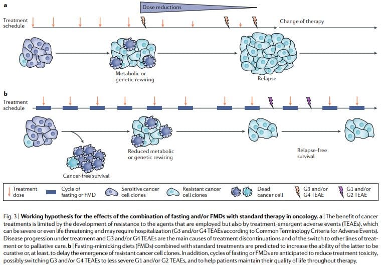
Periodic fasting or FMDs consistently show powerful anticancer effects in mouse cancer models including the ability to potentiate chemoradiotherapy and TKIs and to trigger anticancer immunity. FMD cycles are more feasible than chronic dietary regimens because they allow patients to consume food regularly during the FMD, maintain a normal diet between cycles and do not result in severe weight loss and possibly detrimental effects on the immune and endocrine systems. Notably, as standalone therapies, periodic fasting or FMD cycles would probably show limited efficacy against established tumors. In fact, in mice, fasting or FMDs affect the progression of a number of cancers similarly to chemotherapy, but alone, they rarely match the effect obtained in combination with cancer drugs which can result in cancer-free survival11,59. Thus, we propose that it is the combination of periodic FMD cycles with standard treatments that holds the highest potential to promote cancer-free survival in patients, as suggested by the mouse models11,59 (Fig. 3).
This combination may be particularly potent for several reasons: first, cancer drugs and other therapies can be effective, but a portion of patients do not respond because cancer cells adopt alternative metabolic strategies leading to survival. These alternative metabolic modes are much more difficult to sustain under fasting or FMD conditions because of the deficiencies or changes in glucose, certain amino acids, hormones, and growth factors, as well as in other unknown pathways leading to cell death. Second, fasting or FMDs can prevent or reduce resistance acquisition. Third, fasting or FMDs protect normal cells and organs from the side effects caused by a wide variety of cancer drugs. On the basis of preclinical and clinical evidence of feasibility, safety and efficacy (at reducing IGF1, visceral fat and cardiovascular risk factors), FMDs also appear as a viable dietary approach to be studied in cancer prevention. An important future challenge will be to identify those tumours that are the best candidates to benefit from fasting or FMDs. Even in cancer types that are apparently less responsive to fasting or FMDs, it may still be possible to identify the mechanisms of resistance and to intervene with drugs able to revert that resistance. Conversely, more caution should be adopted with other types of diets, especially if high in calories, as they could lead to exacerbated and not inhibited growth of certain cancers. For example, the KD increases growth of a melanoma model with mutated BRAF in mice123, and it was also reported to accelerate disease progression in a mouse AML model72.
Furthermore, it is essential to apply FMDs with an understanding of the mechanisms of action, since their potency if applied incorrectly could generate negative effects. For example, when rats were fasted and treated with a potent carcinogen before refeeding, this resulted in the growth of aberrant foci in liver, colon and rectum when compared with non-fasted rats151,152. Although the mechanisms involved in this effect are not understood, and these foci may have not resulted in tumours, these studies suggest that a minimum period of 24�48hours between the chemotherapy treatment and the return to the normal diet is important to avoid combining the regrowth signals present during the refeeding after fasting with high levels of toxic drugs such as chemotherapy. The clinical studies of fasting or FMD in patients undergoing chemotherapy support its feasibility and overall safety52,53,58,61. In a small-size randomized trial that enrolled 34 patients, an FMD helped patients maintain their quality of life during chemotherapy and reduced fatigue61. In addition, preliminary data suggest the potential of fasting or FMDs to reduce chemotherapy induced DNA damage in healthy cells in patients52,53.
Ongoing clinical studies of FMDs in patients with cancer63,65�68 will provide more solid answers as to whether prescribing periodic FMDs in combination with conventional anticancer agents helps improve tolerability and activity of the latter. It is important to consider that FMDs will not be effective in reducing the side effects of cancer treatments in all patients and neither will they work to improve the efficacy of all therapies, but they have great potential to do so at least for a portion and possibly for a major portion of patients and drugs. Frail or malnourished patients or patients at risk of malnutrition should not be enrolled in clinical studies of fasting or FMDs, and patient nutritional status and anorexia should be carefully monitored throughout clinical trials. An appropriate intake of proteins, essential fatty acids, vitamins and minerals combined, where possible, with light and/or moderate physical activity aimed at increasing muscle mass should be applied between fasting or FMD cycles in order for the patients to maintain a healthy lean body mass18,19. This multimodal dietary approach will maximize the benefits of fasting or FMDs while at the same time protecting patients from malnutrition.
Although oral devices, such as splints and bite guards, are the most prevalent treatments for facial pain associated with temporomandibular disorders, or TMD, patients have found that these remedies are frequently less effective than self-care techniques, such as jaw exercises or warm compresses, according to a new research study published by researchers at the New York University (NYU) College of Dentistry in New York City.
The research study, published in the journal Clinical Oral Investigations, demonstrates that self-care techniques should primarily be utilized to help treat muscle-related temporomandibular disorders or TMD.
TMD, occasionally known as TMJ after the temporomandibular joint, is a collection of prevalent painful conditions which develop in the jaw joint and its surrounding muscles. Myofascial temporomandibular disorder, or mTMD, is a muscular condition which affects over 10 percent of women. Individuals with TMD often suffer from other chronic pain conditions. Research studies found that 7 to 18 percent of people with TMD also experience fibromyalgia, a condition characterized by widespread pain.
Treatments for TMD and Fibromyalgia
Dentists and patients utilize an assortment of treatments to help manage facial pain, such as oral devices like splints and bite guards, pain medicines, including nonsteroidal anti-inflammatory drugs, and self-care methods like jaw exercises and hot compresses.
Oral devices are a prevalent first-line treatment for TMD, regardless of research study outcome measures regarding their advantages, stated Vivian Santiago, Ph.D., MPH, research study scientist at the Department of Oral and Maxillofacial Pathology, Radiology, and Medicine at NYU College of Dentistry, and the research study’s leading author.
“While oral splints have been discovered to have some benefits, they have yet to be found to be as successful for patients who have widespread pain when treating mTMD,” she explained.
In this research study, the researchers evaluated what non-medication remedies women with mTMD utilized to handle their pain as well as how successful patients perceived these remedies. The researchers interviewed a total of 125 women including 26 women who had fibromyalgia and mTMD, so as to find out whether treatment differed for patients.
The most frequent treatments reported were oral devices (utilized by 59 percent of participants), physical therapy (utilized by 54 percent of participants), and at-home jaw exercises (utilized by 34 percent of participants). The least frequent treatments reported were acupuncture (utilized by 20 percent), chiropractic care (utilized by 18 percent), trigger point injections (utilized by 14 percent), yoga (utilized by 7 percent), and meditation (utilized by 6 percent). Participants frequently used more than one treatment.
Participants reported the most improvement in their pain from well-known self-care techniques, such as jaw exercises, yoga, meditation, massage, and warm compresses, with over 84 percent reporting that these techniques helped reduce painful symptoms. Only 64 percent of participants who used the oral devices reported that they helped improve their pain. About 11 percent of women who used oral devices stated that these made their pain worse, an area which warrants further research studies.
Oral devices failed to outperform self-care techniques in improving facial pain, according to Karen Raphael, Ph.D., professor at the Department of Oral and Maxillofacial Pathology, Radiology, and Medicine at NYU College of Dentistry, and the research study’s co-author.
“Our outcome measures encourage utilizing self-care techniques as the first line of treatment for mTMD before contemplating more costly interventions,” stated Raphael.
The researchers didn’t find substantial differences between the amount of remedies reported by women with and without fibromyalgia. While the use of alternative treatment options for mTMD was reported among women with fibromyalgia, further research studies are still required. Pain relief tended to be greater through the use of self-care techniques in women with and without fibromyalgia.
“While fibromyalgia is diagnosed by a healthcare professional, such as a rheumatologist, TMD is typically diagnosed and treated by a dentist,” said Santiago. “Our research study demonstrates that dentists must ask patients with facial pain if they also have widespread chronic pain because this might provide more information to help plan their treatment.”
Fibromyalgia is a health issue characterized by widespread chronic pain accompanied by fatigue, sleep, memory and mood problems. Fibromyalgia has been associated with a variety of other health issues, such as TMD and/or TMJ. Individuals with this painful disorder may often struggle to engage in their everyday physical activities. As a qualified and experienced chiropractor, I’ve helped treat numerous patients with fibromyalgia. It’s important for patients to know that they are not alone when it comes to treating their painful symptoms. Chiropractic care is an alternative treatment option which can help treat a variety of health issues, including fibromyalgia.
Dr. Alex Jimenez D.C., C.C.S.T. Insight
The scope of our information is limited to chiropractic, spinal health issues, and functional medicine articles, topics, and discussions. To further discuss the subject matter above, please feel free to ask Dr. Alex Jimenez or contact us at 915-850-0900 .
Curated by Dr. Alex Jimenez
Additional Topic Discussion: Acute Back Pain
Back pain is one of the most prevalent causes of disability and missed days at work worldwide. Back pain attributes to the second most common reason for doctor office visits, outnumbered only by upper-respiratory infections. Approximately 80 percent of the population will experience back pain at least once throughout their life. Your spine is a complex structure made up of bones, joints, ligaments, and muscles, among other soft tissues. Injuries and/or aggravated conditions, such as herniated discs, can eventually lead to symptoms of back pain. Sports injuries or automobile accident injuries are often the most frequent cause of back pain, however, sometimes the simplest of movements can have painful results. Fortunately, alternative treatment options, such as chiropractic care, can help ease back pain through the use of spinal adjustments and manual manipulations, ultimately improving pain relief.
XYMOGEN�s Exclusive Professional Formulas are available through select licensed health care professionals. The internet sale and discounting of XYMOGEN formulas are strictly prohibited.
Proudly, Dr. Alexander Jimenez makes XYMOGEN formulas available only to patients under our care.
Please call our office in order for us to assign a doctor consultation for immediate access.
If you are a patient of Injury Medical & Chiropractic Clinic, you may inquire about XYMOGEN by calling 915-850-0900.
For your convenience and review of the XYMOGEN products please review the following link.*XYMOGEN-Catalog-Download
* All the above XYMOGEN policies remain strictly in force.
A heart-healthy diet rich in fruits, vegetables, fish, and nuts which is also low in meat, and full-fat dairy is related to improved middle-aged cognitive function, such as memory and thinking skills, according to a research study published in the journal “Neurology”.
The research study, directed by Claire McEvoy, Ph.D., of Queen’s University Belfast in Northern Ireland, analyzed which dietary patterns, including the Mediterranean diet plan, Dietary Approaches to Stop Hypertension (DASH), and the A Priori Diet Quality Score (APDQS), throughout early adulthood have been correlated with middle age brain function and cognitive performance.
Nutrition and Better Brain Function
Researchers evaluated 2,621 Coronary Artery Risk Development in Young Adults, or CARDIA, study participants with a mean age of 25 years old throughout a 30-year period. The study participants were asked about their diet at the start of the research study, after seven years, and after 20 years. The study participants’ brain function was analyzed when they were 50 and 55 years old.
For every diet, the participants of the research study were separated into one of 3 categories, low, moderate, or higher adherence, dependent on how closely they followed their diet plan. The evaluation demonstrated that the DASH diet wasn’t associated with any alteration in cognitive function. A Mediterranean diet or a APDQS diet also resulted in a lesser decline in cognitive functioning.
Individuals with higher adherence to the Mediterranean diet were 46 percent less likely to experience poor memory and thinking skills than individuals with reduced adherence to this diet plan, according to the research study. Of those individuals in the adherence group, 9 percent had poor memory and thinking skills, compared to 29 percent of those 798 men and women in the adherence group.
Individuals with higher adherence to the APDQS diet were 52 percent less likely to have poor memory and thinking skills than individuals with reduced adherence to the diet plan. Of the 938 men and women in the adherence class, 6 percent had poor memory and thinking skills, compared to 32 percent of those 805 men and women in the adherence group, according to researchers.
The outcome measures of the research study were adjusted for other variables that may affect cognitive functioning, including the degree of education, as well as smoking, physical activity or exercise, and health issues like diabetes, according to the research study.
Numerous research studies have attempted to evaluate the effects of nutrition on brain health. Recent research studies, however, have demonstrated that certain diet plans, such as the Mediterranean diet, the DASH diet, and the APDQS diet, can help decrease the decline of cognitive function during middle age. The research study also demonstrated the effects these diets have on overall brain health. A balanced nutrition is essential towards optimal well-being. Before following any of the diet plans described in the following article, make sure to talk to a healthcare professional about your options.
Dr. Alex Jimenez D.C., C.C.S.T. Insight
Types of Diets for Improved Brain Function
The Mediterranean diet emphasizes the consumption of whole grains, fruits, vegetables, legumes, nuts, healthy unsaturated fats, and fish while restricting the consumption of red meat, poultry, and full-fat dairy. In contrast, the DASH diet emphasizes the consumption of grains, fruits, vegetables, legumes, nuts, and low-fat dairy while restricting the consumption of meat, fish, poultry, total fat, saturated fat, sweets, and sodium. The APDQS diet emphasizes the consumption of fruits, vegetables, legumes, low-fat fish, poultry, and alcohol while restricting the consumption of fried foods, salty snacks, sweets, high-fat dairy, and sugar-sweetened soft drinks.
The Mediterranean diet and the DASH diet regularly rank as some of the top diets for humans, according to the U.S. News and World Report’s yearly evaluation. According to researchers, it’s uncertain why the DASH diet didn’t result in improved cognitive functioning.
Further research studies are required to specify how nutrition, diet, and food contributes towards optimal brain health throughout our life span. According to a statement published by the American Academy of Neurology, “While we do not yet understand the perfect nutritional supplement for brain health, shifting towards a heart-healthy diet might be a relatively safe and effective approach to decrease the danger of developing health issues with memory and thinking skills throughout our middle age.”
The scope of our information is limited to chiropractic, spinal health issues, and functional medicine articles, topics, and discussions. To further discuss the subject matter above, please feel free to ask Dr. Alex Jimenez or contact us at 915-850-0900 .
Curated by Dr. Alex Jimenez
Additional Topic Discussion: Acute Back Pain
Back pain is one of the most prevalent causes of disability and missed days at work worldwide. Back pain attributes to the second most common reason for doctor office visits, outnumbered only by upper-respiratory infections. Approximately 80 percent of the population will experience back pain at least once throughout their life. Your spine is a complex structure made up of bones, joints, ligaments, and muscles, among other soft tissues. Injuries and/or aggravated conditions, such as herniated discs, can eventually lead to symptoms of back pain. Sports injuries or automobile accident injuries are often the most frequent cause of back pain, however, sometimes the simplest of movements can have painful results. Fortunately, alternative treatment options, such as chiropractic care, can help ease back pain through the use of spinal adjustments and manual manipulations, ultimately improving pain relief.
XYMOGEN�s Exclusive Professional Formulas are available through select licensed health care professionals. The internet sale and discounting of XYMOGEN formulas are strictly prohibited.
Proudly, Dr. Alexander Jimenez makes XYMOGEN formulas available only to patients under our care.
Please call our office in order for us to assign a doctor consultation for immediate access.
If you are a patient of Injury Medical & Chiropractic Clinic, you may inquire about XYMOGEN by calling 915-850-0900.
For your convenience and review of the XYMOGEN products please review the following link.*XYMOGEN-Catalog-Download
* All the above XYMOGEN policies remain strictly in force.
Hormones can also play a significant part in the pain. One of the many changes that occur in a pregnant woman�s body is an increase in the production of a hormone called Relaxin. The job of this hormone is to �relax� the ligaments or soften them. This allows the pelvis to spread so that the baby can be born. However, while the body is making these positive and necessary preparations, it can also allow bones to shift or move in directions that they shouldn�t. The result is an impingement or compression on the nerves exiting the spinal canal, causing the tissues in the area to become irritated and inflamed.
Another effect of the increased production of relaxin is that it can lead to intrauterine constraint. This condition causes the pelvic bones to become misaligned which obstructs the baby�s natural movement in utero while it develops. This can keep the baby from moving into the birthing position with the head down. As the bones move out of their natural place, it can stress, pull, and twist the attached ligaments. The baby doesn�t have as much space as it should so it can�t move like a normal fetus can. This can also cause breech births. Stress, poor posture, and overexertion can also cause pregnancy back pain.
Why is pregnancy low back pain so difficult to treat and what treatments are available?
Medications given to the mother will cross over the placenta delivering a dose of the medication to the baby. Because of this, pain medications are generally off limits for pregnant women. In order to protect the baby, natural pain control is preferred. This makes chiropractic a preferred treatment for pregnancy low back pain. The chiropractor may perform a spinal subluxation to bring the spine back into alignment and the body back onto balance.
He or she may also make some recommendations to help the woman manage her pain on her own, including improving her posture, stretching, and special exercises. Other recommendations may include:
Not wearing high heels. They put strain on the back when a woman isn�t pregnant. On a pregnant body, the strain is even worse.
Avoid bending over to pick up things, but instead, squat � or ask for help.
Sleeping on the left side and use pillows under the belly to support it as well as a pillow between her legs.
Rest, lots of rest and elevating her feet.
Regular chiropractic care and following the doctor�s instructions can help greatly decrease low back pain for the mom to be so that she can better enjoy the excitement and joy of her pregnancy.
Lower Back Pain Pregnancy Chiropractic Treatment El Paso, TX
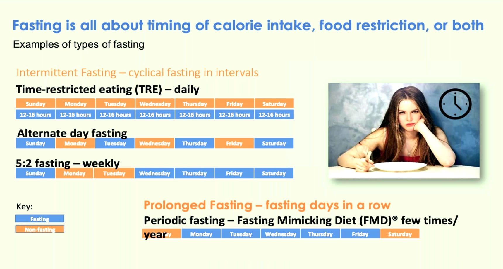
Recent science has unleashed the power of fasting to optimize body aging and dismantle chronic diseases, e.g., obesity, diabetes, cancer, autoimmune disorders, Alzheimer�s and cardiovascular disease. At El Paso Back Clinic, Dr. Jimenez’s mission is to educate the public about the health benefits of fasting. The idea is to introduce it to them in a safe, effective way through the Fasting Mimicking Diet (FMD).
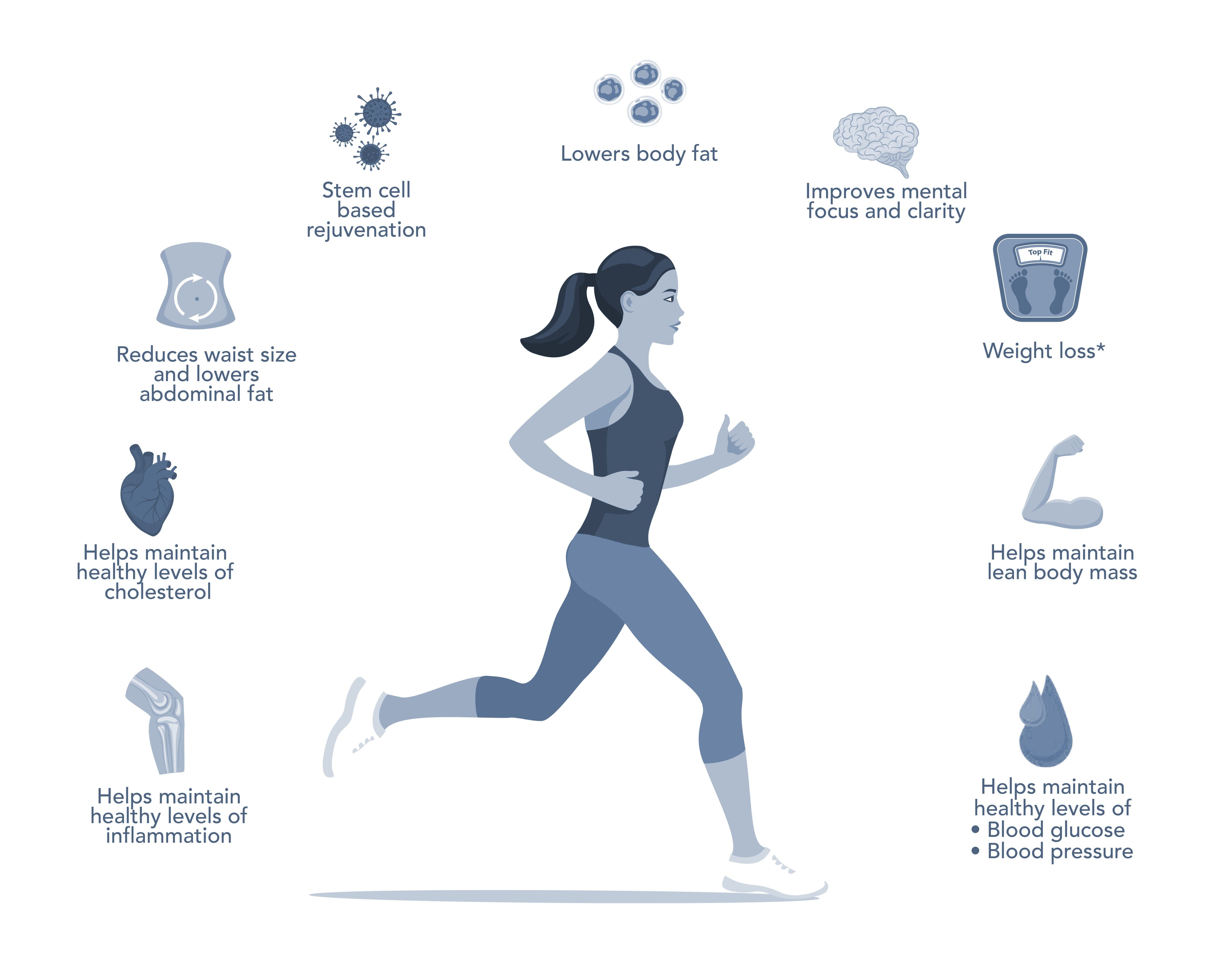
Pre-clinical and clinical studies have proven that periodic fasting, done for several consecutive days, is a very powerful intervention that our bodies learned to naturally cope with by protecting and rejuvenating itself. These two factors are both anti-aging measures that offer additional health benefits. The 5-Day ProLon Fasting Mimicking Diet has been clinically tested and found to promote beneficial effects in a wide variety of conditions ranging from excess weight and fasting blood glucose, to growth factors associated with DNA damage and aging.
Min Wei; Sebastian Brandhorst et al. Fasting?Mimicking Diet and Risk Factors for Aging, Diabetes, Cancer and Cardiovascular Disease.
5-Day Program
The ProLon meal plan is followed 5 days per month. Once an individual has finished the five-day plan, they go back to a normal healthy dietthe last twenty-five days. Fasting with the Prolon� plan follows a low carbohydrate/protein meal and contains the good kind of fatty acids. The FMD� recipe keeps your body on a fasting-type mode, that triggers protection measures that the body has developed. This causes the body to optimize its performance, rejuvenate cells, and thrive.
Unboxing the ProLon Fasting Mimicking Diet
Age-related Disease: A Revolution Is Coming
I just finished reading a groundbreaking book called The Longevity Diet: Discover the New Science Behind Stem Cell Activation and Regeneration to Slow Aging, Fight Disease, and Optimize Weight, which was written by Valter Longo, PhD,1 who is director of the Longevity Institute at the University of Southern California and a principal scientist in the development and study of the fasting mimicking diet (FMD). I have followed Dr. Longo�s career for many years with great admiration. He has been published in top-tier journals. In his book, Dr. Longo writes about some exciting findings regarding the FMD research group consisting of 16-month-old mice, which are described as being the equivalent of a 45-year-old human: �A stem cell-dependent process rejuvenated the immune system. Regeneration also occurred in the liver, muscle, and brain. Levels of several types of stem cells increased.�1 He went on to explain: �The fasting itself destroys many damaged cells and damaged components inside the cells but it also activates stem cells.�1
Jeffrey S. Bland, PhD, FACN, FACB, Associate Editor
Get Your Free Copy At El Paso Back Clinic!
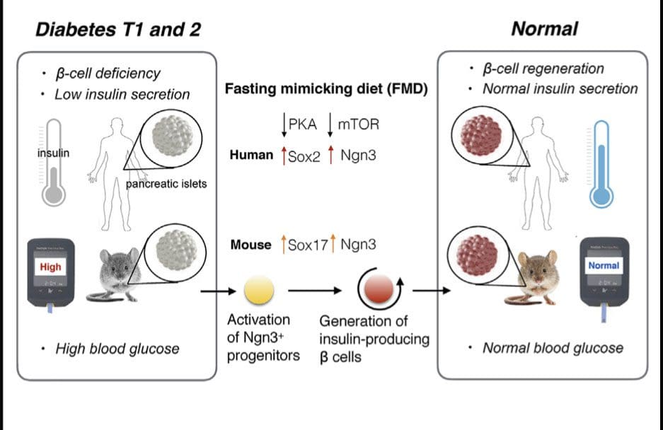
Fasting-Mimicking Diet Promotes b-Cell Regeneration to Reverse Diabetes
Fasting mimicking diet induces prenatal-development gene expression in adult pancreas
FMD promotes Ngn3 expression to generate insulin-producing b cells
Cycles of FMD reverse b-cell failure and rescue mice fromT1D and T2Dd
Inhibition of PKA or mTOR promotes Ngn3-driven b-cell regeneration in human T1D islets
Stem-cell-based therapies can potentially reverse organ dysfunction and diseases, but the removal of impaired tissue and activation of a program leading to organ regeneration pose major challenges. In mice, 4-day fasting mimicking diet (FMD) induces a stepwise expression of Sox17 and Pdx-1, followed by Ngn3-driven generation of insulin-producing b cells, resembling that observed during pancreatic development. FMD cycles restore insulin secretion and glucose homeostasis in both type 2 and type 1diabetes mouse models.
In human type 1 diabetes pancreatic islets, fasting conditions reduce PKAand mTOR activity and induce Sox2 and Ngn3expression and insulin production. The effects of the FMD are reversed by IGF-1 treatment and recapitulated by PKA and mTOR inhibition. These results indicate that an FMD promotes the reprogramming of pancreatic cells to restore insulin generation in islets from T1D patients and reverse both T1D andT2D phenotypes in mouse models.
Chia-Wei Cheng,1,6,7Valentina Villani,2,7Roberta Buono,1,5,7Min Wei,1Sanjeev Kumar,4Omer H. Yilmaz,6Pinchas Cohen,1Julie B. Sneddon,3Laura Perin,2and Valter D. Longo1,4,5,8,*
Individuals getting a very low percentage of their daily calories from carbohydrates, such as fruits, grains, and starchy vegetables, are more likely to develop atrial fibrillation, or AFib. This health issue is one of the most prevalent heart rhythm disorders, according to a new research study being presented at the American College of Cardiology’s 68th Annual Scientific Session.
The research study examined the health records of almost 14,000 people spanning two or more decades. Researchers brought data from Atherosclerosis Risk in Communities, or ARIC, a research study controlled by the National Institutes of Health which was conducted from 1985 to 2016. Of almost 1,900 participants that were diagnosed through a mean of 22 years of follow-up, a majority of them were identified with AFib by researchers. The details of the research study are described below.
AFib and Carbohydrates
Research study participants were requested to report the everyday consumption of 66 distinct food items in a poll. The researchers utilized this information to gauge the percentage of calories which came from carbohydrates from each participant’s calorie intake. Carbohydrates were contained in roughly half of the daily calories consumed by the participants.
Researchers subsequently separated the participants into three separate groups categorized by low, moderate, and high carbohydrate intake, representing diets where carbohydrates consisted less than 44.8 percent of their daily calories, followed by 44.8 to 52.4 percent, and finally where carbohydrates consisted more than 52.4 percent of their daily calories, respectively.
Participants who reporting reduced carbohydrate consumption were the ones who had the highest probability of developing AFib, according to researchers. As the statistics of the research study later demonstrated, these participants were also 18 percent more likely to come up with AFib compared to those with moderate carbohydrate intake and 16 percent more likely to come up with AFib compared to those with high carbohydrate ingestion. Some diets can also help decrease the risk of heart rhythm disorders.
The type of carbohydrates you eat can make a huge difference in your overall health and wellness. Complex carbohydrates are digested more slowly than simple carbohydrates and these release a steady release of sugar, or glucose, into the blood stream. Complex carbohydrates, often referred to as “starchy” foods, include legumes, starchy vegetables, whole grain, and fiber. According to the research study in the following article, consuming low amounts of carbohydrates, which often includes fruits, vegetables, and whole grains, can contribute to cardiovascular diseases, such as atrial fibrillation. When it comes to carbohydrates, it’s important to consume this essential macronutrient for overall health and wellness.
Dr. Alex Jimenez D.C., C.C.S.T. Insight
Nutrition for AFib
Restricting carbohydrates has become a popular weight loss plan. Many diets, such as the Paleo and the ketogenic diet, highlight the consumption of proteins. According to Xiaodong Zhuang, MD, PhD, cardiologist and the research study’s lead author, “The long-term impact of carbohydrate restriction remains controversial, particularly with respect to its own influence on cardiovascular disease.” “Considering the possible effects on arrhythmia, our research study indicates that this popular weight control system ought to be recommended carefully,” he stated in a statement published by the ACC.
The findings complement previous research studies, a number of which have correlated both polyunsaturated and high-carbohydrate diets with a greater probability of death. While previous research studies indicated that this part of the diet affected the outcome measures found, the research study itself didn’t determine these findings. “Low carbohydrate diets have been associated with greater risk of developing AFib irrespective of the type of fat or protein utilized to substitute the carbohydrate,” Zhuang said.
“Several possible mechanisms could explain why limiting carbohydrates may contribute to AFib,” Zhuang said. One is that individuals eating a low-carbohydrate diet often consume fewer fruits, vegetables, and whole grains. Without these foods, individuals may experience more widespread inflammation, which has been connected with AFib. According to the research study, another potential explanation is that eating more fat and protein instead of carbohydrate-rich foods can result in oxidative stress, which has also been connected to AFib. The effect may be associated with an increased risk of other types of cardiovascular disease.
The Longevity Diet Plan, presented in the book by Dr. Valter Longo, eliminates the consumption of processed foods which can cause inflammation, promoting well-being and longevity. While this diet program doesn’t focus on weight loss, the emphasis of the longevity diet plan is on eating healthier. The Longevity Diet Plan has been demonstrated to help activate stem cell-based renewal, reduce abdominal fat, and prevent age-related bone and muscle loss, as well as build resistance to developing cardiovascular disease.
The fasting mimicking diet, or FMD, allows you to experience the benefits of traditional fasting without depriving your body of food. The main difference of the FMD is that instead of completely eliminating all food for several days or even weeks, you only restrict your calorie intake for five days out of the month. The FMD can be practiced once a month to help promote overall health and wellness.
While anyone can follow the FMD on their own, the ProLon� fasting mimicking diet offers a 5-day meal program which has been individually packed and labeled for each day, which serves the foods you need for the FMD in precise quantities and combinations. The meal program is made up of ready-to-eat and easy-to-prepare, plant-based foods, including bars, soups, snacks, supplements, a drink concentrate, and teas. Before starting the ProLon� fasting mimicking diet, 5-day meal program, or any of the lifestyle modifications described above, please make sure to talk to a healthcare professional to find out if this dietary program is right for you.
Furthermore, the research study didn’t monitor participants with asymptomatic AFib, or people who had AFib but were never admitted to a hospital. It didn’t investigate subtypes of AFib, therefore it’s unknown if patients were far more likely to have episodes of persistent or arrhythmia AFib. Zhuang reported that the research study didn’t show cause and effect. A randomized trial could be required to validate the connection between AFib and carbohydrate intake to evaluate the result in a more diverse population.
The scope of our information is limited to chiropractic, spinal health issues, and functional medicine articles, topics, and discussions. To further discuss the subject matter above, please feel free to ask Dr. Alex Jimenez or contact us at 915-850-0900 .
Curated by Dr. Alex Jimenez
Additional Topic Discussion: Acute Back Pain
Back pain is one of the most prevalent causes of disability and missed days at work worldwide. Back pain attributes to the second most common reason for doctor office visits, outnumbered only by upper-respiratory infections. Approximately 80 percent of the population will experience back pain at least once throughout their life. Your spine is a complex structure made up of bones, joints, ligaments, and muscles, among other soft tissues. Injuries and/or aggravated conditions, such as herniated discs, can eventually lead to symptoms of back pain. Sports injuries or automobile accident injuries are often the most frequent cause of back pain, however, sometimes the simplest of movements can have painful results. Fortunately, alternative treatment options, such as chiropractic care, can help ease back pain through the use of spinal adjustments and manual manipulations, ultimately improving pain relief.
XYMOGEN�s Exclusive Professional Formulas are available through select licensed health care professionals. The internet sale and discounting of XYMOGEN formulas are strictly prohibited.
Proudly, Dr. Alexander Jimenez makes XYMOGEN formulas available only to patients under our care.
Please call our office in order for us to assign a doctor consultation for immediate access.
If you are a patient of Injury Medical & Chiropractic Clinic, you may inquire about XYMOGEN by calling 915-850-0900.
For your convenience and review of the XYMOGEN products please review the following link.*XYMOGEN-Catalog-Download
* All the above XYMOGEN policies remain strictly in force.
IFM's Find A Practitioner tool is the largest referral network in Functional Medicine, created to help patients locate Functional Medicine practitioners anywhere in the world. IFM Certified Practitioners are listed first in the search results, given their extensive education in Functional Medicine