Sacroiliac joint dysfunction is characterized as a sharp, stabbing pain which radiates from the pelvis and hips, down into the lower back or lumbar spine and throughout the legs. Patients might experience tingling sensations or numbness. The sacroiliac joint is generally attributed to causing between 15 to 30 percent of chronic low back pain cases. Approximately 80 percent of adults will experience some type of low back pain throughout their lifetimes. Low back pain is also ultimately considered to be one of the most general causes of disability as well as the most common cause of missed workdays. �
What are the Sacroiliac Joints?
The sacroiliac joints are situated where the sacrum and ilium come together. The sacrum is the triangle-shaped bone close to the base of the spine, just over the coccyx or the tailbone. Among the three bones that make up the hip structure, the ilium is at the top of the pelvis. The sacroiliac joints support the weight of the human body, maintaining it around the pelvis. This reduces pressure and functions as a shock absorber. The bones of the sacroiliac joints are all jagged to remain in alignment. �
Gaps between the bones of the sacroiliac joints are filled with fluid for lubrication. These gaps are also filled with free nerve endings which are in charge of transmitting pain signals. It may be debilitating when the sacroiliac joints come out of alignment. All the bones at the sacroiliac joints are connected by muscles and ligaments which promote stability and permit for limited motion. This motion is essential for women to give birth and for people to stay standing vertically. �
What Causes Sacroiliac Joint Dysfunction?
Irritation, swelling, or inflammation of one or more sacroiliac joints is commonly referred to as sacroiliac joint dysfunction, sacroiliac joint disease, or sacroiliitis. Moreover, sacroiliac joint dysfunction or disease may cause sacroiliitis. This can be a health issue which encompasses a variety of other injuries and/or underlying conditions. These include: �
Walking patterns
Injury
Gout
Ankylosing spondylitis
Osteoarthritis
Pregnancy
What are the Symptoms of SI Joint Dysfunction?
Every person experiences symptoms of SI joint dysfunction differently and the signs can vary from person to person, depending on the source of the sacroiliac joint dysfunction. Common signs and symptoms of SI joint dysfunction include: �
low back pain
pain in the buttocks, hips, and pelvis
pain in the groin
painful symptoms in the SI joints
pain when standing from a sitting position
stiffness
burning sensations
weakness
numbness
pain radiating down into the thighs and legs
feeling like the legs may buckle and not support the weight of the body
How is Sacroiliac Joint Dysfunction Diagnosed?
SI joint dysfunction can be hard to diagnose. Because the joints are situated deep within the human body, it often makes it difficult for healthcare professionals to properly diagnose the health issue. Moreover, damage due to trauma or injury to the sacroiliac joints doesn’t appear on imaging tests like CT scans, MRIs, or X-rays. And the signs and symptoms are much like other health issues, such as sciatica, bulging or herniated discs and arthritis of the hip. The healthcare professional may perform a variety of tests so as to diagnose SI joint dysfunction and determine other health issues, including: �
Provocative tests are frequently utilized by healthcare professionals to determine whether the painful symptoms are originating from the SI joint. The maneuvers are utilized to isolate the SI joint as the source of pain.
Injecting a numbing drug and/or medication, such as lidocaine, to the sacroiliac joint. This can ultimately help determine if the patient has an SI joint health issue if the painful symptoms are reduced after a brief period of time.
Imaging tests, including X-rays, MRIs, and CT scans.
Diagnosing SI Joint Disorders – Provocative Testing
How is Sacroiliac Joint Dysfunction Treated?
Physical therapy, chiropractic care, stretches and exercises, such as yoga, and massage can help stabilize and strengthen the SI joints and alleviate painful symptoms. Another treatment suggestion involves the utilization of cold packs for pain relief. Utilize heat with a heating pad or heat wrapping, or a soak in a warm bath after the painful symptoms are more manageable. It is also possible to put on a sacroiliac belt to help support the sacroiliac joint which might help alleviate painful symptoms. �
Medicine and Non-surgical Treatment
If sacroiliac joint dysfunction signs and symptoms can’t be managed with physical therapy, chiropractic care, stretches and exercises, and/or massage, or whether it is brought on by an underlying health issue, your healthcare professional may recommend the utilization of medicine and non-surgical treatment. These treatment approaches can include: �
anti-inflammatory medications, including nonsteroidal, anti-inflammatory drugs (NSAIDs)
muscle relaxants
oral steroids, but only for short-term utilization
tumor necrosis factor inhibitors (TNF inhibitors)
corticosteroid injections
radiofrequency ablation, which utilizes energy to deactivate the nerves which are causing pain and discomfort
Healthcare professionals consider surgery to be the last resort for sacroiliac joint dysfunction if none of the other treatment approaches mentioned above helped reduce painful symptoms. With sacroiliac joint surgery, small plates and screws are utilized to hold the SI joint together so the bones fuse or grow together. The healthcare professional may suggest this surgery if the pain and discomfort become constant and other treatment approaches haven’t been effective. Furthermore, it’s fundamental for patients to receive a diagnosis for them to follow-up with treatment for their SI joint dysfunction. �
Differential Diagnosis of Hip Pain and Discomfort
� �
Sacroiliac, or SI, joint dysfunction is believed to be a common cause of low back pain and hip/thigh/leg pain. Because of the painful symptoms along the lower extremities, SI joint dysfunction may feel similar to sciatica. However, sciatica is caused by the compression or impingement of the sciatic nerve. Accurately diagnosing sacroiliac joint dysfunction can be difficult. A positive diagnosis for SI joint dysfunction is generally determined through the utilization of provocative testing and/or an injection. Proper diagnosis is important for proper treatment. – Dr. Alex Jimenez D.C., C.C.S.T. Insight
Fibromyalgia Magazine
The purpose of the article was to discuss SI joint dysfunction and sciatica. SI joint dysfunction is often confused with the symptoms of sciatica, however, diagnosis and treatment differ for this health issue. The scope of our information is limited to chiropractic, musculoskeletal and nervous health issues as well as functional medicine articles, topics, and discussions. To further discuss the subject matter above, please feel free to ask Dr. Alex Jimenez or contact us at 915-850-0900 . �
Curated by Dr. Alex Jimenez
Additional Topic Discussion: Severe Sciatica
Back pain�is one of the most prevalent causes of disability and missed days at work worldwide. Back pain attributes to the second most common reason for doctor office visits, outnumbered only by upper-respiratory infections. Approximately 80 percent of the population will experience back pain at least once throughout their life. Your spine is a complex structure made up of bones, joints, ligaments, and muscles, among other soft tissues. Injuries and/or aggravated conditions, such as�herniated discs, can eventually lead to symptoms of sciatica, or sciatic nerve pain. Sports injuries or automobile accident injuries are often the most frequent cause of painful symptoms, however, sometimes the simplest of movements can have these results. Fortunately, alternative treatment options, such as chiropractic care, can help ease sciatic nerve pain, or sciatica, through the utilization of spinal adjustments and manual manipulations, ultimately improving pain relief. �
Formulas for Methylation Support
XYMOGEN�s Exclusive Professional Formulas are available through select licensed health care professionals. The internet sale and discounting of XYMOGEN formulas are strictly prohibited.
Proudly,�Dr. Alexander Jimenez makes XYMOGEN formulas available only to patients under our care.
Please call our office in order for us to assign a doctor consultation for immediate access.
If you are a patient of Injury Medical & Chiropractic�Clinic, you may inquire about XYMOGEN by calling 915-850-0900.
�
For your convenience and review of the XYMOGEN products please review the following link.*XYMOGEN-Catalog-Download �
* All of the above XYMOGEN policies remain strictly in force. �
Athletes participate in a variety of exercises and physical activities on a regular basis, however, this can increase the risk of injury. Proper sports injury therapy depends on the correct diagnosis in order for them to be able to return-to-play quickly. Dr. Alexander Jimenez, a chiropractor, helps many athletes recover to optimal performance through the use of chiropractic treatment.
As El Paso�s Chiropractic Rehabilitation Clinic & Integrated Medicine Center,�we passionately are focused on treating patients after frustrating injuries and chronic pain syndromes. We focus on improving your ability through flexibility, mobility and agility programs tailored for all age groups and disabilities.
We want you to live a life filled with more energy, positive attitude, better sleep, less pain, proper body weight and educated on how to maintain this way of life.
We Are Ready To Help Get You Back To Optimal Performance!
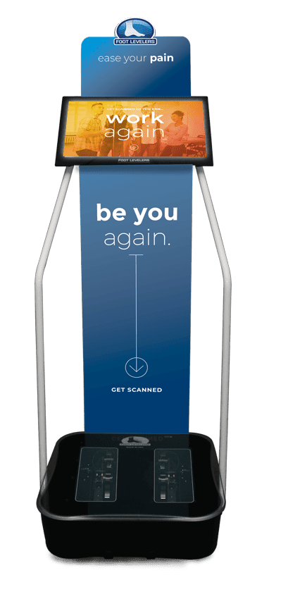
The Foot Levelers Kiosk
The Kiosk helps guide patients in selecting the best custom-made orthotics for their condition and lifestyle. It’s�
Fast: Push the Start button and the scanner begins.
Easy to use: User-friendly easy touch screen.
Engaging: Videos explain the importance of healthy feet and the benefits of custom-made orthotics.
Cloud-based: Results can be securely accessed from anywhere.
Comprehensive: Easily retrieve previous scans to compare them to new scans and see the difference.
The Foot Levelers Kiosk helps you. It saves time so you can spend more time living your life.
Workouts & Working
Our uplifting southwest community surrounded by it infinite beauty is a fantastic place to live and enjoy our families; it is, therefore, our mission to help each of our patients to�live,�to�love,�to�matter�and�to�thrive�pain-free�in this beautiful special place.
When your body is truly healthy, you will arrive at your optimal fitness level proper physiological fitness state. �We want to help you live a new and improved lifestyle. Over the last two decades, while researching and testing methods with thousands of patients, we have learned what works effectively at decreasing pain while increasing human vitality.
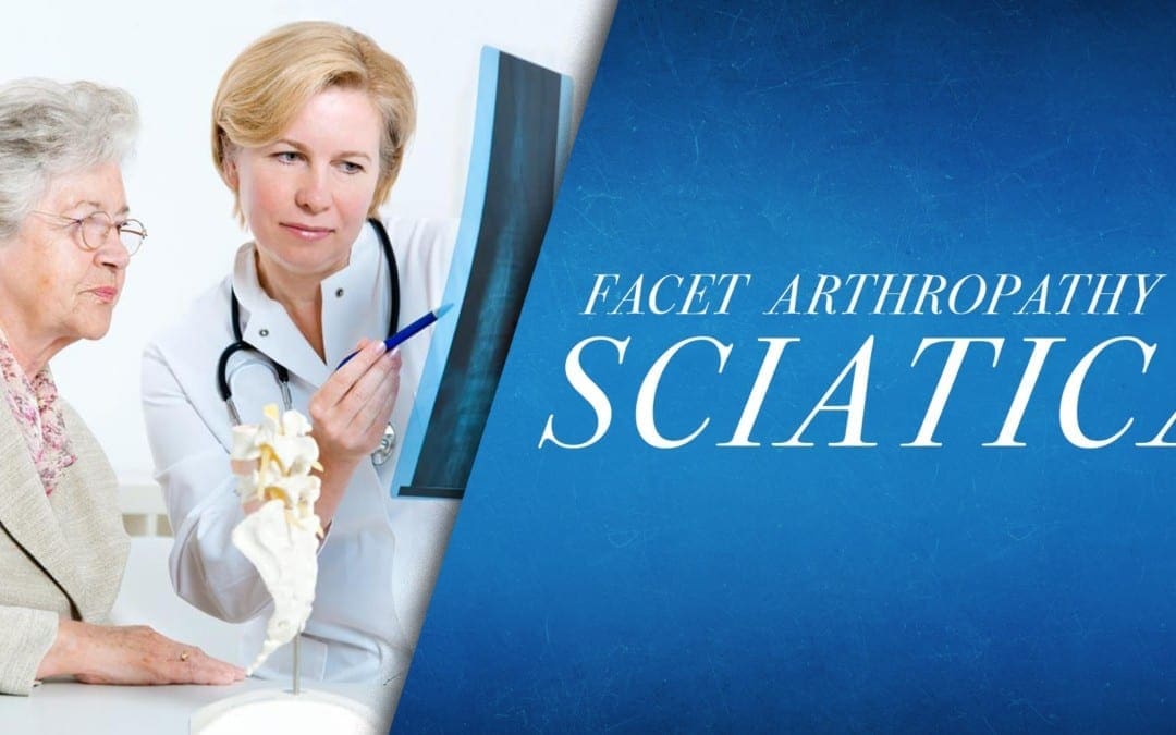
The facet joints are the joints which are found behind the spine to counterbalance the intervertebral discs found between the vertebrae of the spine. These are ultimately essential for restricting the movements of the spine for the vertebrae to maintain proper alignment. Over time, the natural aging process can cause the facet joints to deteriorate or wear down. Facet joint arthritis can also gradually develop over time, as it may in any other joint. This is referred to as arthropathy. �
What are the Symptoms of Facet Arthropathy?
Individuals with facet arthropathy will generally experience low back pain which can often worsen with standing, twisting, or bending backward. The painful symptoms associated with facet joint arthropathy occur in one specific region of the spine. The pain and discomfort are commonly described as a dull ache on one or both sides of the lower back or lumbar spine.� �However, unlike the well-known signs and symptoms of sciatica, caused by the compression or impingement of the sciatic nerve in the lower back, facet arthropathy signs and symptoms generally don’t radiate down the buttocks or into the legs. �
However, the facet joint, in the same way as any other joint which has arthritis, can become enlarged and add pressure on nerve roots, causing pain and discomfort to radiate down the lower extremities. Facet arthropathy symptoms are generally relieved by bending forward. Bending your body forward into a spinal flexion position can help reduce painful symptoms. �
What Causes Facet Arthropathy?
The natural aging process is frequently considered to be one of the most common indirect sources of facet arthropathy. Other health issues which can affect the facet joints and ultimately cause facet arthropathy include: �
Osteoarthritis: Degeneration of joint cartilage and underlying bone, generally during middle age
Facet joint degeneration: Wear and tear on the facet joint brought on over time due to aging
Facet joint injury: Trauma to the facet joints caused by an impact, such as a fall or automobile accident
Synovial cyst: A fluid-filled sac which develops in the spine, generally as a result of aging
How is Facet Arthropathy Diagnosed?
If you’re experiencing chronic low back pain, make an appointment with a healthcare professional to determine a diagnosis and follow-up with the proper treatment. By performing a physical evaluation, your healthcare professional will then be able to analyze the source of your painful symptoms. The doctor will also ask you questions regarding your medical history and your symptoms as well as order several of the following tests to help diagnose facet arthropathy, including: �
CT scan or MRI scan: These imaging tests can show evidence of facet joint degeneration, even mild to moderate cases.
Bone scan: This test shows bone density to demonstrate any source of inflammation on the spine.
Anti-inflammatory steroid injection: An injection into your facet joint can determine facet arthropathy.
X-rays: These will help the healthcare professional evaluate the overall health and wellness of your spine.
Can Facet Arthropathy Cause Other Health Issues?
Facet arthropathy may cause bone spurs, tiny bone outgrowths. Bone spurs can decrease the distance available between nerve roots, causing a health issue known as spinal stenosis. Spinal stenosis may cause pain, weakness, and numbness on the buttocks, hips, and thighs. It’s frequently associated with other health issues which could lead to facet arthropathy. �
Arthritis caused by a variety of other health issues, such as degenerative disc disease, can occur due to the human body’s natural aging process, causing the discs between the vertebrae of the spine to lose their flexibility, elasticity, and capacity to absorb shock from walking and other physical activities. This may ultimately cause painful symptoms to develop. �
How is Facet Arthropathy Treated?
There are numerous treatment approaches to help treat facet arthropathy symptoms. Treatments include: �
Anti-inflammatory drugs and/or medications
Avoidance of movements which cause pain and discomfort, such as repetitive twisting or lifting
Physical therapy
Chiropractic care
Epidural steroid injections
Facet joint ablation or the destruction of the facet nerves with electrical shocks
Spinal surgery when there is nerve-root compression
Differential Diagnosis of Hip Pain and Discomfort
� �
Facet joint arthropathy is a well-known health issue which can commonly occur due to the human body’s natural aging process, however, injury or underlying conditions may also cause facet joint arthropathy. Although facet joint arthropathy may cause pain, discomfort, and numbness in the lower back, the symptoms are different from sciatica in which these don’t radiate down the buttocks, legs, and/or feet. Diagnosis is essential for facet joint arthropathy to follow-up with treatment. – Dr. Alex Jimenez D.C., C.C.S.T. Insight
Fibromyalgia Magazine
The purpose of the article was to discuss degenerative disc disease and sciatica. Degenerative disc disease is often associated with pain, tingling sensations, and numbness, similar to the symptoms of sciatica. The scope of our information is limited to chiropractic, musculoskeletal and nervous health issues as well as functional medicine articles, topics, and discussions. To further discuss the subject matter above, please feel free to ask Dr. Alex Jimenez or contact us at 915-850-0900 . �
Curated by Dr. Alex Jimenez �
Additional Topic Discussion: Severe Sciatica
Back pain�is one of the most prevalent causes of disability and missed days at work worldwide. Back pain attributes to the second most common reason for doctor office visits, outnumbered only by upper-respiratory infections. Approximately 80 percent of the population will experience back pain at least once throughout their life. Your spine is a complex structure made up of bones, joints, ligaments, and muscles, among other soft tissues. Injuries and/or aggravated conditions, such as�herniated discs, can eventually lead to symptoms of sciatica, or sciatic nerve pain. Sports injuries or automobile accident injuries are often the most frequent cause of painful symptoms, however, sometimes the simplest of movements can have these results. Fortunately, alternative treatment options, such as chiropractic care, can help ease sciatic nerve pain, or sciatica, through the utilization of spinal adjustments and manual manipulations, ultimately improving pain relief. �
�
�
Formulas for Methylation Support
XYMOGEN�s Exclusive Professional Formulas are available through select licensed health care professionals. The internet sale and discounting of XYMOGEN formulas are strictly prohibited.
Proudly,�Dr. Alexander Jimenez makes XYMOGEN formulas available only to patients under our care.
Please call our office in order for us to assign a doctor consultation for immediate access.
If you are a patient of Injury Medical & Chiropractic�Clinic, you may inquire about XYMOGEN by calling 915-850-0900.
�
For your convenience and review of the XYMOGEN products please review the following link.*XYMOGEN-Catalog-Download �
* All of the above XYMOGEN policies remain strictly in force. �
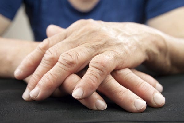
Rheumatoid arthritis (RA) is a condition that causes considerable discomfort if diagnosed with this autoimmune disease. This is when your immune system starts attacking your joints, instead of foreign invaders, which then causes inflammation.
The worse the RA, the more severe the symptoms become. If things get bad enough you can lose mobility altogether, which is why it is so important to get treatment. Fortunately, chiropractic is excellent for reducing inflammation and improving mobility. Chiropractic treatment can do a lot to ease your pain and get you back to moving the way you are supposed to.
RA and Chiropractic
Chiropractic is an effective RA treatment for a number of reasons. Chiropractic treatment:
Individual Treatment
The image many people have of a chiropractic adjustment is one of popping backs and hard, jerking motions. While adjustments can certainly include these things, they do not have to. According to the Arthritis Foundation, chiropractors have more than 150 techniques they can use to adjust your body.
They strive to give treatments specific to the needs of each patient, which means adjusting the body as gently as necessary to produce the desired result. If your joints are swollen and painful the chiropractor will carefully work to realign the joint�which reduces inflammation and improves movement�while minimizing any pain or discomfort you feel from the adjustment.
Reduced Inflammation
With RA typically the worst symptoms are the result of inflammation. Chiropractic treatment may not be able to change the way your immune system is malfunctioning, but it can do a lot to help the painful areas become less inflamed.
The treatment you get from your chiropractor will ensure that your joints are moving as properly as possible given your condition. By putting the body back in alignment, chiropractic improves the way the nervous system operates and lessens inflammation.
Improved Mobility
One of the most difficult things for many with RA is the loss of mobility that comes when their joints swell. When the pain becomes more substantial it is normal for RA sufferers to avoid movement because it hurts.
But it is important to remember that movement, even when it hurts, is necessary to maintain joint mobility. The longer you avoid moving a joint the more likely it is that you will lose function.
A useful aspect of chiropractic is that you can get help with moving, so you are not all alone with the daunting prospect of moving your joints so they start working better. Your chiropractor is your partner in movement, helping to guide your body so that it moves as well as possible. Results are different for everyone based on their unique situations, but you can be sure that chiropractic will serve as a powerful tool to keep your body working as well as possible.
Hand Deformed From Rheumatoid Arthritis
Drug-Free and Non-invasive
In chiropractic, the focus is to help the body heal itself, which means avoiding surgery and prescription medications as much as possible. Surgery and medication often have unwanted side-effects�sometimes worse than the problem they were intended to solve. With chiropractic negative side-effects are unlikely. You can get a lot of relief from gentle, effective treatments that will not make you feel worse than when you started.
Please contact our office to schedule an appointment. We look forward to speaking with you!
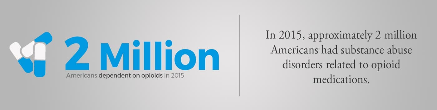
Opioid Addiction Alternative
Opioids (such as hydrocodone, oxycodone, codeine, and morphine) mask symptoms and do nothing to address the cause of pain.
There is an opioid crisis raging.� A sensible and safe alternative to opioids: Custom-made orthotics help relieve low back pain as well as hip and neck pain by removing imbalances in the musculoskeletal system, which originate in the feet.
Before considering taking an opioid for pain control, give Chiropractic care and foot orthotics a try. The combination of Chiropractic and orthotics is proven in clinical studies.
In 2015, about 2 million Americans had substance abuse disorders related to opioid medications.
In 2012, 80 out of 100 Americans were prescribed opioids. That’s about 259 million prescriptions � more than enough to give every American adult their own bottle of pills.
Less Pain & More Comfort
Custom orthotics help more than your feet! Stabilizing orthotics bring health and healing to the whole body by balancing the musculoskeletal system.
El Paso Back Clinic
Here are some videos that discuss how chiropractic care can help with arthritis, fibromyalgia, seniors and whole body wellness.
NCBI Resources
Here are some articles to check out for extended information on arthropathies.
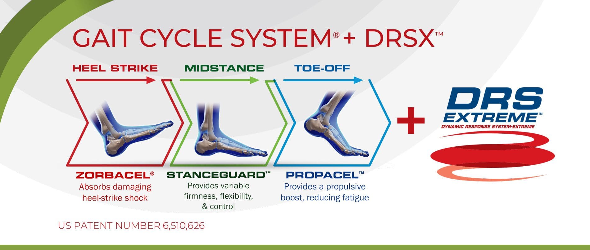
You need to understand your feet! Flat feet, foot pronation, and foot imbalances can lead to all kinds of pain:
Knee pain
Hip pain
Back pain
Yes even Shoulder pain
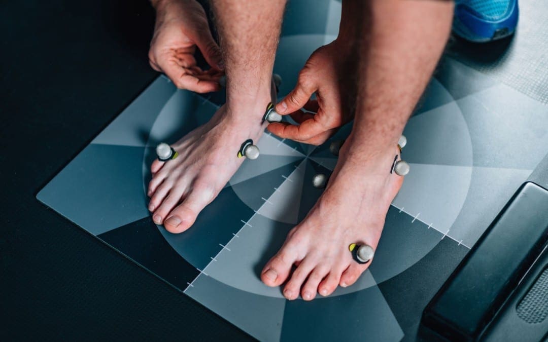
If you are considering custom orthotics, a health care professional such as Doctor Jimenez and Injury Medical Chiropractic Clinic can perform a foot scan to show you what imbalances in the feet can lead to. The foot scan will show how the orthotics can help. Following the foot scan, a report will provide the caregiver a Pronation/Stability Index, Foot Assessment, and Body Assessment.
Pronation & Stability
The Pronation/Stability Index� score appears on the clinical report that is produced when your feet are scanned by our 3D machine.
The Pronation/Stability Index� is an algorithm based on 16 different measurements. Taken from a laser scan of the feet, the index indicates the amount of arch collapse. The higher the number, the more collapse.
The index reveals the severity of pronation/stability of your feet, which can be anywhere from optimal to severe. An index of 102, for example, is a moderate pronation/instability. Untreated imbalances at any level can lead to chronic problems.
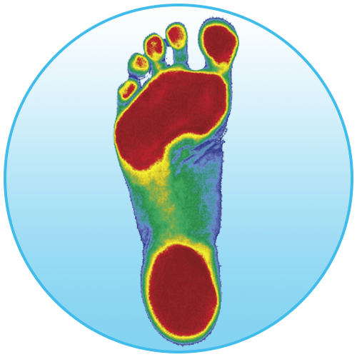
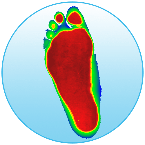
Optimal Feet
This image shows what the optimal foot looks like.
The red areas represent where pressure on your foot should be:
The toes
The balls of the foot
The heel
Unfortunately, 99% of feet do not look like this.
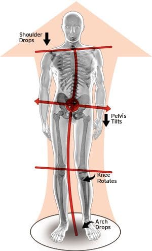
Flat Feet
This image shows what flat feet look like. Here, all three arches of the foot have collapsed. Once the arches collapse it throws the rest of the body off. So when you move there is a higher risk for pain. This includes pain in the feet, knees, hips, back, and neck.
The Body
The body image shows imbalances in the feet and how they can lead to knee rotation, pelvic tilt and shoulder drop.
Throughout our lives, we need some type of support for our feet. After all, they take all of our weight, so let’s treat them right and understand your feet.
El Paso Back Clinic
NCBI Resources
Effective footwear can make a huge difference. Especially when it comes to being able to stand and walk in a comfortable manner.
Degenerative disc disease is a health issue which occurs when one or more of the discs found between the vertebrae of the spine break down, causing painful symptoms and other problems. Common symptoms may include pain, tingling sensations, weakness, and numbness. Despite its name, degenerative disc disease is actually not a disease, but rather, a natural process which occurs with aging. The rubbery discs found between the vertebrae function like shock absorbers, allowing the back to flex and bend accordingly.� When they wear out, however, they no longer provide as much protection as before. �
Causes
The intervertebral discs also referred to as spinal discs or intervertebral fibrocartilage, provide the padding required between the vertebrae of the spine. The intervertebral discs are an elastic structure made from fibrocartilage tissue. The outer part of the disc is referred to as the annulus fibrosus. The annulus fibrosus is tough and it’s made up of many overlapping layers. The inner part of the disc is referred to as the nucleus pulposus. The nucleus pulposus is soft and gelatinous. The intervertebral discs cushion the stress of the spine, bears weight, and also helps the spine bend and flex. �
As people age, repeated daily stresses on the backbone and occasional trauma and/or injuries, including minor, undetected health issues, may ultimately damage the intervertebral discs in the back. Changes caused by damage may include: �
Decreased fluid: The intervertebral disks of a healthy young adult are made up of around 90 percent fluid. With age, the fluid material decreases, causing the disc to become thinner. The distance between each vertebra, in turn, becomes smaller and it makes them even less effective to function as a cushion or shock-absorber.
Disc structure: Small tears or cracks can become larger in the outer layer of the disc. The soft and gelatinous material from the inner part may push through the disc, causing a bulging or ruptured disc. The disc may break into fragments.
If the vertebrae have less padding between them, the backbone also becomes less stable. To compensate, the human body builds osteophytes, or bone spurs, small bony structures which develop along the edge of bones. These structures can compress or impinge the spinal cord or nerve roots. Other health issues caused by degenerative disc disease includes the breakdown of cartilage or the tissue which cushions the joints, a bulging disc, known as a herniated disc, and a narrowing of the spinal cord, also referred to as spinal stenosis. These changes can cause painful symptoms and lead to fatigue. �
Symptoms
Degenerative disc disease can either cause no symptoms or the pain and discomfort may be so severe, it can tremendously affect an individual’s quality of life. This health issue generally aggravates due to injury or trauma to the backbone, however, symptoms can also affect other parts of the human body, depending on the direct location of the degenerative disc disease. The pain and discomfort can range from mild to severe and it may often be debilitating. It may ultimately result in osteoarthritis, with pain and discomfort along with stiffness in the back. Fatigue can generally accompany other symptoms. �
If degenerative disc disease affects the low back or the lumbar spine, the pain and discomfort may radiate down the buttocks, hips, and thighs, into the knees and feet. There might also be tingling sensations and numbness, a collection of symptoms known as sciatica, caused due to the compression or impingement of the sciatic nerve. If degenerative disc disease affects the neck or the cervical spine, the pain and discomfort may radiate into the shoulders, arms, and hands. The painful symptoms may worsen when sitting, bending, twisting or lifting. Rest may help provide some pain relief. �
Diagnosis
A healthcare professional will ask the patient about their symptoms, including where and when the pain developed as well as whether there are tingling sensations or numbness. The doctor may also need to know which circumstances cause the most painful symptoms and if the patient suffered any injuries and/or aggravated conditions. A physical evaluation may examine pain and discomfort in response to touch or movement, muscle strength, flexibility, and performance, as well as nerve structure and function. The healthcare professional may also order diagnostic tests, such as MRI or CT scans. �
Treatment
Treatment for degenerative disc disease might include occupational therapy, physical therapy, chiropractic care, exercise or physical activity, drugs and/or medications, weight loss, and surgery. Medical options include injecting the joints next to the broken disc with steroids and a local anesthetic. Medicines include pain relief medicine, such as Tylenol, and non-steroidal anti-inflammatory medications, or NSAIDs, including ibuprofen. Muscle relaxers and steroids may also be prescribed. �
A corset or brace may also offer back support. Patients who don’t respond well to conservative treatment options might require surgery. Furthermore, a patient who develops osteoarthritis, a herniated disc, or spinal stenosis may require a combination of different types of treatment approaches to achieve pain relief. It’s fundamental for a healthcare professional to provide a patient with the proper diagnosis of their symptoms to follow-up with the most appropriate treatment. �
Differential Diagnosis of Hip Pain and Discomfort
�
Degenerative disc disease is characterized as the normal, gradual deterioration of the intervertebral discs with age, which may occasionally cause a variety of painful symptoms. Common symptoms associated with degenerative disc disease can include pain and discomfort, tingling sensations, and numbness, similar to sciatica. Painful symptoms may also cause fatigue and other health issues. Nearly everyone’s intervertebral discs will break down over time, however, not everyone will develop painful symptoms. – Dr. Alex Jimenez D.C., C.C.S.T. Insight
Fibromyalgia Magazine
The purpose of the article was to discuss degenerative disc disease and sciatica. Degenerative disc disease is often associated with pain, tingling sensations, and numbness, similar to the symptoms of sciatica. The scope of our information is limited to chiropractic, musculoskeletal and nervous health issues as well as functional medicine articles, topics, and discussions. To further discuss the subject matter above, please feel free to ask Dr. Alex Jimenez or contact us at 915-850-0900 . �
Curated by Dr. Alex Jimenez �
Additional Topic Discussion: Severe Sciatica
Back pain�is one of the most prevalent causes of disability and missed days at work worldwide. Back pain attributes to the second most common reason for doctor office visits, outnumbered only by upper-respiratory infections. Approximately 80 percent of the population will experience back pain at least once throughout their life. Your spine is a complex structure made up of bones, joints, ligaments, and muscles, among other soft tissues. Injuries and/or aggravated conditions, such as�herniated discs, can eventually lead to symptoms of sciatica, or sciatic nerve pain. Sports injuries or automobile accident injuries are often the most frequent cause of painful symptoms, however, sometimes the simplest of movements can have these results. Fortunately, alternative treatment options, such as chiropractic care, can help ease sciatic nerve pain, or sciatica, through the utilization of spinal adjustments and manual manipulations, ultimately improving pain relief. �
Formulas for Methylation Support
XYMOGEN�s Exclusive Professional Formulas are available through select licensed health care professionals. The internet sale and discounting of XYMOGEN formulas are strictly prohibited.
Proudly,�Dr. Alexander Jimenez makes XYMOGEN formulas available only to patients under our care.
Please call our office in order for us to assign a doctor consultation for immediate access.
If you are a patient of Injury Medical & Chiropractic�Clinic, you may inquire about XYMOGEN by calling 915-850-0900.
�
For your convenience and review of the XYMOGEN products please review the following link.*XYMOGEN-Catalog-Download �
* All of the above XYMOGEN policies remain strictly in force. �
Automobile accidents are unfortunate conditions which can tremendously influence an individual’s quality of life, health, and wellbeing. Car crash injuries, such as whiplash-associated ailments, neck pain, and back pain, can finally cause debilitating symptoms which can greatly limit an individual’s capacity to take part in their regular responsibilities.
Patients describe how Dr. Alex Jimenez, chiropractor or doctor of chiropractic in El Paso, TX, has helped them regain their quality of life in addition to their general health and wellness. Chiropractic care is a safe and effective, alternative treatment option that focuses on the identification, therapy, and prevention of a variety of health problems, including car crash injuries. Patients highly recommend Dr. Alex Jimenez since the non-surgical choice for auto accident accidents.
El Paso Back Clinic
We are blessed to present to you�El Paso�s Premier Wellness & Injury Care Clinic.
As El Paso�s Chiropractic Rehabilitation Clinic & Integrated Medicine Center,�we passionately are focused on treating patients after frustrating injuries and chronic pain syndromes. We focus on improving your ability through flexibility, mobility and agility programs tailored for all age groups and disabilities.
We want you to live a life filled with more energy, positive attitude, better sleep, less pain, proper body weight and educated on how to maintain this way of life.
We Are Ready To Help You!
If you have enjoyed this video and we have helped you in any way, please feel free to�subscribe�and�recommend�us.
Simply defined, fibromyalgia syndrome (FMS) can be said to be a debilitating illness, characterized primarily by musculoskeletal pain, fatigue, sleep disturbances, depression and stiffness (Yunus & Inanici 2002). There is no single cause, or cure, for its widespread and persistent symptoms (however, as will become clear, there do seem to exist distinct subsets of individuals with different aetiologies to their conditions, such as thyroid imbalance and whiplash injuries).
NCBI Resources
After being involved in an automobile accident, injuries inflicted to the spine can be a common complication for many individuals. From herniated discs to compression fractions, the force of an auto collision can place great amounts of stress on the complex structures of the spine, often leading to damage, injuries and even aggravate an existing condition.
IFM's Find A Practitioner tool is the largest referral network in Functional Medicine, created to help patients locate Functional Medicine practitioners anywhere in the world. IFM Certified Practitioners are listed first in the search results, given their extensive education in Functional Medicine
 �
�