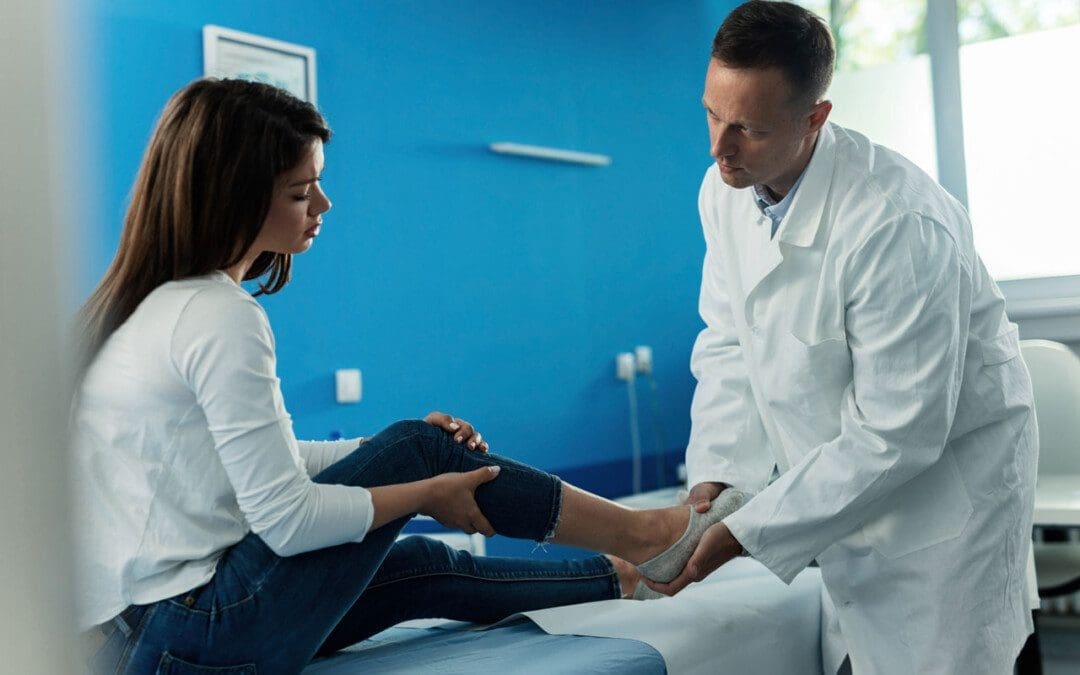
Progress can be challenging for individuals in post total ankle replacement surgery. How can physical therapy help in recovery and restoring leg function?
Total Ankle Replacement Post Surgery Physical Therapy
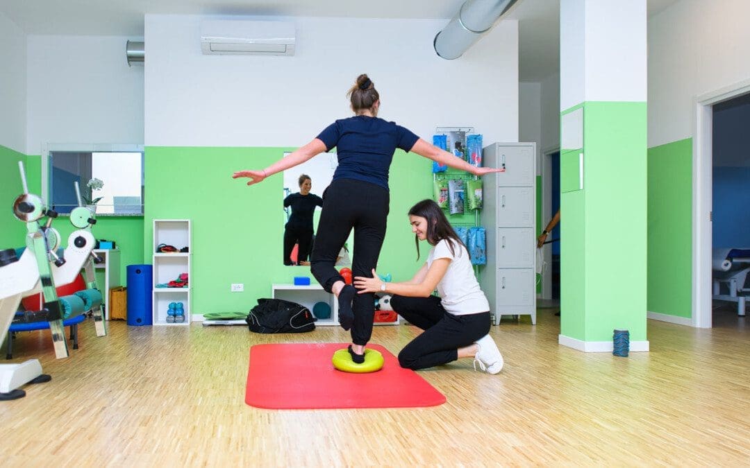
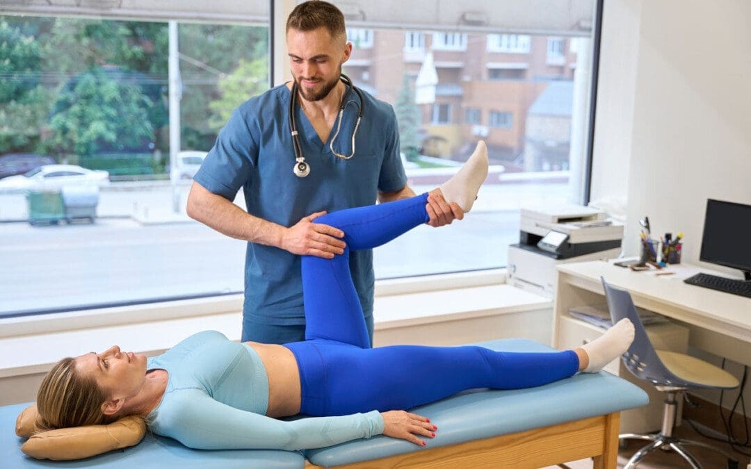
Total ankle replacement surgery is a major procedure that takes time to recover. A total ankle replacement surgery or arthroplasty can benefit individuals with chronic ankle pain or disability. This procedure can significantly improve an individual’s overall pain and function with time. Physical therapy is essential to regaining movement in the ankle and restoring full mobility. A physical therapist will work with the individual to control pain and swelling, restore the ankle’s range of motion, train on walking gait and balance, and rebuild strength in the leg. This will help maximize the chances of a successful outcome after surgery.
Total Ankle Replacement
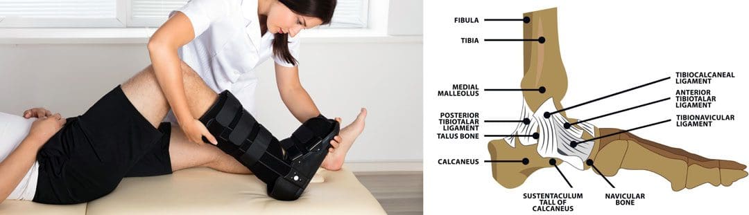
The ankle joint is the section of the lower leg where the shinbone/tibia meets the talus bone on the top of the foot. What can happen is the slippery surface/articular cartilage that coats the ends of these bones begins to thin or deteriorate. As the deterioration progresses, it can lead to significant pain, disability, and difficulty walking. (Cleveland Clinic. 2021) This is where a specialist may recommend total ankle replacement for the best results. Various conditions can be helped by this procedure, including:
During an ankle replacement procedure, an orthopedic surgeon removes the damaged ends of the tibia and talus bones and replaces them with an artificial covering. A polyethylene component is also secured between the two structures to support the smooth movement of the new joint endings. (Massachusetts General Hospital. N.D.) Following the procedure, individuals are typically placed in a protective boot or splint. The healthcare provider will recommend staying off the leg for 4 to 8 weeks to allow healing.
Physical Therapy
Outpatient physical therapy is usually initiated several weeks after the ankle operation. (UW Health Orthopedics and Rehabilitation. 2018) Physical therapy can last for five months or more, depending on the severity of the condition and injury. The physical therapist will focus on different areas to get the best results. (Cort D. Lawton et al., 2017)
Pain and Swelling Control
Post-operative pain and swelling are normal after a total ankle replacement. It is not unusual for an ankle to be swollen for even six to 12 months after the operation. (UW Health Orthopedics and Rehabilitation. 2018) The surgeon will normally prescribe medication to help manage discomfort early on, and physical therapy also plays an important role in addressing the symptoms. Treatments used can include:
Electrical stimulation – mild electrical pulses applied to the muscles.
Ice
Vasopneumatic compression, where an inflatable sleeve is used to create pressure around the area, is commonly utilized at the beginning of physical therapy to reduce pain or swelling.
Other modalities, such as stretching and targeted exercises, are combined with other treatments.
Range of Motion
Early after the procedure, the ankle will be very stiff and tight. This is due to several factors, including the inflammation and swelling after surgery and the time spent immobilized in a boot.
The physical therapist will employ various techniques to improve the ankle joint’s range of motion to rotate and flex.
The physical therapist may employ passive stretching induced by an outside force such as the therapist or a resistance band) to help improve mobility.
After multiple weeks of reduced movement and lack of bearing any weight on the ankle, the muscles that surround the ankle have often atrophied/weakened, which can impact balance.
When the individual can begin placing weight on the leg, the therapist will apply proprioceptive/sense of body position training to improve overall stability. (UW Health Orthopedics and Rehabilitation. 2018)
Balance exercises will be added to the home program and will progress from week to week.
Strength
The muscles in the leg, ankle, and foot become weak from the surgery and the time spent in a splint or boot. These structures have a significant role in balance, the ability to stand, walk, and go up or down the stairs.
Regaining the strength and power of these muscles is a critical goal of rehabilitation.
In the first weeks, the physical therapist will focus on gentle strengthening exercises.
Isometrics lightly activate the muscles but avoid irritating the surgical site.
As time passes and weight-bearing is allowed, these gentle moves are replaced with more challenging ones, like resistance bands and standing exercises, to accelerate strength gains.
Lawton, C. D., Butler, B. A., Dekker, R. G., 2nd, Prescott, A., & Kadakia, A. R. (2017). Total ankle arthroplasty versus ankle arthrodesis-a comparison of outcomes over the last decade. Journal of orthopaedic surgery and research, 12(1), 76. doi.org/10.1186/s13018-017-0576-1
Individuals in post-surgery recovery or dealing with illness or an injury can experience weakened muscles and endurance that can cause temporary loss of sleeping mobility and not being able to move around normally because of weakness, decreased range of motion, or pain. Can they benefit from physical therapy to help get back to normal functional mobility?
Sleeping Mobility
For individuals who are hospitalized or homebound from injury, illness, or surgical recovery, a physical therapist will assess various areas of functional mobility. These include transfers – from sitting to standing positions, walking, and sleeping mobility. Sleeping mobility is the ability to perform specific motions while in bed. A therapist can assess sleeping or bed mobility and recommend strategies and exercises to improve movements. (O’Sullivan, S. B., Schmitz, T. J. 2016) A therapist may have the individual use specific devices, like an over-the-bed trapeze or a sliding board, to help move around.
All of these movements require strength in different muscle groups. By checking out individual motions in sleeping mobility, a therapist can work out specific muscle groups that may be weak and require targeted exercises and stretches to restore mobility to normal. (O’Sullivan, S. B., Schmitz, T. J. 2016) Individuals visiting a therapist in an outpatient clinic or rehabilitation area may have the individual work on sleeping mobility on a treatment table. The same motions on the treatment table can be done in the bed.
Importance
The body is meant to move.
For individuals who cannot move comfortably on their bed, the body may suffer disuse atrophy or the wasting away of muscular strength, which can lead to increased difficulties. Not being able to move can also lead to pressure ulcers, especially for individuals who are severely deconditioned and/or remain in one position for a long period. Skin health may start to break down, leading to painful wounds that require specialized care. Being able to move around in bed can help prevent pressure ulcers. (Surajit Bhattacharya, R. K. Mishra. 2015)
Improvement
A physical therapist can prescribe specific exercises to strengthen muscle groups and improve sleeping mobility. The muscles include:
Shoulder and rotator cuff muscles.
Triceps and biceps in the arms.
Gluteus muscles of the hips.
Hamstrings
Quadriceps
Calf muscles
The shoulders, arms, hips, and legs work together when moving the body around the bed.
Various Exercises
To improve bed movement, physical therapy exercises can include:
Physical therapists are trained to assess these motions and functions and prescribe treatments to improve body movement. (O’Sullivan, S. B., Schmitz, T. J. 2016) Maintaining appropriate physical fitness can help the body stay active and mobile. Performing mobility exercises prescribed by a physical therapist can keep the right muscle groups working properly, and working with a physical therapist can ensure the exercises are correct for the condition and are performed properly.
Bhattacharya, S., & Mishra, R. K. (2015). Pressure ulcers: Current understanding and newer modalities of treatment. Indian journal of plastic surgery : official publication of the Association of Plastic Surgeons of India, 48(1), 4–16. doi.org/10.4103/0970-0358.155260
For individuals having difficulty moving or functioning normally due to injury, surgery, or illness, can a chiropractic and physical therapy team help expedite recovery?
Friction Massage
Individuals may develop scar tissue or tissue adhesions that limit normal motion after injury or surgery. A pain management team may use various treatments and modalities and may incorporate friction massage as part of a rehabilitation treatment plan. Friction massage, also known as transverse friction or cross friction massage, is a technique used to help improve scar tissue and adhesion mobility to move better and decrease the negative effects. The therapist uses their fingers to massage the scar in a direction that is at right angles to the scar line. It is a specialized technique that breaks up tissue adhesions that are limiting normal movement in the skin and underlying tissues. (Haris Begovic, et al., 2016)
Scar Tissue and Adhesions
For individuals who require surgery due to an injury or an orthopedic condition, their doctor will cut into the skin, tendons, and muscle tissue during the operation. Once sutured and healing has begun, scar tissue forms. Healthy tissue is made up of collagen that is comprised of cells that are arranged in a regular pattern. Healthy collagen is strong and can resist forces when tissues are pulled and stretched. (Paula Chaves, et al., 2017)
During the healing process after an injury, the collagen cells are laid down in a haphazard pattern and form scar tissue. The random accumulation of cells becomes tight and does not react well to tension and stretching forces. (Qing Chun, et al., 2016) The body can form scar tissue after a soft tissue injury, like a muscle or tendon strain. (Qing Chun, et al., 2016)
If a muscle or tendon gets strained the body will generate new collagen during the healing. The new collagen is laid down in a random fashion, and scar tissue or tissue adhesions can form that can limit the normal range of motion. Healthy tissue stretches and glides as the body moves. Scar tissue is rigid. At the site of the scar tissue, there can be some movement, but it is tight, less pliable, and can be painful. If scar tissue or adhesions are limiting motion, cross-friction massage can improve tissue gliding and sliding. This process is referred to as remodeling.
Massage Objectives
The objectives and goals of friction massage to adhesions or scar tissue may include:
Stimulation of nerve fibers to decrease and relieve pain.
The entire area of scar tissue or adhesion should be treated.
If the scar tissue is in a muscle, it should be relaxed.
If the scar tissue is in a tendon sheath, that tendon should be slightly stretched during the procedure.
The therapist places two or three fingers over the scar or adhesion and moves their fingers perpendicular to the scar to smooth the collagen fibers down.
The fingers and underlying tissues move together.
The massage should feel deep and uncomfortable but not painful.
There may be some pain, but should remain within the individual’s tolerance.
If the massage is too painful, less pressure may be used.
After several minutes the therapist will assess the tissue mobility.
Specific stretches may be done to elongate the scar tissue or adhesions.
At-home exercises and stretches may be prescribed to maintain flexibility.
Contraindications
There are situations where friction massage should not be used and can include: (Paula Chaves, et al., 2017)
Around an active open wound.
If there is a bacterial infection.
Areas with decreased sensation.
If calcification is present in the muscle or tendon tissue.
The therapist will explain the procedure and inform of the goals and risks associated with it.
Adhesive capsulitis in the shoulder/frozen shoulder.
Joint contracture.
Ligament tears.
Scar tissue buildup after surgery or trauma.
Friction massage is a popular technique used in physical therapy, but some research suggests it is not any more effective than other rehabilitation techniques. One study found that static stretches and exercises were more effective than massage in improving tissue length and strength in uninjured soccer players. Other studies have supported this, but individuals may find that the massage helps improve injured tissues’ movement as well. (Mohammed Ali Fakhro, et al. 2020)
The main goal of any treatment in physical therapy is to help the individual regain movement and flexibility. Friction massage, combined with targeted stretches and exercises, can help individuals expedite recovery and get back to normal.
Chiropractic Care After Accidents and Injuries
References
Begovic, H., Zhou, G. Q., Schuster, S., & Zheng, Y. P. (2016). The neuromotor effects of transverse friction massage. Manual therapy, 26, 70–76. doi.org/10.1016/j.math.2016.07.007
Chaves, P., Simões, D., Paço, M., Pinho, F., Duarte, J. A., & Ribeiro, F. (2017). Cyriax’s deep friction massage application parameters: Evidence from a cross-sectional study with physiotherapists. Musculoskeletal science & practice, 32, 92–97. doi.org/10.1016/j.msksp.2017.09.005
Chun, Q., ZhiYong, W., Fei, S., & XiQiao, W. (2016). Dynamic biological changes in fibroblasts during hypertrophic scar formation and regression. International wound journal, 13(2), 257–262. doi.org/10.1111/iwj.12283
Fakhro, M. A., Chahine, H., Srour, H., & Hijazi, K. (2020). Effect of deep transverse friction massage vs stretching on football players’ performance. World journal of orthopedics, 11(1), 47–56. doi.org/10.5312/wjo.v11.i1.47
For individuals experiencing pelvis pain symptoms and associated problems, can integrating pelvic floor physical therapy exercises help with treatment and prevention?
Pelvic Floor Physical Therapy
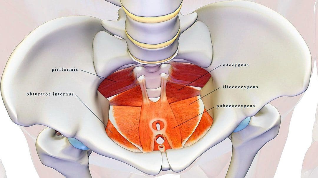
The pelvic floor muscles are located at the base of the pelvis and protect the pelvic organs like the vagina, cervix, uterus, bladder, urethra, and rectum. (U.S. Food and Drug Administration. 2019)
When the muscles fail to function correctly, individuals can experience symptoms like:
Painful intercourse
Prolapse – when an organ or tissue drops or shifts out of place.
Urinary incontinence
Constipation problems
These conditions are common in pregnant individuals or older women.
These symptoms can be treated with pelvic floor physical therapy to alleviate discomfort. Pelvic floor physical therapy can help women and individuals with vaginas:
Alleviate issues like painful sex, urinary leakage, and prolapse.
In physical therapy, individuals work on breathing, relaxation, and lengthening and strengthening techniques to train their muscles to function optimally.
Causes of Pelvic Floor Issues
Pelvic floor dysfunction tends to happen with age, during pregnancy, or in combination with events like the postpartum period and menopause, which can lower hormone levels.
Individuals who are pregnant are especially prone to pelvic floor issues but might not know they have a problem.
The pregnancy weight of a uterus can pressure and strain the muscles.
If left untreated, these symptoms can worsen over time.
Pelvic Floor Physical Therapy
An individual will meet with a specialist to discuss symptoms and undergo a physical examination that includes:
Pelvic floor exam.
Evaluation of posture, mobility, and core strength.
Once the initial exams and evaluation are complete, the practitioner will go over pelvic floor exercises and provide a treatment plan.
Recommended exercises vary based on symptoms but focus on relaxing, stretching, and/or strengthening muscles.
Muscle Relaxation
To relax the muscles, a therapist may recommend breathing exercises.
For pregnant individuals, this means timing breaths with contractions.
For individuals experiencing constipation, breathing exercises can help the body relax and reduce strain.
Stretching Muscles
Stretching can help relieve muscle tightness and stiffness.
A therapist may help stretch the pelvic floor through various therapy modalities.
This type of physical therapy can help loosen tight muscles or help gently reset dislocated organs back into place.
Strengthening Muscles
After the pelvic floor is loose and relaxed, the focus typically switches to strengthening the muscles.
Strength work may target abdominal muscles or the pelvic floor muscles themselves.
With time, commitment, and targeted treatment, individuals can use pelvic floor physical therapy to loosen tissues, strengthen muscles, and restore function.
Sartori, D. V. B., Kawano, P. R., Yamamoto, H. A., Guerra, R., Pajolli, P. R., & Amaro, J. L. (2021). Pelvic floor muscle strength is correlated with sexual function. Investigative and clinical urology, 62(1), 79–84. doi.org/10.4111/icu.20190248
Raizada, V., & Mittal, R. K. (2008). Pelvic floor anatomy and applied physiology. Gastroenterology clinics of North America, 37(3), 493–vii. doi.org/10.1016/j.gtc.2008.06.003
Soave, I., Scarani, S., Mallozzi, M., Nobili, F., Marci, R., & Caserta, D. (2019). Pelvic floor muscle training for prevention and treatment of urinary incontinence during pregnancy and after childbirth and its effect on urinary system and supportive structures assessed by objective measurement techniques. Archives of gynecology and obstetrics, 299(3), 609–623. doi.org/10.1007/s00404-018-5036-6
Individuals who have gone through recent low back surgery, like a lumbar laminectomy and discectomy, could they benefit from physical therapy for full recovery? (Johns Hopkins Medicine. 2008)
Rehabilitation Exercise Program
A lumbar laminectomy and discectomy is a surgical procedure performed by an orthopedic or neurologic surgeon to help decrease pain, relieve associated symptoms and sensations, and improve flexibility and mobility. The procedure involves cutting away disc and bone material that presses against, irritates, and damages the spinal nerves. (Johns Hopkins Medicine. 2023)
Post-Surgery
The therapist will work with the individual to develop a rehabilitation exercise program. The objective of a rehabilitation exercise program is to help the individual:
Relax their muscles to prevent muscle tensing and becoming over-cautious
Regain full range of motion
Strengthen their spine
Prevent injuries
A guide on what to expect in physical therapy.
Postural Retraining
After back surgery, individuals have to work to maintain proper posture when sitting and standing. (Johns Hopkins Medicine. 2008)
Postural control is important to learn as it maintains the lower back in the optimal position to protect and expedite the healing of lumbar discs and muscles.
A physical therapist will teach the individual how to sit with proper posture and use lumbar support.
Attaining and maintaining proper posture is one of the most important things to help protect the back and prevent future back problems.
Walking helps to improve cardiovascular health and blood circulation throughout the body.
This helps to provide added oxygen and nutrients to the spinal muscles and tissues as they heal.
It is an upright exercise that puts the spine in a natural position, which helps to protect the discs.
The therapist will help set up a program tailored to the individual’s condition.
Prone Press Up
One of the exercises to protect the back and lumbar discs is prone press-ups. (Johns Hopkins Medicine. 2008) This exercise helps keep the spinal discs situated in the proper position. It also helps to improve the ability to bend back into lumbar extension.
To perform the exercise:
Lie facing down on a yoga/exercise mat and place both hands flat on the floor under the shoulders.
Keep the back and hips relaxed.
Use the arms to press the upper part of the body up while allowing the lower back to remain against the floor.
There should be a slight pressure in the lower back while pressing up.
Hold the press-up position for 2 seconds.
Slowly lower back down to the starting position.
Repeat for 10 to 15 repetitions.
Sciatic Nerve Gliding
Individuals who had leg pain coming from the back prior to surgery may have been diagnosed with sciatica or an irritation of the sciatic nerve. Post-surgery, individuals may notice their leg feels tight whenever straightening it out all the way. This could be a sign of an adhered/trapped sciatic nerve root, a common problem with sciatica.
After lumbar laminectomy and discectomy surgery, a physical therapist will prescribe targeted exercises called sciatic nerve glides to stretch and improve how the nerve moves. (Richard F. Ellis, Wayne A. Hing, Peter J. McNair. 2012)
Nerve glides can help free the stuck nerve root and allow for normal motion.
To perform the exercise:
Lie on the back and bend one knee up.
Grab underneath the knee with the hands.
Straighten the knee while supporting it with the hands.
Once the knee is fully straightened, flex and extend the ankle about 5 times.
Return to the starting position.
Repeat the sciatic nerve glide 10 times.
The exercise can be performed several times to help improve how the nerve moves and glides in the lower back and leg.
Supine Lumbar Flexion
After surgery, gentle back flexion exercises can help safely stretch the low-back muscles and gently stretch the scar tissue from the surgical incision. Supine lumbar flexion is one of the simplest exercises to improve lumbar flexion range of motion.
To perform the exercise:
Lie on the back with the knees bent.
Slowly lift the bent knees towards the chest and grasp the knees with both hands.
Gently pull the knees toward the chest.
Hold the position for 1 or 2 seconds.
Slowly lower the knees back to the starting position.
Perform for 10 repetitions.
Stop the exercise if experiencing an increase in pain in the lower back, buttocks, or legs.
Hip and Core Strengthening
Once cleared, individuals can progress to an abdominal and core strengthening program. This involves performing specific motions for the hips and legs while maintaining a pelvic neutral position. Advanced hip strengthening exercises help generate strength and stability in the muscles that surround the pelvic area and lower back. A physical therapist can help decide which exercises are recommended for the specific condition.
Return-to-Work and Physical Activities
Once individuals have gained an improved lumbar range of motion, hip, and core strength, their doctor and therapist may recommend working on specific activities to help them return to their previous level of work and recreation. Depending on job occupation, individuals may need to:
Work on proper lifting techniques.
Require an ergonomic evaluation if they spend time sitting at a desk or workstation.
Some surgeons may have restrictions on how much an individual can bend, lift, and twist from two to six weeks after surgery.
Low-back surgery can be difficult to rehab properly. Working with a healthcare provider and physical therapist, individuals can be sure to improve their range of motion, strength, and functional mobility to return to their previous level of function quickly and safely.
Ellis, R. F., Hing, W. A., & McNair, P. J. (2012). Comparison of longitudinal sciatic nerve movement with different mobilization exercises: an in vivo study utilizing ultrasound imaging. The Journal of orthopaedic and sports physical therapy, 42(8), 667–675. doi.org/10.2519/jospt.2012.3854
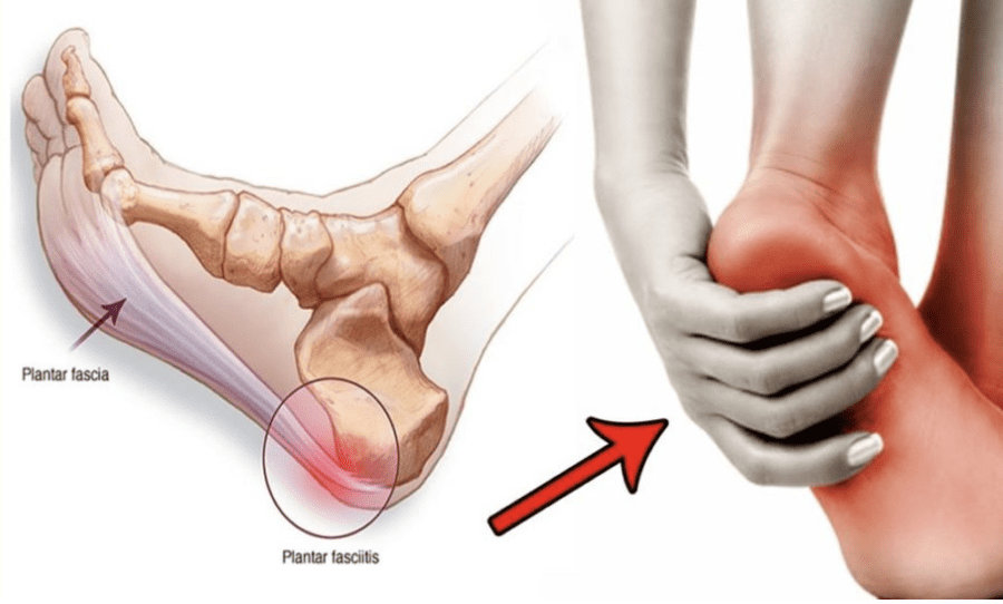
Individuals with plantar fasciitis may experience consistent flare-ups. Can knowing the causes help to find pain relief?
Plantar Fasciitis Flare-Up
Plantar fasciitis is a common cause of heel and foot pain. The plantar fascia is a band of tissue that runs along the bottom of the foot and becomes inflamed. Certain factors can cause plantar fasciitis flare-ups, including:
Increased levels of physical activity.
Not stretching regularly.
Wearing shoes without proper support.
Weight gain.
Causes
A plantar fasciitis flare-up is often triggered by physical activity. (MedlinePlus. U.S. National Library of Medicine. 2022) It can also be brought on by underlying conditions, like increased body weight, arthritis, or the shape of the foot. (Johns Hopkins Medicine. 2023) Despite the root cause, there are activities and experiences that can contribute to and/or worsen the condition.
New Exercise Routine
Being highly physically active can exacerbate plantar fasciitis symptoms.
High heels, boots, or shoes that raise the heel above the toes.
Worn-out shoes like exercise workout shoes.
Not Stretching Properly or At All
Tight calves can increase pressure on the plantar fascia.
Stretching the calves, Achilles tendon/heel, and the bottom of the feet is highly recommended to help treat and prevent the condition. (Johns Hopkins Medicine. 2023)
Not stretching thoroughly or skipping stretches can worsen symptoms.
Individuals with plantar fasciitis are recommended to stretch before and after physical activities, exercise, before going to bed, and after waking up.
Working Through the Pain
Individuals may try to continue physical activities during a flare-up.
This is not recommended as doing so can cause more pain and worsen the condition.
When pain presents, it’s recommended to:
Stop all activities that strain the feet
Stay off the feet for at least a week.
Tearing the Plantar Fascia
The plantar fascia rarely tear completely from repeated stress known as a plantar fascia rupture.
Pascoe, S. C., & Mazzola, T. J. (2016). Acute Medial Plantar Fascia Tear. The Journal of orthopaedic and sports physical therapy, 46(6), 495. doi.org/10.2519/jospt.2016.0409
It can be difficult for individuals and athletes to stay motivated, manage stress and prevent becoming overwhelmed. Can mental toughness and a positive attitude help increase potential and performance levels?
Mental Toughness
Athletes and fitness enthusiasts work on conditioning, skills training, and perfecting techniques. Physical training can take individuals far but another necessary part of maximizing athletic potential is building mental toughness and having the right attitude. Like anything, mental training takes time, effort, and regular adjustments to find ways to shift a losing or bad attitude into a positive one that can bring out the best.
Attitude Is Important
If negativity begins to set in like dealing with an injury, getting rid of self-limiting beliefs can be difficult, as well as generate optimism to rise up and succeed. For athletes or individuals who enjoy competitive sports, developing a positive mental attitude will help with:
Emotions that can affect cognitive functioning strategies.
Energy levels.
Other aspects of physical performance.
Mental Strategies
Mood Improvement
Individuals frustrated by a pessimistic perspective tend to dwell on problems or issues. To shift into a positive mood do something to lift your spirits, even if you don’t think it will help.
Listen to your favorite or uplifting music.
Watch an inspirational movie.
Read a sports psychology book.
Get together or call a teammate or friend that are cheerful and upbeat.
Play different games just for fun.
Take a break, go to the park, walk around, and meditate.
Get into hobbies.
Relax with a therapeutic massage.
Positive Self Talk
Continuing sports psychology research shows that practicing positive self-talk can improve athletic performance. (Nadja Walter, et al., 2019) Sports psychologists describe this through the idea that thoughts create beliefs, that drive actions.
Positive self-talk can take different forms.
For some reciting a specific phrase, sentence, or a single word can effectively manage thoughts, push out the negativity, and focus on taking care of business. Anything that inspires can include:
Focus
Remember the fundamentals!
You know what to do!
You can do it!
You got this!
Research shows that positive self-talk reduces anxiety and increases self-confidence, optimization, efficacy, and performance. (Nadja Walter, et al., 2019) However, self-talk needs to be practiced and part of a regular routine to be effective.
Visualization
Another strategy is using visualization exercises.
This could be using all the senses to imagine the venue where the tournament is taking place, the sound of the crowd, the smells, how the ground or court feels, and/or how the ball or specific sports object feels.
The wisdom is if you can think it, you can do it, once that is determined apply strategies to get there.
Sports Injury Rehabilitation
References
Walter, N., Nikoleizig, L., & Alfermann, D. (2019). Effects of Self-Talk Training on Competitive Anxiety, Self-Efficacy, Volitional Skills, and Performance: An Intervention Study with Junior Sub-Elite Athletes. Sports (Basel, Switzerland), 7(6), 148. doi.org/10.3390/sports7060148
Reiser, M., Büsch, D., & Munzert, J. (2011). Strength gains by motor imagery with different ratios of physical to mental practice. Frontiers in psychology, 2, 194. doi.org/10.3389/fpsyg.2011.00194
IFM's Find A Practitioner tool is the largest referral network in Functional Medicine, created to help patients locate Functional Medicine practitioners anywhere in the world. IFM Certified Practitioners are listed first in the search results, given their extensive education in Functional Medicine