About 6 million people in the United States have Alzheimer�s disease (AD) and about 50 million people worldwide have dementia. There aren’t many treatments to treat these neurological diseases. Scientists in a 2018 research study on red light therapy and mice described that �treatment for Alzheimer�s disease and dementia has not been effective for more than 100 years.� Another research study described that there is currently “no treatment to prevent brain health issues. �
However, research studies on red light therapy as a treatment for Alzheimer�s disease and dementia have been positive over the last few years in laboratory settings with rodent models. Based on this lab data, researchers recommend red light therapy and near-infrared light therapy in human patients with AD and dementia. In this article, we will look at what the initial human research studies on red light therapy and Alzheimer�s disease/dementia have shown over the last few years. �
Red Light Therapy for Alzheimer�s and Dementia
The first few double-blinded, placebo-controlled human trials on red light therapy for AD, dementia, and other neurological diseases published in 2017 had very positive results. The data showed that red light therapy caused changes in executive function, clock drawing, immediate recall, memory, visual attention, and task switching, among other positive results. One research study showed that patients treated with transcranial light therapy experienced improvements, such as: �
Increased cognitive function
Better sleep
Fewer angry outbursts
Less anxiety
Less wandering
The research study noted that there were �no negative side-effects� on transcranial light therapy for neurological diseases. The research study concluded that transcranial light therapy shows potential for the treatment of brain health issues. �
More Human Trials with Red Light Therapy in Progress
The results of these initial human trials are encouraging for Alzheimer’s disease and dementia patients and families looking for better treatment options, especially natural and non-invasive treatments with no drugs/medications or side effects. �
In early 2019, three more human trials on red light therapy and AD/dementia have been in progress at the University of California and a hospital system in France. With the previous positive results, more and larger research studies and human trials are being organized. Scientists hope that in the following years, the base of positive evidence will be large enough to recommend red light therapy as a treatment for Alzheimer�s disease and dementia, among other neurological diseases. �
The results from human trials over the last few years have established a much bigger base of similarly positive results from research studies of rodent brains in Alzheimer�s disease and dementia models, both of which are outlined below. �
Red Light Therapy Reduces Oxidative Stress and Improves Memory
A 2018 research study of mice in an age-related AD/dementia model showed that red light therapy considerably reduced oxidative stress levels and restored memory function. The researchers also praised red light therapy for being a non-invasive treatment option as well as having a high rate of tissue penetration and low phototoxicity. The researchers additionally found that red light therapy not only prevented early-stage memory decline but also recovered late-stage memory deficits. �
Researchers in a similar 2015 research study with a mouse AD/dementia model utilized near-infrared (NIR) light instead of red light therapy. The NIR treatments also reduced oxidative stress in the cerebellar cortex. The researchers concluded that NIR treatments had the ability to prevent brain degeneration in every region of the mouse brain. The research studies concluded that light therapy opens a promising opportunity to translate LED-therapy into treatments for patients. �
Red Light Therapy Prevents Brain Degeneration
Several research studies have shown that red light therapy can suppress the buildup of Beta-amyloid (A?), a protein which causes senile plaques in people with Alzheimer�s disease and dementia. Synaptic dysfunction, due to the disruptive binding of (A?) in the brain, is one of the symptoms of AD and dementia responsible for causing initial cognitive decline. Preventing synaptic dysfunction can be an effective treatment for AD and dementia, helping to regulate and manage symptoms. �
Red Light Therapy Improves Memory, Motor Skills, and Recognition
Research studies in 2017 evaluated the hippocampus of rat brains in an Alzheimer�s model with light therapy. Both research studies considerably reduced A? plaques in the rats treated with light therapy. Both research studies also tested the subjects and found that treatments reduced hippocampal neurodegeneration and improved spatial memory, recognition, and basic motor skills in the light therapy groups. Another research study also showed considerable A? reduction and noted that NIR light can reduce synaptic dysfunction from A?, showing that NIR light therapy is a viable treatment for AD and dementia. �
Red Light Therapy Shows Promise for Neurological Diseases
The initial research studies on red light therapy for Alzheimer�s disease and dementia have ultimately been encouraging for researchers. Red light therapy is not FDA-approved for the treatment of Alzheimer�s Disease or dementia, however, there is hope that more positive results in human trials will show that light therapy is fundamental for AD and dementia treatment. �
Based on the available base of positive evidence, however, red light therapy shows promise as a natural, non-invasive, drug/medication-free treatment for brain degeneration where pharmacological solutions have long failed. �
By reducing oxidative stress and preventing the accumulation of the Beta-amyloid which causes brain plaques and synapse dysfunction, red light therapy offers hope towards delaying the onset of Alzheimer�s disease and dementia symptoms as well as hopefully even reversing or preventing brain degeneration and cognitive function decline. Researchers, patients, and families affected by AD and dementia will be watching closely in the following years as more positive results emerge. �
Research studies have demonstrated positive results on red light therapy for Alzheimer’s disease and dementia. Initial research studies on mice and rat models have shown the effects of light therapy on neurological diseases. Although, more human trials are still necessary to establish the effectiveness of red light therapy for AD and dementia, the base positive results are promising. Many healthcare professionals can help treat the symptoms associated with a variety of neurological diseases, among other health issues. – Dr. Alex Jimenez D.C., C.C.S.T. Insight
Research studies on red light therapy for AD and dementia have been positive over the last years. The initial human research studies on red light therapy and Alzheimer�s disease/dementia have been promising. The scope of our information is limited to chiropractic, musculoskeletal and nervous health issues as well as functional medicine articles, topics, and discussions. To further discuss the subject matter above, please feel free to ask Dr. Alex Jimenez or contact us at 915-850-0900 . �
Curated by Dr. Alex Jimenez �
Additional Topic Discussion: Chronic Pain
Sudden pain is a natural response of the nervous system which helps to demonstrate possible injury. By way of instance, pain signals travel from an injured region through the nerves and spinal cord to the brain. Pain is generally less severe as the injury heals, however, chronic pain is different than the average type of pain. With chronic pain, the human body will continue sending pain signals to the brain, regardless if the injury has healed. Chronic pain can last for several weeks to even several years. Chronic pain can tremendously affect a patient’s mobility and it can reduce flexibility, strength, and endurance.
Neural Zoomer Plus for Neurological Disease
Dr. Alex Jimenez utilizes a series of tests to help evaluate neurological diseases. The Neural ZoomerTM Plus is an array of neurological autoantibodies which offers specific antibody-to-antigen recognition. The Vibrant Neural ZoomerTM Plus is designed to assess an individual�s reactivity to 48 neurological antigens with connections to a variety of neurologically related diseases. The Vibrant Neural ZoomerTM Plus aims to reduce neurological conditions by empowering patients and physicians with a vital resource for early risk detection and an enhanced focus on personalized primary prevention. �
Formulas for Methylation Support
XYMOGEN�s Exclusive Professional Formulas are available through select licensed health care professionals. The internet sale and discounting of XYMOGEN formulas are strictly prohibited.
Proudly,�Dr. Alexander Jimenez makes XYMOGEN formulas available only to patients under our care.
Please call our office in order for us to assign a doctor consultation for immediate access.
If you are a patient of Injury Medical & Chiropractic�Clinic, you may inquire about XYMOGEN by calling 915-850-0900.
�
For your convenience and review of the XYMOGEN products please review the following link.*XYMOGEN-Catalog-Download �
* All of the above XYMOGEN policies remain strictly in force.
Central nervous system, or CNS, infections can be life-threatening if they are not diagnosed and treated early. Because CNS infections are non-specific, determining an accurate diagnosis can be challenging. The nucleic acid in vitro amplification-based molecular methods are starting to be utilized for routine microbial diagnosis. These molecular methods have improved beyond conventional diagnostic techniques with increased sensitivity and specificity. Moreover, molecular methods utilized on cerebrospinal fluid samples are considered the new standard for diagnosis of CNS infections caused by pathogens. �
Molecular methods for the diagnosis of CNS infections offers a variety of monoplex and multiplex PCR assays to diagnose several types of health issues. Pan-omic molecular platforms can also help diagnose CNS infections. Although molecular methods are utilized for the diagnosis of CNS infections, the outcome measures for these diagnostic techniques must be carefully identified by healthcare professionals. The following article discusses conventional diagnostic techniques and molecular methods utilized for the diagnosis of central nervous system infections, their application, and future approaches. �
Molecular Methods in the Diagnosis of CNS Infections
Because of increased sensitivity and specificity, nucleic acid in vitro amplification-based molecular methods has tremendously improved the ability to diagnose CNS infections in a reasonable and effective time frame. Several PCR-derived techniques have also ultimately increased the flexibility and rigor of currently available diagnostic techniques. �
Reverse transcriptase, or RT,-PCR was developed to increase RNA targets. Its utilization plays a fundamental role in the diagnosis of RNA-virus infections as well as managing their reaction to treatment. Timely access to enterovirus RT-PCR outcome measures has demonstrated shorter hospital stays, reduced unnecessary antibiotic utilization, and decreased ancillary laboratory evaluations and tests. Broad-range rRNA PCR techniques, which utilize a single pair of primers targeting conserved regions of genes, have been utilized to diagnose bacterial pathogens and herpes viruses in the CSF. Isothermal amplification-based techniques. including loop-mediated isothermal amplification or LAMP, have been developed to offer a diagnosis within several minutes to hours. Table 2 demonstrates commercial molecular in vitro diagnostic devices, or IVD, which have been cleared by the US Food and Drug Administration, or FDA, for diagnosis of microbial pathogens in CSF. �
Monoplex Assays
A conventional molecular method involves three phases: sample extraction, target nucleic acid amplification, and amplicon detection. One of the first molecular assays successfully utilized for the diagnosis of CNS infections was utilized for the diagnosis of HSV in cerebrospinal fluid or CSF. PCR became the test of choice when research studies demonstrated that CSF PCR was similar to culture of brain tissue for diagnosis of HSV encephalitis and meningitis. Many PCR based methods for the diagnosis of herpes and enteroviruses have become available with increased sensitivity compared to viral culture. �
Real-time PCR with nucleic acid amplification and amplicon detection further improved the transition to molecular methods in clinical laboratories. Unlike conventional PCR, the real-time system is a �closed� system and it overcomes the fundamental problem of carryover contamination. At the time of manuscript preparation, three molecular assays utilized to help diagnose HSV and enteroviruses in CSF have ultimately been approved by the FDA as demonstrated in Table 2 of the previous article. � Real-time PCR-based methods are the main diagnostic technique utilized to help diagnose the Zika virus, which was first reported in Uganda in 1947, and is now a worldwide concern after the virus spread widely in Brazil and Central America. Research studies developed a one-step RT-PCR assay utilized to diagnose the Zika virus in human serum with a limited detection of 7.7pfu/reaction. Along with plasma, the Zika virus RNA can be diagnosed through urine and plasma within the first 2 weeks after symptoms have manifested. In March 2016, the FDA approved a trioplex-PCR assay under emergency use authorization for the simultaneous diagnosis of Zika, Chikungunya, and Dengue viruses in serum, urine, CSF and amniotic fluid. The RT-PCR assay utilizes dual labeled hydrolysis probes with a LOD of 1.54�10 4 GCE/ ml of Zika virus in serum. �
Introduction of real-time PCR based diagnostic assays have affected early and effective diagnosis of several bacterial infections. Isothermal amplification-based molecular assays have excellent performance characteristics and they don’t require any specialized equipment. These assays are fundamental for the utilization of on or near point-of-care testing. LAMP-based methods have been utilized to diagnose Neisseria meningitis, Streptococcus pneumoniae, Haemophilus influenzae type b, M. tuberculosis, and JEV in the CSF. The Xpert MTB/RIF assay has tremendously improved regulation of tuberculosis by offering an integrated and automated system which allows quick clinical decision making in a POC or near-care context. Several research studies have utilized the Xpert MTB/RIF to evaluate the diagnosis of M. tuberculosis in CSF from TB meningitis. In a meta-analysis of thirteen research studies, the pooled sensitivity of the Xpert assay was 80.5 percent, or 95 percent CI 59.0 percent to 92.2 percent, against culture and 62.8 percent, or 95 percent CI 47.7 percent to 75.8 percent, against composite standard. Utilizing a large volume of sample, of at least 8�10 ml, is necessary for testing CSF and centrifugation can cause considerable improvements in yield. Despite the lack of standardization for sample processing, WHO has allowed testing CSF with the automated Xpert MTB/RIF assay as the first-line test over conventional microscopy. �
Multiplex Assays
Simplicity makes multiplex molecular assays fundamental for the diagnosis of a panel of microbial targets. Several multiplex PCR assays have been developed to diagnose bacterial pathogens in CSF targeting the most common causes of meningitis: S. pneumoniae, N. meningitis, H. influenzae, L. monocytogenes, S. agalactiae, S. aureus, E. coli, and M. pneumoniae. A multiplex PCR followed by Luminex suspension array can simultaneously diagnose eight bacterial and viral pathogens in CSF, including N. meningitis, S. pueumoniae, E. coli, S. aureus, L. monocytogenes, S. agalactiae, HSV-1/2, and VZV, among others. �
Considering the variety of pathogens involved in CNS infection, application of comprehensive molecular panels with multiple bacterial and viral targets have improved the efficiency of diagnosis. The BioFire FilmArray Meningitis/Encephalitis panel is currently the only FDA cleared multiplex assay utilized for the diagnosis of six bacterial, such as Escherichia coli K1, Haemophilus influenzae, Listeria monocytogenes, Neisseria meningitides, Streptococcus agalactiae and Streptococcus pneumoniae, seven viral, such as cytomegalovirus, enterovirus, HSV-1, HSV-2, human herpesvirus 6 or HHV-6, human parechovirus and VZV, as well as a single fungal, such as Cryptococcus neoformans/gattii, target in CSF as demonstrated in Table 2. The integrated FilmArray system takes about an hour, with only 2 minutes of hands-on time. During the preparation of the manuscript, two research studies demonstrated the performance of this assay. Utilizing 48 samples from gram stain negative CSF samples from suspected cases of meningitis, research studies demonstrated that this system diagnosed more viral pathogens, such as EBV. Four cases of WNV and a single case of Histoplasma were not diagnosed by this assay. Among HIV infected patients in Uganda, the test performance demonstrated increased sensitivity and specificity for the diagnosis of Cryptococcus. Although the FilmArray Meningitis/Encephalitis panel offers a quick diagnosis of CNS infections, further research studies are needed to determine its performance for a variety of targets and other high-risk populations. �
Co-infections are frequently found among immunocompromised patients and can ultimately be challenging to diagnose for clinicians. The multiplex design allows simultaneous diagnosis of multiple targets on the same sample. One research study utilized a panel of monoplex and multiplex molecular assays to conduct a prospective cohort research study in Uganda to comprehensively evaluate the etiology of meningitis among HIV-infected adults. Among the 314 HIV-infected patients with meningitis, EBV co-infection was diagnosed with Cryptococcus, M. tuberculosis, or other viral pathogens. EBV in CSF in these settings is not completely understood although a single research study associated increased EBV viral load as a marker of poor outcome measures in patients with bacterial meningitis and EBV co-infection/ reactivation, among others. �
Pan-Omic Molecular Assays
Technological improvements in metagenomic deep sequencing have increased its utilization for clinical diagnosis of CNS infections. Several research studies have demonstrated its ability to solve diagnostic technique problems which challenge the limits of traditional laboratory testing. Due to sterile status and protection by BBB, CSF and brain biopsies are fundamental to further explore the utilization of this technology for pathogen diagnosis. Metagenomics was successfully utilized to establish a diagnosis of neuroleptospirosis in a 14-year-old boy with severe combined immunodeficiency who also suffered from recurrent bouts of fever, headache, and coma. Similarly, high-throughput RNA sequencing performed on brain biopsy from an 18-month-old boy with encephalopathy diagnosed a new Astrovirus as the cause. Despite the utilization of metagenomics for the diagnosis of infectious disease, there are many technological and practical concerns which need to be addressed before this form of diagnostic testing can become mainstream and part of the clinical standard of care. �
Other promising advances have occurred in transcriptomics, proteomics and metabolomics. Host and microbial microRNA or miRNA, profiles have been utilized for a variety of inflammatory and infectious diseases. Two miRNAs, miR-155 and miRNA-29b, were reported as potential biomarkers for JEV infection and treatment targets for anti-JEV therapy. Host neural epidermal growth factor, including 2 and apolipoprotein B in CSF, was able to diagnose tuberculous meningitis with 83.3 percent to 89.3 percent sensitivity and 75 percent to 92 percent specificity. CSF metabolite profiling has been reported to be useful in the classification, diagnosis, epidemiology, and treatment assessment of CNS infections in HIV patients. CSF metabolic profile analysis demonstrated bioenergetic adaptation in regulating shifts of HIV-infected patients. �
Outcome Measures Associated with Diseases
Diagnosis of an etiologic agent in patients with CNS infections needs consideration of the most common causative organisms, the available diagnostic techniques and molecular methods for these agents, and the highest-yield clinical specimens for evaluation and testing. Knowledge of the epidemiology and clinical presentation of specific agents is fundamental in selecting which diagnostic methods are appropriate for patients. Animal or vector exposures, geographic location, recent travel history, season of the year, exposure of ill contacts, and occupational exposures should be considered. �
When selecting appropriate pathogen-specific molecular diagnostic methods, the following factors should be considered. CSF is the optimal specimen for PCR testing for patients with meningitis or meningoencephalitis. While indirect evidence can be determined by testing other specimen types, attempts should be made to obtain CSF samples early before treatment can compromise yield. Time of testing from the manifestation of symptoms is fundamental to understand and rule out false-negative results and recommend retesting within a certain time frame. By way of instance, HSV PCR can commonly render false-negative results if CSF sample is obtained very early or late in the process of HSE infection. Host health is also known to affect test performance characteristics. Immunocompromised patients are at risk for infection by a variety of opportunistic pathogens, by way of instance HHV-6, JC virus, Toxoplasma encephalitis in bone marrow transplant recipients and patients with HIV. Often, infection can be more severe, such as WNV, and challenging to diagnose in this population. Table 3 below demonstrates practical recommendations on application and pitfalls of molecular test for the diagnosis of CNS infections. �
Furthermore, a positive nucleic acid amplification testing results are considered to be complicated by the fact that some viruses survive latently in macrophages or neurologic tissues even if they’re incidentally diagnosed by sensitive molecular techniques without an actual pathogenic role which can potentially lead to overtreatment. Utilization of adjunctive biomarkers which help determine active replication is being explored to overcome this drawback in research studies. �
Historically, the diagnosis of microbiologic agents in patients with CNS infections has been hindered by the low yield of CSF culture for viral and fastidious bacterial organisms, delays in CNS production of organism-specific antibodies, and challenges in determining optimum samples for testing. The nucleic acid in vitro amplification-based molecular diagnostic methods and techniques have a wider and better application in clinical microbiology practice. The monoplex assay will likely be the main platform utilized for urgent, random-access, low throughput assays. Multiplex assays have the additional benefit of diagnosing multiple targets and mixed infections. As the volume of CSF sample retrieved is often small, multiplex assays enable comprehensive diagnostic analysis with a low amount of sample, obviating the need for repeated lumbar punctures. The clinical relevance and cost-effectiveness of simultaneous multi-pathogen diagnosis strategies need further research studies. Application of pan-omic techniques in challenging to diagnose CNS infections is the new exciting frontier, the technology is promising but routine implementation is expected to be slow due to various challenges, such as lack of applicable regulatory guidelines and adaptation in the clinical setting, although the outcome measures are promising. �
As previously mentioned, central nervous system, or CNS, infections can be life-threatening health issues if they are not accurately diagnosed and properly treated. However, determining a diagnosis of CNS infections can be challenging for many clinicians. Fortunately, a variety of diagnostic techniques and molecular methods can ultimately help determine the source of CNS infections and other health issues. These diagnostic techniques and molecular methods have tremendously improved over the years, as previously mentioned, and more of these evaluations are being utilized in clinical settings to accurately diagnose CNS infections for proper treatment. – Dr. Alex Jimenez D.C., C.C.S.T. Insight
In part 2 of our “Diagnosis of Central Nervous System Infections” article, we discussed the molecular methods and the pan-omic molecular assays which are utilized in the diagnosis of CNS infections as well as how specific testing outcome measures have ultimately been associated with clinical diseases and health issues. The scope of our information is limited to chiropractic, musculoskeletal and nervous health issues as well as functional medicine articles, topics, and discussions. To further discuss the subject matter above, please feel free to ask Dr. Alex Jimenez or contact us at 915-850-0900 . �
Curated by Dr. Alex Jimenez �
Additional Topic Discussion: Chronic Pain
Sudden pain is a natural response of the nervous system which helps to demonstrate possible injury. By way of instance, pain signals travel from an injured region through the nerves and spinal cord to the brain. Pain is generally less severe as the injury heals, however, chronic pain is different than the average type of pain. With chronic pain, the human body will continue sending pain signals to the brain, regardless if the injury has healed. Chronic pain can last for several weeks to even several years. Chronic pain can tremendously affect a patient’s mobility and it can reduce flexibility, strength, and endurance.
Neural Zoomer Plus for Neurological Disease
Dr. Alex Jimenez utilizes a series of tests to help evaluate neurological diseases. The Neural ZoomerTM Plus is an array of neurological autoantibodies which offers specific antibody-to-antigen recognition. The Vibrant Neural ZoomerTM Plus is designed to assess an individual�s reactivity to 48 neurological antigens with connections to a variety of neurologically related diseases. The Vibrant Neural ZoomerTM Plus aims to reduce neurological conditions by empowering patients and physicians with a vital resource for early risk detection and an enhanced focus on personalized primary prevention. �
Formulas for Methylation Support
XYMOGEN�s Exclusive Professional Formulas are available through select licensed health care professionals. The internet sale and discounting of XYMOGEN formulas are strictly prohibited.
Proudly,�Dr. Alexander Jimenez makes XYMOGEN formulas available only to patients under our care.
Please call our office in order for us to assign a doctor consultation for immediate access.
If you are a patient of Injury Medical & Chiropractic�Clinic, you may inquire about XYMOGEN by calling 915-850-0900.
�
For your convenience and review of the XYMOGEN products please review the following link.*XYMOGEN-Catalog-Download �
* All of the above XYMOGEN policies remain strictly in force.
The central nervous system, or CNS, plays a fundamental role in the pathogenesis of infection. The CNS is regulated by the blood-brain barrier or BBB, however, it can still be exposed to a microbial invasion from a contiguous focus, hematogenous dissemination, or intraneural passage of organisms. A variety of environmental or commensal bacteria, viruses, fungi, protozoa, or parasites can enter the CNS and cause a variety of infections and health issues. Central nervous system infections can ultimately cause headache, stiff neck, vomiting, fever, photophobia, and focal neurological symptoms. �
What are Central Nervous System Infections?
CNS infections are characterized according to their affected region. Infection of the brain, spinal cord, and meninges results in meningitis, encephalitis, brain abscess, and myelitis. Infections can affect single or multiple regions of the brain, such as meningoencephalitis and encephalomyelitis. Moreover, CNS infections are characterized as acute, sub-acute, chronic, or recurrent based on their duration. Meningitis can cause headache, neck stiffness, fever, and photophobia over a period of hours to days. Encephalitis can cause brain parenchymal inflammation which can ultimately cause lethargy to coma. Last but not least, Myelitis can cause inflammation of the spinal cord including headache, fever, and paraparesis or paralysis. �
One of the most fatal CNS infections, acute bacterial meningitis, with three to five cases for every 100,000 people in the United States, has a mortality rate of 6 percent to 26 percent. Approximately 4,000 cases of acute bacterial meningitis occur in the U.S. every year with about 500 deaths. The frequent cause of acute bacterial meningitis includes Streptococcus pneumoniae, group B Streptococcus, Neisseria meningitides, Haemophilus influenzae, and Listeria monocytogenes. �
CNS infections caused by viruses are more common and are mostly mild and self-limited. However, these can manifest as meningitis and/or encephalitis. CNS infections caused by viruses can vary due to region and season. Non-polio enteroviruses are responsible for the majority of meningitis and/or encephalitis cases from late spring to fall. CNS infections due to herpes simplex viruses, or HSV, are associated with sporadic encephalitis and meningitis with severe sequelae if left untreated. �
Diagnosis of CNS Infections
Diagnosis of microbial pathogens is fundamental for treatment. Methods and techniques utilized in clinical microbiology laboratories include direct microscopic examination, and culture techniques as well as antigen and antibody detection assays. However, each method and technique has several essential limitations. By way of instance, direct microscopic examination of CSF restricted sensitivity and specificity. The sensitivity of culture for enteroviruses is between 65 percent to 75 percent with average retrieval time of 3.7 to 8.2 days. Moreover, several serotypes of enteroviruses, especially Coxsackievirus A strains, are well-known to be non-cultivable and frequently grow poorly. Because enteroviruses are missing a common antigen found throughout a variety of serotypes, universal antigen and/or antibody diagnosis is impossible. Diagnosis of CNS HSV infections through methods and techniques utilized to determine culture sensitivity from CSF is tremendously poor. The presence of HSV IgG antibodies in CSF can ultimately be utilized to determine a diagnosis, however, the production is delayed until day 10 or day 12 after infection and it is not considered ideal for early diagnosis.
Diagnostic techniques, especially PCR based amplification, have developed a variety of mainstay tools to help determine the diagnosis of microbial pathogens in CSF. Molecular methods have demonstrated greater diagnosis rates than other diagnostic techniques. One research study demonstrated that 16S rRNA PCR-based assays were able to diagnose the causative organism in 65 percent of banked CSF samples compared to 35 percent when utilizing culture and microscopy. In another research study, diagnosis based on diagnostic techniques like molecular methods were utilized to optimize antibiotic treatment of patients with infectious meningitis when conventional methods and techniques demonstrated a negative outcome measure. Molecular methods and diagnostic techniques utilized on CSF samples are a fundamental standard when compared to the culture standard in the diagnosis of CNS infections caused by viruses which are challenging to diagnose. �
The diagnosis of CNS infections has tremendously changed over the last several years. PCR-based molecular methods have become a fundamental element in the clinical microbiology laboratory, providing tools for an accurate diagnosis. As demonstrated in Table 2, a variety of commercial molecular assays have been cleared by the Food and Drug Administration, or FDA, for the diagnosis of microbial pathogens. The approved assays for pathogen detection in the CNS are shown below. �
However, there are still several challenges in molecular diagnostic techniques and methods. Utilizing a combination of conventional diagnostic techniques and molecular methods, research studies demonstrated that in approximately 62 percent of patients with encephalitis, an etiologic organism could not be identified. Researchers have started to focus on developing advanced techniques and methods. In the following series of articles, we will demonstrate an update on the current conventional and molecular platforms utilized for the diagnosis of CNS infections. We will also demonstrate a preview on the potential clinical application of future technologies, including pan-omic assays. The emphasis of the following series of articles is to demonstrate optimal test selection in the clinical scenario for the diagnosis of CNS infection. �
Conventional Microbiology Methods and Techniques
Microscopic Examination
A positive CSF Gram stain confirms the diagnosis of bacterial meningitis. The sensitivity of the Gram stain for the diagnosis of bacterial meningitis is approximately 60 percent to 80 percent in patients not on antimicrobial treatment and approximately 40 percent to 60 percent in patients on antibacterial treatment. In one research study, Gram stain diagnosed as much as 90 percent Streptococcus pneumoniae and 50 percent Listeria monocytogenes in CSF collected from patients with bacterial meningitis confirmed by PCR 26 techniques and methods. Two organisms which are frequently diagnosed by microscopy include Mycobacterium tuberculosis by acid-fast bacillus, or AFB, staining and Cryptococcus neoformans by India ink or Gram stain. However,� the sensitivities of these techniques and methods are poor and culture is generally utilized instead. �
Culture
Culture of brain tissue can demonstrate a positive diagnosis of CNS infections, however, getting biopsies are tremendously invasive and frequently avoided unless a clinician determines that they are absolutely necessary. CSF sampling is most commonly performed to diagnose CNS infection. CSF viral, bacterial, including mycobacterial, and fungal cultures are fundamental in the diagnosis of infectious meningitis. However, CSF cultures in these cases are extremely low. Another disadvantage of CSF bacterial culture is that it generally requires up to 72 hours to determine a final diagnosis. A recent research study demonstrated that CSF mycobacterial culture had a sensitivity of 22 percent and a specificity of 100 percent in the diagnosis of tuberculosis meningitis. For viruses, utilizing monoclonal antibodies through culture increased the speed and specificity. However, due to time and sensitivity, CSF viral culture is frequently unable to determine a diagnosis. �
Rapid Antigen Detection
Cryptococcal antigen is the most commonly utilized antigen assay for CNS infections. The test utilizes Cryptococcus capsular polysaccharide antigens in CSF through enzyme immunoassay to determine a diagnosis. In a single research study which evaluated patients less than 35 years of age with CNS cryptococcosis, overall sensitivity and specificity of 93 percent to 100 percent and 93 percent to 98 percent were shown. Cryptococcus is a neurotropic fungus. Polysaccharide serum antigen titers with host immune status are frequently utilized to determine the need for a lumbar puncture to evaluate the patient for CNS health issues. The baseline peak titer of polysaccharide antigen in serum or CSF has demonstrated fundamental prognostic significance with an increased titer, or peak titer less than 1:1024, associated with antifungal therapy failure. �
The diagnosis of galactomannan, or GM, antigen and 1,3 ?-D-glucan, or BDG, in CSF, can help in the diagnosis of CNS aspergillosis or other invasive fungal infection such as fusariosis. Increased BDG in serum and CSF is associated with fungal infections. Measuring the levels of BDG is a beneficial biomarker in the evaluation of fungal CNS infection. It was recently demonstrated that patients receiving effective antifungal therapy demonstrated a decrease in CSF BDG concentrations with less than 31pg/ml and for this reason, BDG titers in CSF are a beneficial biomarker when monitoring response to treatment. �
For acute bacterial meningitis, a rapid antigen assay can help diagnose for a pneumococcal capsular antigen. Several research studies have demonstrated the utilization of M. tuberculosis-specific antigens in CSF for the diagnosis of tuberculosis meningitis. M. tuberculosis Early Secreted Antigenic Target 6, or ESAT-6, has been utilized for tuberculosis meningitis. �
Serology
Serological diagnosis of CNS infections is determined by identifying IgM antibodies or by demonstrating an increase in neutralizing antibody titers between acute- and convalescent-phase CSF. Due to a delay in antibody response when symptoms have manifested, a negative antibody test cannot be utilized to rule out infections and retesting may be required. Moreover, in specific populations, such as immunocompromised patients, the tests may not offer optimum sensitivity. In most instances, nucleic acid amplification tests have surpassed antibody-based detection as the test of choice. For several CNS infections, these assays play a fundamental role. CSF IgM is the most commonly utilized test for West Nile virus, or WNV, infections. Antibodies may manifest in as soon as 3 days and may continue for up to 3 months. However, its accuracy is challenged by high cross-reactivity with other flaviviruses and associated vaccines. Antibodies in recombinant WNV E proteins can determine where cross-reacting viruses co-circulate or determine which patients have been immunized. �
Fundamental serological assays for CNS infections are utilized for the diagnosis of neurosyphilis. Neurosyphilis is determined by a positive CSF venereal disease research laboratory, or VDRL, test. Diagnosis of varicella-zoster virus, or VZV, IgG in CSF is the most common technique and/or method for the diagnosis of VZV associated with CNS infection. �
Central nervous system, or CNS, infections can ultimately be life-threatening health issues if they are not diagnosed and treated early. Determining an accurate diagnosis of CNS infections can be challenging. Fortunately, a variety of diagnostic techniques and molecular methods can help determine the source of CNS infections. These diagnostic techniques and molecular methods have tremendously improved over the years and more and more of these evaluations are being utilized in clinical settings to accurately diagnose CNS infections for early treatment. – Dr. Alex Jimenez D.C., C.C.S.T. Insight
In part 2 of our “Diagnosis of Central Nervous System Infections” article, we will ultimately discuss the molecular methods and the pan-omic molecular assays which are utilized in the diagnosis of CNS infections as well as discuss how specific testing outcome measures are associated with clinical diseases and health issues. The scope of our information is limited to chiropractic, musculoskeletal and nervous health issues as well as functional medicine articles, topics, and discussions. To further discuss the subject matter above, please feel free to ask Dr. Alex Jimenez or contact us at 915-850-0900 . �
Curated by Dr. Alex Jimenez �
Additional Topic Discussion: Chronic Pain
Sudden pain is a natural response of the nervous system which helps to demonstrate possible injury. By way of instance, pain signals travel from an injured region through the nerves and spinal cord to the brain. Pain is generally less severe as the injury heals, however, chronic pain is different than the average type of pain. With chronic pain, the human body will continue sending pain signals to the brain, regardless if the injury has healed. Chronic pain can last for several weeks to even several years. Chronic pain can tremendously affect a patient’s mobility and it can reduce flexibility, strength, and endurance.
Neural Zoomer Plus for Neurological Disease
� �
Dr. Alex Jimenez utilizes a series of tests to help evaluate neurological diseases. The Neural ZoomerTM Plus is an array of neurological autoantibodies which offers specific antibody-to-antigen recognition. The Vibrant Neural ZoomerTM Plus is designed to assess an individual�s reactivity to 48 neurological antigens with connections to a variety of neurologically related diseases. The Vibrant Neural ZoomerTM Plus aims to reduce neurological conditions by empowering patients and physicians with a vital resource for early risk detection and an enhanced focus on personalized primary prevention. �
Formulas for Methylation Support
XYMOGEN�s Exclusive Professional Formulas are available through select licensed health care professionals. The internet sale and discounting of XYMOGEN formulas are strictly prohibited.
Proudly,�Dr. Alexander Jimenez makes XYMOGEN formulas available only to patients under our care.
Please call our office in order for us to assign a doctor consultation for immediate access.
If you are a patient of Injury Medical & Chiropractic�Clinic, you may inquire about XYMOGEN by calling 915-850-0900.
�
For your convenience and review of the XYMOGEN products please review the following link.*XYMOGEN-Catalog-Download �
* All of the above XYMOGEN policies remain strictly in force.
Mitochondria are the “energy factory” of the human body. Several thousand mitochondria can be found in nearly every cell. Mitochondria also play several fundamental roles in the body, such as converting chemicals from the foods we eat into energy as well as to process oxygen. Mitochondria produce 90 percent of the energy the human body requires to function accordingly. The purpose of the following article is to describe an overview of mitochondrial disease and well-being. �
What are Mitochondrial Diseases?
Mitochondrial diseases are characterized as chronic, genetic, and often inherited health issues which ultimately occur when mitochondria fail to produce enough energy for the human body to function properly. Mitochondrial diseases may develop from birth however they can frequently develop at any age. Mitochondrial disease can affect any region of the human body, including the cells of the brain, muscles, heart, liver, kidneys, pancreas, eyes, ears, and nerves, among other structures. �
When the mitochondria don’t function as well as they should because of another health issue, mitochondrial dysfunction occurs. Furthermore, many health issues can cause secondary dysfunction and result in other neurological diseases, such as Alzheimer’s disease, Lou Gehrig’s disease, and muscular dystrophy. People with secondary dysfunction don’t have genetic mitochondrial disease and do not need to be concerned about the ongoing development or worsening of symptoms. �
What are the Symptoms of Mitochondrial Disease?
Symptoms of mitochondrial disease depend on which cells of the human body are affected. Symptoms can develop at any age, involve one or more organs, and may range from mild to severe. Even patients within the same household, having the exact same mitochondrial disease can have gaps in symptoms, severity, and age of onset or beginning of symptoms. �
Symptoms of mitochondrial diseases can include: �
Poor growth
Muscle pain, muscle weakness, exercise intolerance, low muscle tone
Vision and/or hearing problems
Learning disabilities, delays in development, mental retardation
In many people, primary mitochondrial disease is a genetic health issue which can be inherited in several ways. To understand inheritance types, it is helpful to find out more about genes and DNA. Genes are substances which provide us our traits, like brown eyes or blue eyes. Genes contain DNA, which is the “blueprint” which gives each person their distinctive make-up. �
In normal circumstances, a child inherits one gene from the father and one gene from the mother. A child with a mitochondrial disease doesn’t receive the pair of genes from the parents. The gene has mutated or has become defective. Learning how the mitochondrial disease is inherited helps predict the prospect of passing the disease(s) to children. �
Inheritance types of mitochondrial disease are: �
Autosomal recessive inheritance: The child receives one mutated copy of a gene from each parent. There is a 25 percent chance that each child in the family will inherit a mitochondrial disease.
Autosomal dominant inheritance: The child receives one mutated copy of a gene from either parent. There is a 50 percent chance that each child in the family will inherit a mitochondrial disease.
Mitochondrial inheritance: In this unique type of inheritance, the mitochondria contain their own DNA. Only mitochondrial disorders caused by mutations in the mitochondrial DNA are exclusively inherited from mothers. There is a 100 percent chance that each child in the family will inherit a mitochondrial disease.
Random mutations: Occasionally, genes develop a mutation of their own which is not inherited from a parent.
How are Mitochondrial Diseases Diagnosed?
Mitochondrial diseases can be difficult to diagnose by a healthcare professional because mitochondrial diseases can ultimately affect a variety of organs and tissues in the human body and patients can also have a variety of symptoms. There is currently no single lab test or diagnostic test which can confirm the identification of mitochondrial disease. That is why a referral to a medical facility with healthcare professionals who focus on these diseases is essential to making the diagnosis. �
Diagnosis begins with a series of evaluations and tests which may include: �
A review of a patient�s family history
A complete physical evaluation
A neurological evaluation
A metabolic evaluation which includes blood and urine tests, and, if needed, a cerebral spinal fluid test
Other evaluations, determined by the regions of the human body and the patient’s symptoms which may include: �
Magnetic resonance imaging (MRI) or spectroscopy (MRS) for neurological symptoms
Retinal exam or electroretinogram (ERG) for vision symptoms
Electrocardiogram (EKG) or echocardiogram for symptoms of heart disease
Audiogram or auditory-brainstem evoked responses (ABER) for hearing symptoms
Blood test to detect thyroid dysfunction if the patient has thyroid problems
Blood test to perform genetic DNA testing
Testing may include biochemical testing. Biopsies of skin and muscle tissue may also be utilized for diagnosis. �
How are Mitochondrial Diseases Treated?
Unfortunately, there is no cure for mitochondrial disease, however, treatment can help reduce symptoms or slow the decline of overall well-being. Treatment varies from patient to patient and depends on the severity and the mitochondrial disease characterized. There is absolutely no way to predict a patient’s reaction or forecast how that person will be affected in the long-term. No two people respond the same way to the same treatment even if they have the same mitochondrial disease. �
Treatments for mitochondrial disease may include: �
Vitamins and supplements, including Coenzyme Q10; B complex vitamins, such as thiamine (B1) and riboflavin (B2), Alpha lipoic acid, L-carnitine (Carnitor), Creatine, and L-Arginine.
Exercise and physical activity, including endurance exercises and resistance/strength training to increase muscle strength. Endurance exercises include walking, running, swimming, dancing, cycling and others. Resistance/strength training includes exercises such as sit-ups, arm curls, knee extensions, weight lifting and others.
Conserving energy. Don�t try to do too much in a short period of time. Pace yourself.
Other treatments including speech therapy, respiratory therapy, physical therapy, and chiropractic care, among others.
Avoid situations which can make the health issue worse. This includes exposure to cold and/or warmth, starvation, lack of sleep, stressful situations, and usage of alcohol, smokes and monosodium glutamate or MSG, a flavor enhancer commonly added to Chinese foods, canned vegetables, soups, as well as processed meats, among other processed foods. �
Mitochondrial diseases are long-term, genetic, and frequently inherited health issues which occur when the mitochondria fail to produce enough energy for the human body to function accordingly. According to research studies, approximately one in 5,000 people has a genetic mitochondrial disease. Chiropractic care is an alternative treatment option which can help relieve symptoms associated with a variety of health issues, including mitochondrial diseases. Many chiropractors are qualified and experienced in the treatment of neurological diseases. – Dr. Alex Jimenez D.C., C.C.S.T. Insight
The purpose of the article above is to describe mitochondrial disease and its effect on overall health and wellness. Neurological diseases are associated with the brain, the spine, and the nerves. The scope of our information is limited to chiropractic, musculoskeletal and nervous health issues as well as functional medicine articles, topics, and discussions. To further discuss the subject matter above, please feel free to ask Dr. Alex Jimenez or contact us at 915-850-0900 . �
Curated by Dr. Alex Jimenez �
Additional Topic Discussion: Chronic Pain
Sudden pain is a natural response of the nervous system which helps to demonstrate possible injury. By way of instance, pain signals travel from an injured region through the nerves and spinal cord to the brain. Pain is generally less severe as the injury heals, however, chronic pain is different than the average type of pain. With chronic pain, the human body will continue sending pain signals to the brain, regardless if the injury has healed. Chronic pain can last for several weeks to even several years. Chronic pain can tremendously affect a patient’s mobility and it can reduce flexibility, strength, and endurance.
Formulas for Methylation Support
XYMOGEN�s Exclusive Professional Formulas are available through select licensed health care professionals. The internet sale and discounting of XYMOGEN formulas are strictly prohibited.
Proudly,�Dr. Alexander Jimenez makes XYMOGEN formulas available only to patients under our care.
Please call our office in order for us to assign a doctor consultation for immediate access.
If you are a patient of Injury Medical & Chiropractic�Clinic, you may inquire about XYMOGEN by calling 915-850-0900.
�
For your convenience and review of the XYMOGEN products please review the following link.*XYMOGEN-Catalog-Download �
* All of the above XYMOGEN policies remain strictly in force. �
Cerebral perfusion pressure, or CPP, is the net pressure gradient which carries oxygen to brain tissue. It is measured by the difference between the mean arterial pressure, or MAP, and the Intracranial Pressure, or ICP,� which is measured in millimeters of mercury (mm Hg). Regulating CPP is fundamental in the treatment of patients with intracranial pathology, including shock, hemodynamic distress, and traumatic brain injury. �
Although the average CPP is generally between 60 and 80 mm Hg, these values may change to the left or to the right depending on individual physiology. MAP and ICP has to be measured together because CPP is a calculated measure. Regulating CPP at hemodynamically unstable conditions with abnormal ICP or in cases of intracranial pathology will reduce the chance of ischemic brain injury. �
CPP = MAP – ICP
Cerebral Perfusion Pressure Physiology
CPP and ICP
At its own average range of 60 to 80 mm Hg, the CPP is determined by the ICP and the mean arterial pressure. Under regular standards, the ICP is between 5 and 10 mm Hg which has a reduced effect on the CPP than the MAP in clinical circumstances not associated with intracranial pathology. ICP is generally measured through intracranial pressure transduction.
Physiologically, the ICP is a function of intracranial compliance. Intracranial compliance is the relationship between the ICP and the volume of the intracranial cavity including cerebrospinal fluid, or CSF, brain tissue as well as arterial and venous blood volume. Because the skull is a fixed and rigid anatomic space, the ICP can increase if the intracranial volume increases while intracranial compliance decreases. As the ICP increases or intracranial compliance decreases, CPP also decreases. �
Several processes determine that ICP continues to stay within the average range for the longest extended period of time possible, especially throughout periods of affected intracranial volume and compliance. As volume adds to the intracranial space, CSF can shift into the spinal subarachnoid space, causing the ICP to continue significantly unchanged. As volume increases due to a growing space-occupying lesion, brain tissue edema or blood, this process ultimately becomes overwhelming, and ICP begins to increase substantially. �
Cerebral blood flow, or CBF, is also a fundamental factor in ICP homeostasis. Cerebral auto-regulation makes sure that steady blood flow is maintained in the brain over a wide range of physiologic alterations. When blood pressure decreases, auto-regulation causes cerebral vasodilation and an increase in CBF and cerebral blood volume, maintaining ICP and CPP. However, when blood pressure increases, auto-regulation causes cerebral vasoconstriction and a decrease in CBF with a decrease in cerebral blood volume, also regulating ICP and CPP. Too many changes outside of average CBF ranges can cause brain ischemia and injury. �
CPP and MAP
Because ICP in its average ranges is a considerably small number, the CPP generally depends on the mean arterial pressure. MAP is the normal blood pressure during one cardiac cycle which can be measured through invasive hemodynamic monitoring or calculated by the systolic blood pressure, plus two times the diastolic blood pressure, divided by three. The average range of MAP is 70 to 100 mm Hg. �
The average arterial pressure can be affected due to everyday activities, such as rest, stress, and exercise or physical activities. However, if the ICP continues to stay the same, the average arterial pressure can change across its significantly wide range without tremendously decreasing or increasing the CPP. As a matter of fact, CPP and CBF will continue to stay considerably unchanged across a wider range of MAP (50 � 150 mm Hg) than normal due to cerebral auto-regulation and vasoconstriction or vasodilation of cerebral vasculature. �
For patients with hypertension, the auto-regulation setpoint changes, decreasing the average arterial pressure associated with the patient�s normal arterial pressure, which causes vasodilation to increase CBF. Patients with lower than normal average arterial pressure at baseline will have auto-regulatory vasoconstriction as a reaction to an increase in their significant average MAP, to return CBF to baseline. When looking at CBF and CPP in the context of the patient�s average MAP, it is clinically significant based on the regulation of intracranial pathology and hemodynamic derangements. �
Cerebral Perfusion Pressure Complications
Diagnosing and treating cerebral perfusion pressure complications necessitates measuring both the ICP and the MAP. The MAP may be quantified through the utilization of invasive hemodynamic processes, most frequently cannulation of a peripheral artery such as the radial or femoral artery. The MAP may also be measured with a non-invasive blood pressure cuff by applying the formula mentioned above utilizing the systolic and diastolic blood pressures. � Intracranial pressure is generally measured through an intracranial pressure transduction device. The most common and most accurate method or technique is utilizing an intraventricular monitor. The intraventricular dimension of ICP is the normal standard. An intraventricular catheter is inserted into a hole drilled in the skull and into the lateral ventricle to gauge the pressure of the CSF. The benefit of an intraventricular catheter is that CSF could be eliminated, if needed, to decrease ICP. Considerable complications for the ICP include a possibility of bleeding, infection, and difficulty with proper placement. Options include sub-dural and intra-parenchymal monitors. �
The ICP can be measured non-invasively through several methods and techniques, including transcranial Doppler ultrasonography or TCD. TCD utilizes a temporal window to evaluate the speed of blood flow through the middle cerebral artery. Systolic and diastolic average flow velocity is utilized to determine a pulsatility index. The pulsatility index was determined to be closely associated with ICP in several research studies as well as be associated with ICP in other research studies. Therefore, it is not suggested to use TCD as a substitute for direct ICP dimension. Invasive diagnosis and treatment of the MAP through an arterial cannula and the ICP through an intraventricular catheter will give a continuous and accurate calculation of CPP. �
Cerebral Perfusion Pressure Clinical Significance
Two general types of pathologic health issues can ultimately occur where the regulation of the CPP is fundamental, such as intracranial pathology, where ICP regulation is essential and hemodynamic instability/shock where MAP regulation is the most essential. Intracranial pathology involves space-occupying lesions, such as tumors, epidural and subdural hematoma or severe intraparenchymal hemorrhage and cerebral edema as seen after ischemic injury, traumatic brain injury or acute hepatic encephalopathy. In these circumstances, average CPP depends on decreasing the ICP into a normal range as soon as possible while regulating the MAP. When CPP is normal, it’s fundamental to keep in mind that every individual’s brain tissue has a CPP that is “normal” in the context of that individual patient’s physiology, which may be affected by other health issues, such as hypertension or cardiovascular disease. Moving towards a more dynamic direction of the average CPP utilizing the patient’s personal auto-regulatory capacity. These diagnosis and treatment approaches involve more frequent and sophisticated monitoring and might not be readily available for widespread utilization. �
In the instance of considerable traumatic brain injury, significant cerebral edema can decrease intracranial compliance and CSF, developing an increased ICP or intracranial hypertension. Auto-regulatory mechanisms and techniques may or may not function normally and when ICP continues to be elevated, CPP will decrease causing further injury through an ischemic process. In circumstances such as these, together with starting the measures for decreasing the ICP, it is essential to prevent hypotension (MAP – ICP = CPP) and in some instances, allowing hypertension to reasonably occur. �
In circumstances of instability, the ICP is considerably stable as cerebral auto-regulation is undamaged. In the instance of hypotension, the MAP decreases due to blood loss, or hemorrhagic shock, intravascular leak, or distributive shock, and decreased cardiac output, or cardiogenic shock, and the CPP also decreases. It’s the association between MAP and CPP which carries resuscitation guidelines to recommend regulating a MAP greater than or equal to 65 mm Hg. With a normal ICP, this threshold must make sure that a CPP of 55 to 60, the minimum necessary to stop cerebral ischemic injury, is ultimately maintained. As in the circumstance of ICP and cerebral auto-regulation, the goal of MAP is to be within the context of an individual patient’s evaluation hemodynamic function. Patients with untreated hypertension must have increased MAP goals to maintain proper CBF and CPP. �
As previously mentioned in the following article, cerebral perfusion pressure, or CPP, is the net pressure gradient which affects cerebral blood flow to the brain, also known as brain perfusion. According to healthcare professionals, the CPP, or cerebral perfusion pressure, must be constantly regulated within a specific limit because too little pressure or too much pressure could potentially cause a variety of brain health issues. Cerebral perfusion pressure may be associated with a variety of neurological diseases. – Dr. Alex Jimenez D.C., C.C.S.T. Insight
The purpose of the article is to discuss cerebral perfusion pressure and its association with neurodegenerative diseases. Neurological diseases are associated with the brain, the spine, and the nerves. The scope of our information is limited to chiropractic, musculoskeletal and nervous health issues as well as functional medicine articles, topics, and discussions. To further discuss the subject matter above, please feel free to ask Dr. Alex Jimenez or contact us at 915-850-0900 . �
Curated by Dr. Alex Jimenez �
Additional Topic Discussion: Chronic Pain
Sudden pain is a natural response of the nervous system which helps to demonstrate possible injury. By way of instance, pain signals travel from an injured region through the nerves and spinal cord to the brain. Pain is generally less severe as the injury heals, however, chronic pain is different than the average type of pain. With chronic pain, the human body will continue sending pain signals to the brain, regardless if the injury has healed. Chronic pain can last for several weeks to even several years. Chronic pain can tremendously affect a patient’s mobility and it can reduce flexibility, strength, and endurance.
Formulas for Methylation Support
XYMOGEN�s Exclusive Professional Formulas are available through select licensed health care professionals. The internet sale and discounting of XYMOGEN formulas are strictly prohibited.
Proudly,�Dr. Alexander Jimenez makes XYMOGEN formulas available only to patients under our care.
Please call our office in order for us to assign a doctor consultation for immediate access.
If you are a patient of Injury Medical & Chiropractic�Clinic, you may inquire about XYMOGEN by calling 915-850-0900.
�
For your convenience and review of the XYMOGEN products please review the following link.*XYMOGEN-Catalog-Download �
* All of the above XYMOGEN policies remain strictly in force. �
You have probably heard about the “gray matter” of the brain which is made up of cells known as neurons, however, a lesser-known type of brain cell is ultimately what makes up the “white matter” of the brain.� These are known as glial cells. �
Glial cells, also known as glia or neuroglia, were only considered to simply offer structural support. The term “glia” literally translates to “neural adhesive.” However, relatively recent research studies have demonstrated that they play a variety of roles in the brain and the nerves which run throughout the entire human body. However, there is more left to find out. �
Types of Glial Cells
Glial cells commonly offer support to the neurons. Without them, several of the most fundamental roles would never be achieved although they may not perform these roles themselves. Glial cells come in numerous forms, each of which performs certain functions to keep the brain functioning properly or not, in case of a neurological disease which affects the glial cells. �
The central nervous system, or CNS, is made up of the brain, the spinal cord, and the nerves. Five types of glial cells include: �
Astrocytes
Oligodendrocytes
Microglia
Ependymal cells
Radial glia
Moreover, there are also glial cells on the peripheral nervous system, or PNS, which is made up of the nerves in the upper and lower extremities, away from the spine. The two types of glial cells found in the peripheral nervous system include: �
Schwann cells
Satellite cells
� �
Astrocytes
The most common type of glial cell in the central nervous system is the astrocyte, also known as astroglia. The “astro” part of the name refers to how they look like stars with projections coming out all over the glial cell. Protoplasmic astrocytes have thick projections with lots of branches. Fibrous astrocytes have long, slender arms. The fibrous ones are found in the white matter while others are found among neurons in the gray matter.� Astrocytes play several major roles, including: �
Developing the blood-brain barrier or BBB. The BBB is similar to a strict security system which only allows substances which are supposed to be in the brain. This filtering system is essential for maintaining brain health.
Regulating the substances around neurons. Neurons communicate utilizing chemical messengers known as neurotransmitters. Once a chemical has transmitted a message to a cell, it essentially stays there cluttering things up until an astrocyte recycles it through a process known as reuptake. The reuptake process is generally the main target of numerous medications, including anti-depressants. Astrocytes also clean up what’s left behind when a neuron dies, as well as excess potassium ions, which are chemicals that play a fundamental role in nerve function.
Regulating blood flow to the brain. For the brain to process information accordingly, it needs a certain amount of blood to flow throughout all of its different regions. An active region receives more blood flow than an inactive one.
Synchronizing the activity of axons. Axons are characterized as long, thread-like elements of the neurons and the nerve cells which ultimately conduct electricity to help transmit messages from one cell to another.
Astrocyte dysfunction has been potentially connected to a wide variety of neurological diseases, including: �
Amyotrophic lateral sclerosis (ALS or Lou Gehrig’s disease)
Huntington’s chorea
Parkinson’s disease
Animal models of astrocyte-related disorders are helping researchers learn more about these neurological diseases. �
Oligodendrocytes
Oligodendrocytes develop from stem cells. The term is made up of Greek words which, all together, mean “cells with several branches.” Their main role is to help information move faster. Oligodendrocytes appear like white spikey balls. Their purpose is to make a protective layer, similar to the plastic insulation on electric wires. This layer is known as the myelin sheath. �
The myelin sheath is not constant. There is a gap between each membrane which is known as the”node of Ranvier,” and it is the node which helps electrical signals move effectively along neural cells. The signal is transmitted from one node to the next, which increases the velocity of the nerve conduction whilst also reducing how much energy it takes to transmit it. �
Messages along myelinated nerves may travel as fast as 200 miles per second. At birth, you only have a few myelinated axons, and the quantity of these keeps growing until you’re about 25 to 30 years old. Myelination is thought to play an important role in intelligence. Oligodendrocytes also supply stability and transmit energy from blood cells into the axons. �
The expression “myelin sheath” may be familiar to you because of its association with multiple sclerosis. In multiple sclerosis, it is believed that the human body’s immune system attacks the myelin sheaths, which leads to the breakdown of these neurons and ultimately causes impaired brain functioning. Spinal cord injuries may also cause damage to these structures. � Other neurological diseases believed to be associated with oligodendrocyte dysfunction include: �
Leukodystrophies
Tumors known as oligodendrogliomas
Schizophrenia
Bipolar disorder
Several research studies suggest that oligodendrocytes may become affected by the neurotransmitter glutamate, which, among other functions, stimulates regions of the brain so that you’re able to focus and learn new information. Nonetheless, in high levels, glutamate can be considered an “excitotoxin,” which means that it may overstimulate cells until they die. �
Microglia
Microglia are tiny glial cells. They act as the brain’s dedicated immune system, which is necessary since the BBB isolates the brain from the rest of the human body. Microglia are attentive to indications of disease and injury. If they find a problem, they are in charge of taking care of it, even if it ultimately means clearing away dead cells or getting rid of a toxin or pathogen. �
If they respond to an injury, microglia cause inflammation as part of the recovery process. In some cases, such as in Alzheimer’s disease, they might become hyper-activated and cause too much inflammation. That is thought to cause amyloid plaques and other health issues connected with the neurological disease, among a variety of other brain health issues. � Along with Alzheimer’s disease, other neurological diseases which may be associated with microglial malfunction include: �
Fibromyalgia
Chronic neuropathic pain
Autism spectrum disorders
Schizophrenia
Microglia have been thought to play many fundamental roles beyond that, including learning-associated plasticity and guiding the development of the brain. The brain produces many connections between neurons which allow them to pass information back and forth. The brain produces a lot more of these than we need, which is not always efficient. �
Microglia detect unnecessary synapses and they clean them out. Microglial research has really taken off in recent decades, leading to an ever-increasing comprehension of their roles in both health and disease in the central nervous system. �
Ependymal Cells
Ependymal cells are primarily known for creating a membrane known as the ependyma, and it can be described as a thin membrane lining the central canal of the spinal cord and the ventricles or passageways of the brain. They also create cerebrospinal fluid. Ependymal cells are extremely small and they lineup closely together to make the membrane. �
Inside the ventricles, are the cilia, which look like small hairs which move back and forth to help circulate the cerebrospinal fluid. Cerebrospinal fluid provides nutrients and removes waste products in the brain. Additionally, it serves as a cushion and shock absorber between the skull and the brain. It’s also essential for homeostasis in the brain, regulating its temperature along with other attributes which keep its potential and functioning. Ependymal cells are also included in the BBB. �
Radial Glia
Radial glia are believed to be a type of stem cell, which means that they create other types of cells. In the developing brain, they’re the”parents” of neurons, astrocytes, and oligodendrocytes. They also supply scaffolding for developing neurons, thanks to long fibers which direct young brain cells into position as the brain forms in a human embryo. Their role as stem cells, especially as founders of neurons, is ultimately what makes them the focus of research studies regarding how to repair brain damage from injury or illness. Later in life, the radial glia perform important roles in neuroplasticity as well. �
Schwann Cells
Schwann cells are known after the physiologist Theodor Schwann, who discovered them. They function a lot like oligodendrocytes in which they supply myelin sheaths for axons, but they develop in the peripheral nervous system, or PNS, rather than in the central nervous system or CNS. However, Schwann cells form spirals directly across the axon. �
Ranvier’s nodes are found between the membranes of oligodendrocytes and these help in neural transmission in precisely the same exact way. Schwann cells can also be part of the PNS’s immune system. They ultimately have the ability to consume the axons of the nerve and give a protected path for a brand new axon to develop when another nerve cell is damaged. Neurological diseases involving abnormal Schwann cells include: �
Guillain-Barre’ syndrome
Charcot-Marie-Tooth disorder
Schwannomatosis
Chronic inflammatory demyelinating polyneuropathy
Leprosy
Several research studies on bronchial Schwann cells for spinal cord injury and other types of peripheral nerve damage have been promising. Schwann cells are implicated in certain types of chronic pain. Their activation following nerve damage may contribute to dysfunction in a type of nerve fiber known as nociceptors, which feel external factors like heat and cold. �
Satellite Cells
Satellite cells get their name due to the way they surround certain neurons, with several satellites forming a sheath around the cellular surface. Researchers have only just started to learn about these cells but they’re believed to be similar to astrocytes. The main role of satellite cells is believed to be the regulation of the surroundings around the nerves. �
The nerves which have satellite cells make up something known as ganglia, which are clusters of nerve cells in the autonomic nervous system and sensory apparatus. The autonomic nervous system regulates internal organs, even while the sensory system is what enables people to see, hear, taste, touch, and smell. Satellite cells provide nourishment to the neuron and absorb heavy metal toxins, such as lead and mercury, to stop them from damaging the nerves and other structures. �
They are also believed to assist transport several neurotransmitters and other substances, including: �
Glutamate
GABA
Norepinephrine
Adenosine triphosphate
Substance P
Capsaicin
Acetylcholine
Much like microglia, satellite cells detect and respond to injury and inflammation. However, their role in repairing cell damage isn’t yet fully well understood. Satellite cells have been connected to chronic pain between peripheral tissue injury, nerve damage, and a systemic heightening of pain, or hyperalgesia, which can ultimately result from chemotherapy. �
Glial cells, also known as glia or neuroglia, are characterized as non-neuronal cells which are ultimately found in the central nervous system, or CNS, and the peripheral nervous system, or PNS. There are various types of glial cells, including astrocytes, oligodendrocytes, microglia, ependymal cells, and radial glia in the CNS and Schwann cells and satellite cells in the PNS. The glial cells play many fundamental roles in the human nervous system. – Dr. Alex Jimenez D.C., C.C.S.T. Insight
The purpose of the article is to discuss the types of glial cells associated with the brain and neurodegenerative diseases. Neurological diseases are associated with the brain, the spine, and the nerves. The scope of our information is limited to chiropractic, musculoskeletal and nervous health issues as well as functional medicine articles, topics, and discussions. To further discuss the subject matter above, please feel free to ask Dr. Alex Jimenez or contact us at 915-850-0900 . �
Curated by Dr. Alex Jimenez �
Additional Topic Discussion: Chronic Pain
Sudden pain is a natural response of the nervous system which helps to demonstrate possible injury. By way of instance, pain signals travel from an injured region through the nerves and spinal cord to the brain. Pain is generally less severe as the injury heals, however, chronic pain is different than the average type of pain. With chronic pain, the human body will continue sending pain signals to the brain, regardless if the injury has healed. Chronic pain can last for several weeks to even several years. Chronic pain can tremendously affect a patient’s mobility and it can reduce flexibility, strength, and endurance.
Formulas for Methylation Support
XYMOGEN�s Exclusive Professional Formulas are available through select licensed health care professionals. The internet sale and discounting of XYMOGEN formulas are strictly prohibited.
Proudly,�Dr. Alexander Jimenez makes XYMOGEN formulas available only to patients under our care.
Please call our office in order for us to assign a doctor consultation for immediate access.
If you are a patient of Injury Medical & Chiropractic�Clinic, you may inquire about XYMOGEN by calling 915-850-0900.
�
� For your convenience and review of the XYMOGEN products please review the following link.*XYMOGEN-Catalog-Download �
* All of the above XYMOGEN policies remain strictly in force. �
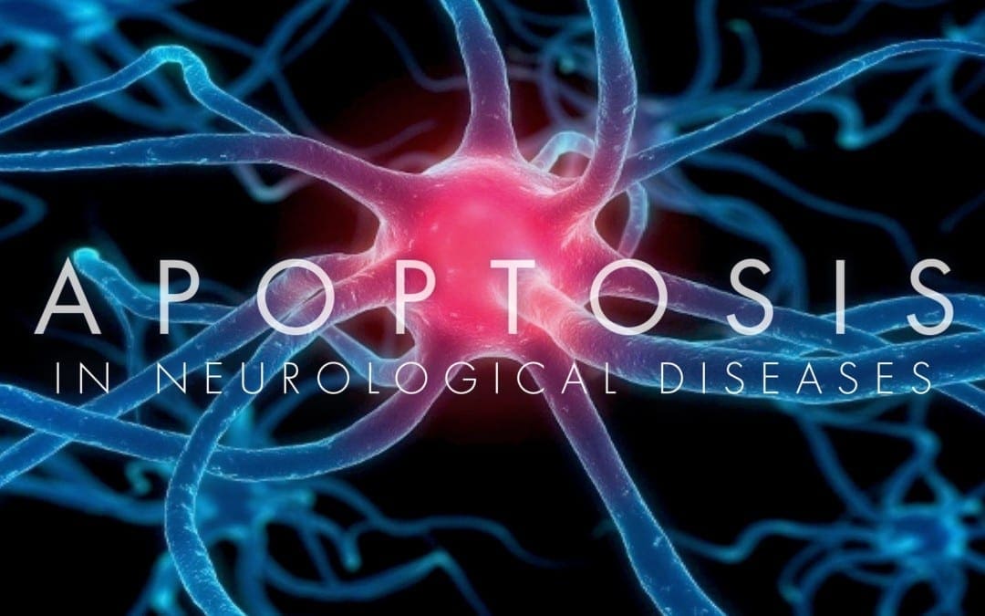
Neural cell death can occur both during the development and throughout the pathophysiology of the nervous system. Two different types of cell death, known as necrosis and apoptosis, are involved in pathological neuronal loss, however, apoptosis is the process of programmed cell death during development. All types of cells will go through apoptosis. This mechanism controls neuronal growth where an excess of neurons is produced and only those which form connections with the target structures will receive enough survival factors. The remaining neurons will then ultimately go through death and removal. �
Apoptosis continues throughout life and it is the main process involved in the elimination of surplus, unwanted, damaged or aged cells. Dysregulation of apoptosis is demonstrated after damage or injury as well as in neurodegeneration and in tumorigenesis. Treatment approaches which influence the apoptotic pathway offer valuable therapeutic options in a wide variety of pathological states. The purpose of the article is to describe the significance of apoptosis in neurological diseases. �
What is Apoptosis?
Apoptosis is the well-conserved and highly controlled process of cell death involved in the removal of unnecessary, surplus, aged or damaged cells. Dysregulation of apoptosis can ultimately develop mutated cells which can result in malformations, autoimmune diseases, and even cancer. Abnormal apoptosis can also result in the elimination of healthy cells which can occur in health issues such as infection, hypoxic-ischaemic injury, neurodegenerative or neuromuscular diseases, and AIDS. �
Apoptosis is different from necrotic cell death. In necrosis, cell death is caused by an external factor and involves the early loss of tissue, damage to organs, and the leakage of cytoplasmic contents, leading to the recruitment of phagocytes which can cause an acute inflammatory reaction. In contrast, apoptosis is often considered cell suicide. According to research studies, cells which die due to apoptosis retain membrane and organelle structure and function until late in the process while still developing plasma membrane blebbing, reduced cytoplasmic volume, chromatin condensation, and nuclear fragmentation. �
In the final phases, cell fragments wrapped in plasma membrane pull away as apoptotic bodies which are then phagocytosed by healthy cells. The removal of cell debris also occurs in the absence of an inflammatory response, and this silent, quick, and efficient elimination of apoptotic cells mean that apoptosis can be difficult to find in cells. However, as many as 50 percent of the cells in developing adulthood may go through apoptosis where less than 1 percent of cells are apoptotic at any one time. �
Apoptosis in the Nervous System
Programmed cell death by apoptosis occurs in several developmental processes, such as body sculpting and removal of self-reacting resistant cells as well as sexual organ growth and gamete formation. The general principle of growth in multicellular organisms involves the development of excess numbers of cells, where the excess or unwanted cells are then removed by apoptosis through the development of functional organs. In the developing nervous system, apoptosis has been demonstrated to occur in neural tube formation and continues throughout terminal differentiation of the neural system. �
A growing number of neurotrophic factors, such as nerve growth factor family, including both the neurokines and development factors like insulin-like growth variables (IGF-I and IGF-II), encourage the survival of several types of neurons. Targeted disruption of genes encoding these factors or their receptors demonstrate that neurotrophic factors are significant for the development of specific neuronal populations. Neurotrophic factors function by binding to specific receptors in the cell membrane. Moreover, the effects of NGF offer a good illustration of the subtle command the system permits. �
The nerve growth factor receptor has high and low affinity components. It will function as a survival factor if it binds to the high-affinity trkA receptor but it will also cause apoptosis of retinal neurons or oligodendrocytes once it binds to the low-affinity receptor p75 in the absence of trkA. Nerve growth factor in the extracellular environment is consequently able to control neural development by both boosting the growth of several types of cells as well as the removal of other cells. �
In some cases, however, concentrated genetic disruption of neurotrophic factors or their receptors may leave the central nervous system seemingly unaffected, demonstrating that these variables can ultimately become biased. According to research studies, it has now become evident that the control of neuronal survival does not only depend on the supply of trophic molecules by the targets but also on activity, humoral factors, and trophic support from glia or glial cells. �
Furthermore, neurons don’t simply undergo programmed cell death during differentiation. Apoptosis appears to regulate cell numbers in systems as diverse as the disappearance of the germinal layer during the third trimester of pregnancy, the sexual differentiation of the medial preoptic nucleus where apoptosis is controlled by testosterone, lineages throughout the olfactory epithelium, oligodendrocyte development in the optic nerve, and the development of Schwann cells in the peripheral nervous system. Programmed cell death occurs in a variety of other processes in the developing nervous system. �
Apoptosis in Nervous System Injuries & Diseases
Although apoptosis is a fundamental process involved in the developing nervous system, apoptosis can ultimately be involved in a variety of nervous system injuries and diseases. In most cases, the connection between a specific mutation or trauma as well as the activation of apoptotic cascades remains evasive. An overview of a developing list of neurological diseases in which apoptosis has been implicated as a significant pathological mechanism is provided below. �
Neuronal Injury
Cerebral hypoxic-ischaemic injury is a cause of neurological injury and death. Magnetic resonance spectroscopy studies have demonstrated that transient hypoxia-ischemia contributes to a biphasic disturbance of cerebral energy metabolism. Related to the biphasic energy collapse, two waves of cell death appear to follow hypoxic-ischaemic injury in the developing brain. Immediate neuronal death is most likely due to necrosis resulting from the accumulation of calcium ions. �
Delayed cell death caused by hypoxic-ischemic injury appears to involve further mechanisms with increasing data which demonstrates that in the delayed phase, cell death occurs by apoptosis. The amount of apoptosis is directly associated with the magnitude of ATP depletion during hypoxia-ischemia. Apoptosis can occur in the brains of newborn babies following birth asphyxia and sudden intrauterine death. Apoptosis can also be notable in white matter injury in newborn babies. �
Apoptosis may continue for months after an hypoxic-ischaemic injury due to constant changes in cerebral energy metabolism in infants during the months after birth asphyxia. Following focal neural injury, apoptosis has been discovered in remote regions from the initial damage. After severe spinal cord injury in reptiles, apoptosis of oligodendrocytes occurs in distant degenerating fiber tracts and after forebrain injury in rats, apoptosis was demonstrated in the cerebellum. �
The apoptotic loss of oligodendrocytes could consequently be a potential source of secondary demyelination in paraplegia and in the chronic degeneration related to multiple sclerosis. Further research studies must be performed in order to provide further evidence on the role of apoptosis in this type of injury which begins from the report of which Bcl-2 expression boosts the growth and regeneration of retinal axons. Apoptosis in neuronal injury can be demonstrated in a variety of ways. �
Neural Cancers
A connection between apoptosis and the cell cycle is demonstrated in carcinogenesis where proto-oncogenes, such as c-fos, c-jun, and c-myc, can activate apoptosis and promote cell division while inactivation of the pro-apoptotic p53 tumor suppressor gene is a frequent mark of human neoplasia. By way of instance, in a number of gliomas, the reduction of wild p53 activity was connected to tumor progression, possibly leading to resistance to chemotherapy and radiotherapy. �
Although there have been reports of Bcl-2 overexpression in glioma cell lines, the correlation between the anti-apoptotic effect of this gene and malignancy is not yet clear. However, a homolog of Bcl-2, the brain associated apoptosis gene (BRAG-1), is found predominantly in the brain, and it is upregulated in human gliomas as a rearranged transcript. As demonstrated above, the process of apoptosis can also be significant in the development of neural cancers, according to research studies. �
Infectious Disease
Apoptosis may play a role in HIV encephalopathy. In the brain, the virus reproduces primarily in microglia which it enters through the CD4 receptor. Although the activation of microglia is believed to be the main reason for adrenal loss and demyelination, neurons die by apoptosis in HIV encephalopathies because of HIV mediated alterations in astrocyte function and aberrant stimulation of NMDA receptors or due to nitric oxide from the activation of inducible nitric oxide synthase. �
In subacute sclerosing panencephalitis, widespread apoptotic death was demonstrated to develop in the brain, although no correlation was observed between viral load, lymphocyte infiltration, and the number of apoptotic cells. DNA fragmentation indicative of apoptosis was detected in scrapie-infected sheep and mice brains, suggesting a function associated with cell death in spongiform encephalopathies. Apoptosis may also ultimately be involved in another infectious disease. �
Neurodegeneration
Spinal muscular atrophy is associated with mutations in the survival of motor neuron and neuronal apoptosis inhibitory protein (NAIP) enzymes. NAIP is closely related to the baculovirus inhibitor of apoptosis protein and inhibits apoptosis in many cell types. This implies that mutations in NAIP could deregulate apoptosis in spinal motor nerves, causing their death. Recent studies emphasize the importance of anti-apoptotic genes in cerebral protection which can rescue neurons. �
Apoptosis has also been implicated in retinal dystrophies such as retinitis pigmentosa. In this case, apoptosis results from mutations in the three photoreceptor genes, rhodopsin, peripherin, and the ?-subunit of cyclic guanosine monophosphate di esterase, resulting in photoreceptor degeneration. The absence of c-fos prevents apoptosis in those cells is unknown. Moreover, defined neurotrophins and growth factors injected intraocularly in animal models of retinal degeneration improve photoreceptor survival, suggesting that the apoptotic cascade can be obstructed by supplying exogenous survival signs. �
The mutation underlying Huntington’s disease is an expanded trinucleotide which is fundamental for normal development and can be regarded as a cell survival gene. Transgenic models demonstrated increased apoptosis in the neurons of an embryonic neuroectoderm. During apoptosis, caspase-3 (apopain) is improved by a gain of function associated with the triplet expansion. This is supported by the overexpression of specific trinucleotide repeats in transgenic mice. �
Most cerebellar ataxias are associated with neuronal loss. Ataxia-telangiectasia, caused by mutations in the ATM gene, is considered to have an apoptotic component. ATM shares extensive and significant homology with the DNA dependent protein kinases involved in DNA damage responses at different cell cycle checkpoints and is downregulated in most patients with ataxia-telangiectasia. The simple fact that inappropriate p53 mediated apoptosis is the major cause of death in ataxia-telangiectasia cells suggests that the mutation causes improper triggering of apoptosis by otherwise non-lethal DNA injury. �
From the familial form of amyotrophic lateral sclerosis gain of function, mutations in the gene encoding copper-zinc superoxide dismutase (sod-1) develop a dominant pro-apoptotic sign. Although cell harm by the accumulation of free radicals can trigger apoptosis, these mutants can induce apoptosis both in nerve cells in culture and in transgenic mice. Mental retardation in Down’s syndrome has also been associated with abnormal apoptosis. Although cortical neurons from fetal Down’s syndrome brains are different, they then degenerate and undergo apoptosis, according to research studies. �
Degeneration is blocked by treatment with free radical scavengers, suggesting that a defect in the metabolism of reactive oxygen species is the trigger for apoptosis. In Parkinson’s disease, the death of dopaminergic neurons in the substantia nigra was demonstrated to occur through apoptosis and may be obstructed by delivery of glial-derived neurotrophic factor. Alzheimer’s disease is associated with the progressive accumulation of ?-amyloid protein which is the fundamental component of neural plaques. The ?-amyloid peptide can cause neurons to undergo apoptosis in vitro research studies. �
Inherited Metabolic Disease
Furthermore, few data suggest that the acute encephalopathy associated with maple syrup urine disease is because of the induction of apoptosis by an accumulating metabolite of leucine, ?-keto isocaproic acid. This compound is a potent inducer of apoptosis in central nervous system glial cells and the result is significantly enhanced in the presence of leucine. Phenylalanine and leucine do not induce apoptosis in this system, suggesting that this result is ultimately unique. �
There are two ways in which a cell can die, necrosis and apoptosis. While necrosis occurs due to an external factor which harms the cell, apoptosis follows a controlled, predictable routine. Apoptosis is generally known as programmed cell death. Apoptosis, or programmed cell death, has many fundamental functions in the developing structures of the human body, however, research studies have demonstrated that abnormal apoptosis can be associated with the development of a variety of neurological diseases. – Dr. Alex Jimenez D.C., C.C.S.T. Insight
The purpose of the article above is to discuss the process of apoptosis, or cell death, in neurodegenerative diseases. Neurological diseases are associated with the brain, the spine, and the nerves. The scope of our information is limited to chiropractic, musculoskeletal and nervous health issues as well as functional medicine articles, topics, and discussions. To further discuss the subject matter above, please feel free to ask Dr. Alex Jimenez or contact us at 915-850-0900 . �
Curated by Dr. Alex Jimenez �
Additional Topic Discussion: Chronic Pain
Sudden pain is a natural response of the nervous system which helps to demonstrate possible injury. By way of instance, pain signals travel from an injured region through the nerves and spinal cord to the brain. Pain is generally less severe as the injury heals, however, chronic pain is different than the average type of pain. With chronic pain, the human body will continue sending pain signals to the brain, regardless if the injury has healed. Chronic pain can last for several weeks to even several years. Chronic pain can tremendously affect a patient’s mobility and it can reduce flexibility, strength, and endurance.
Formulas for Methylation Support
XYMOGEN�s Exclusive Professional Formulas are available through select licensed health care professionals. The internet sale and discounting of XYMOGEN formulas are strictly prohibited.
Proudly,�Dr. Alexander Jimenez makes XYMOGEN formulas available only to patients under our care.
Please call our office in order for us to assign a doctor consultation for immediate access.
If you are a patient of Injury Medical & Chiropractic�Clinic, you may inquire about XYMOGEN by calling 915-850-0900.
�
For your convenience and review of the XYMOGEN products please review the following link.*XYMOGEN-Catalog-Download �
* All of the above XYMOGEN policies remain strictly in force. �
IFM's Find A Practitioner tool is the largest referral network in Functional Medicine, created to help patients locate Functional Medicine practitioners anywhere in the world. IFM Certified Practitioners are listed first in the search results, given their extensive education in Functional Medicine
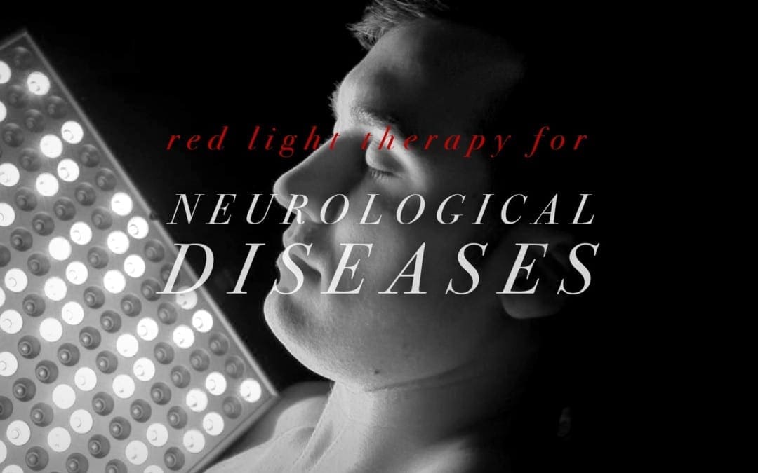


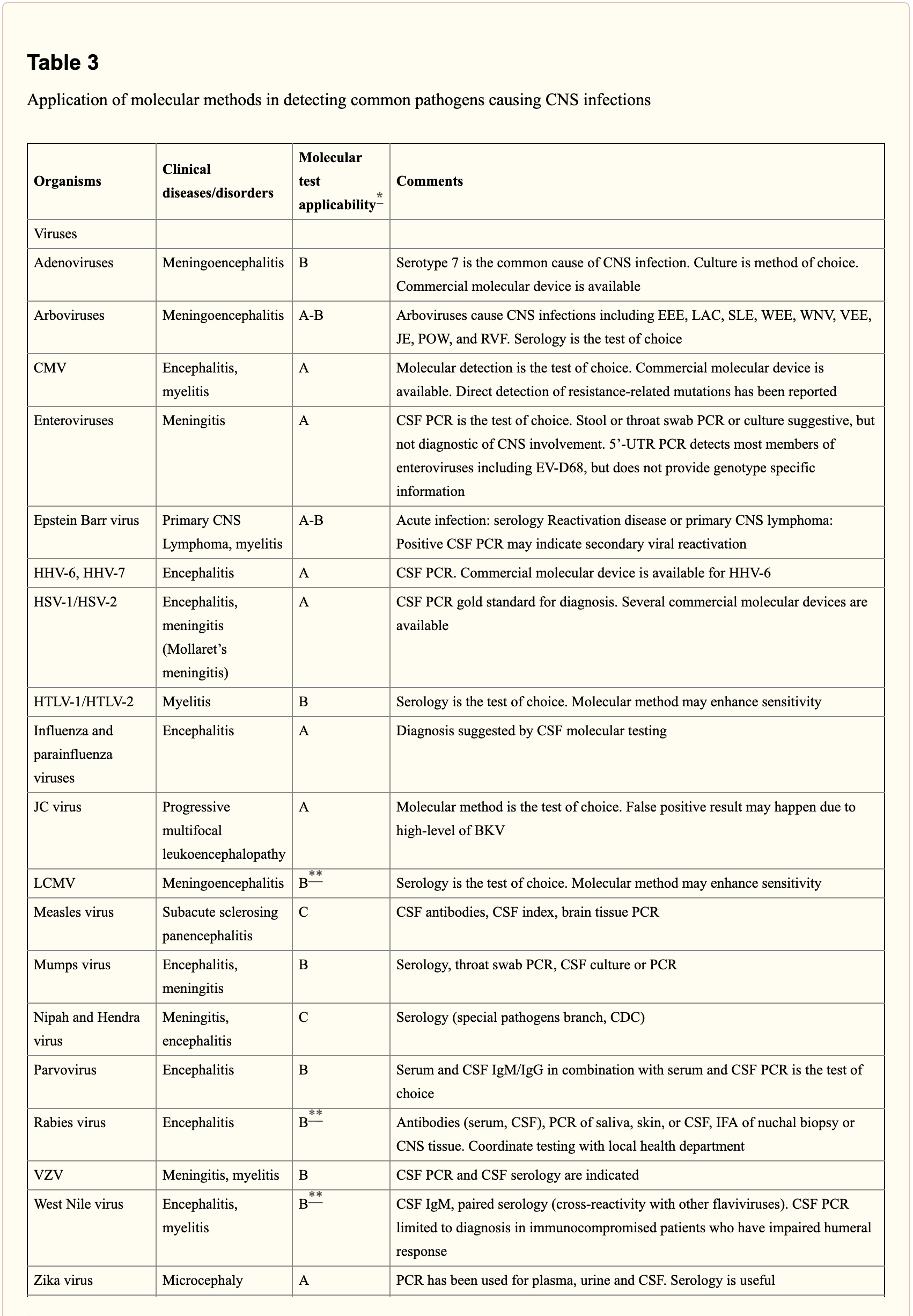
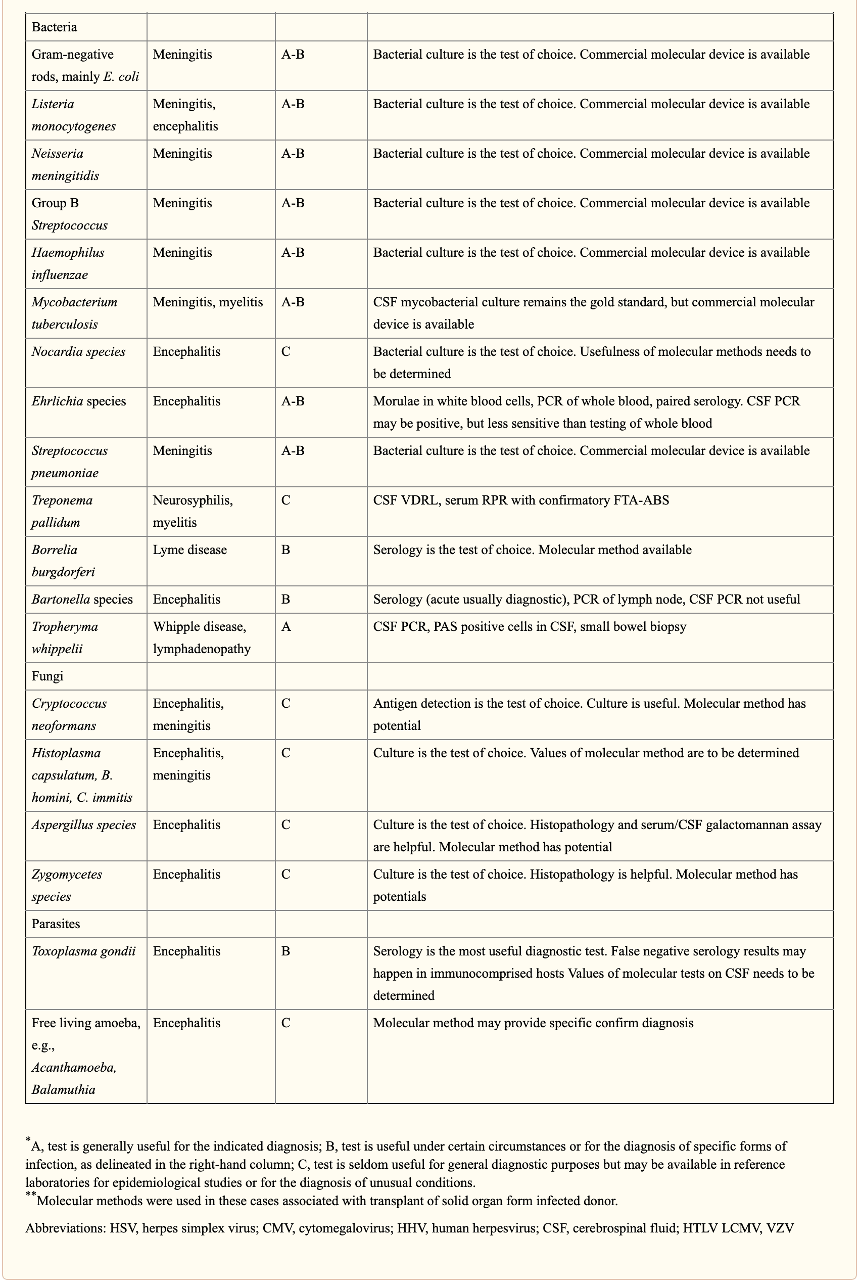
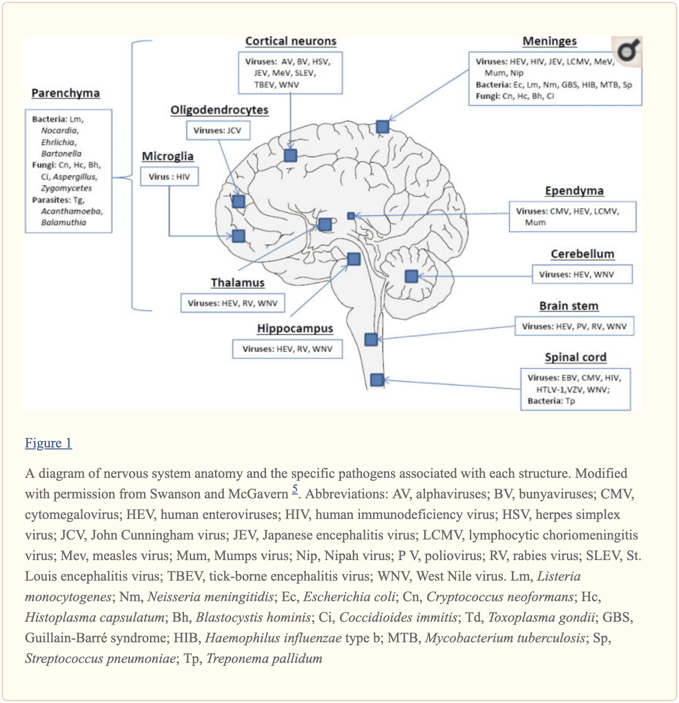
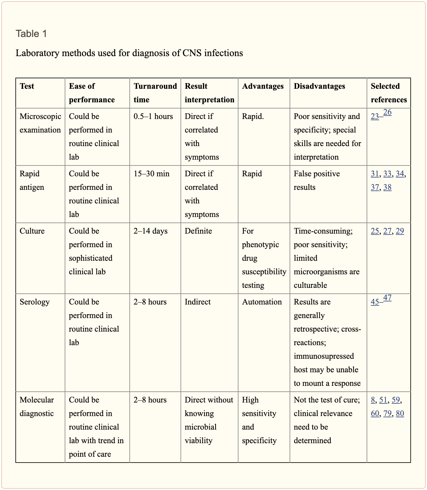
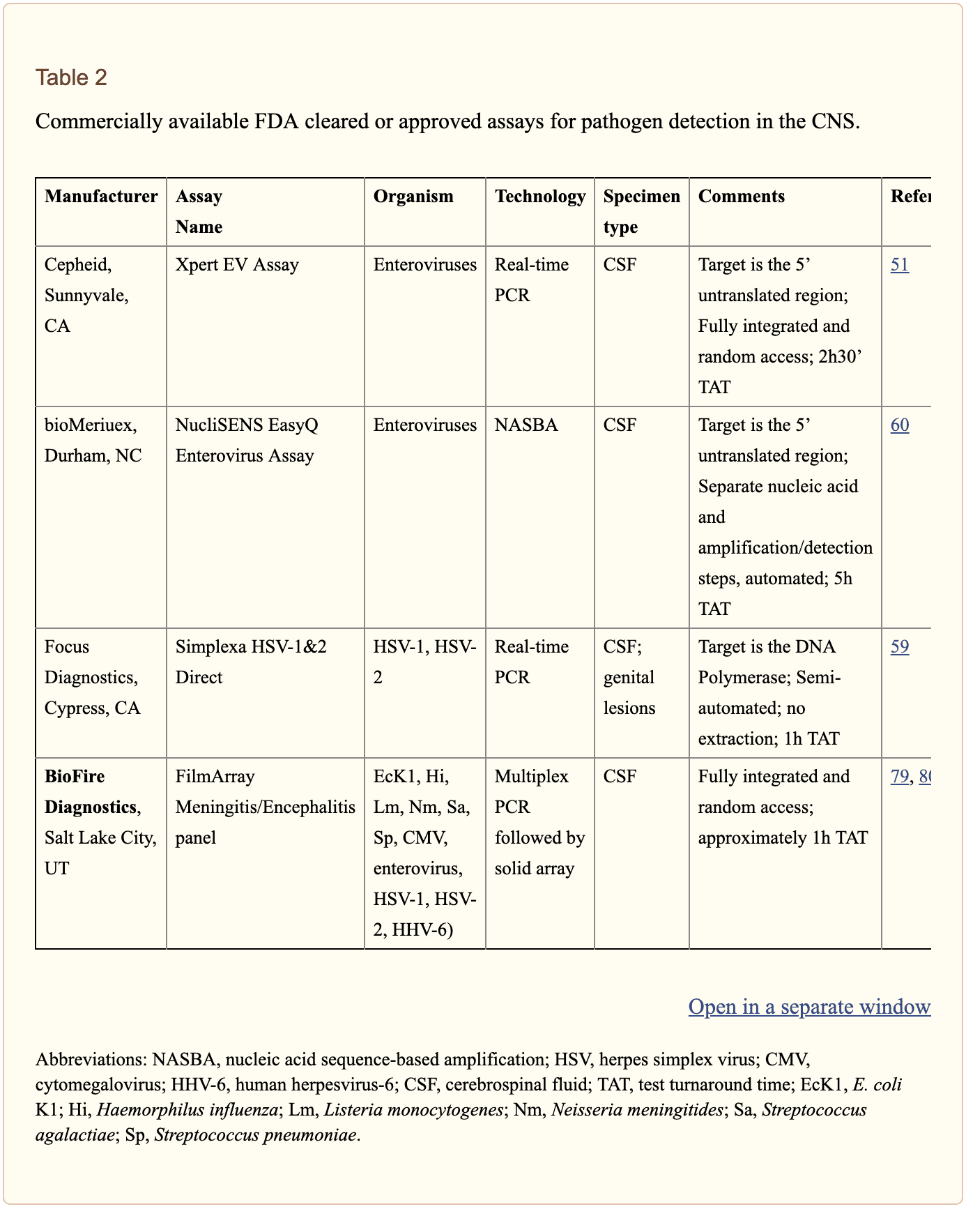
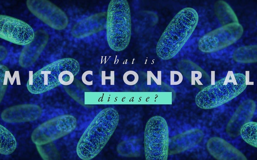
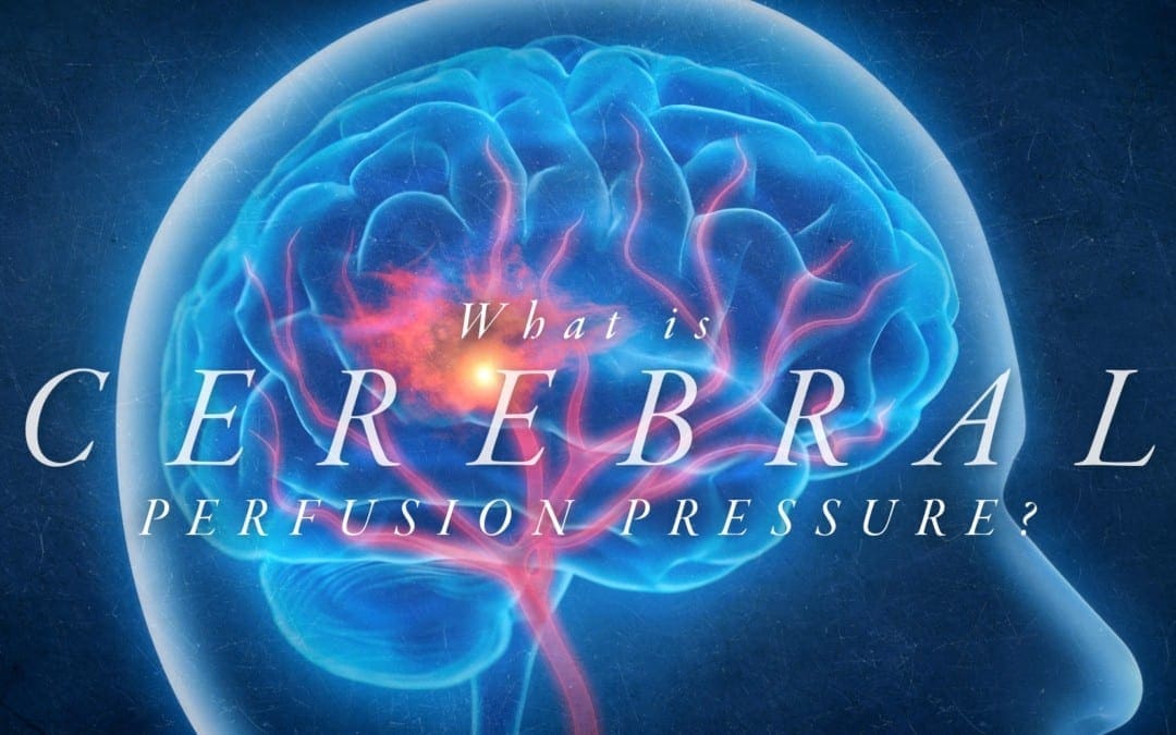
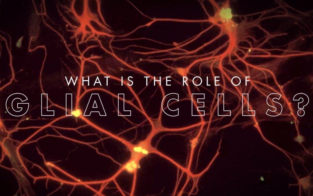
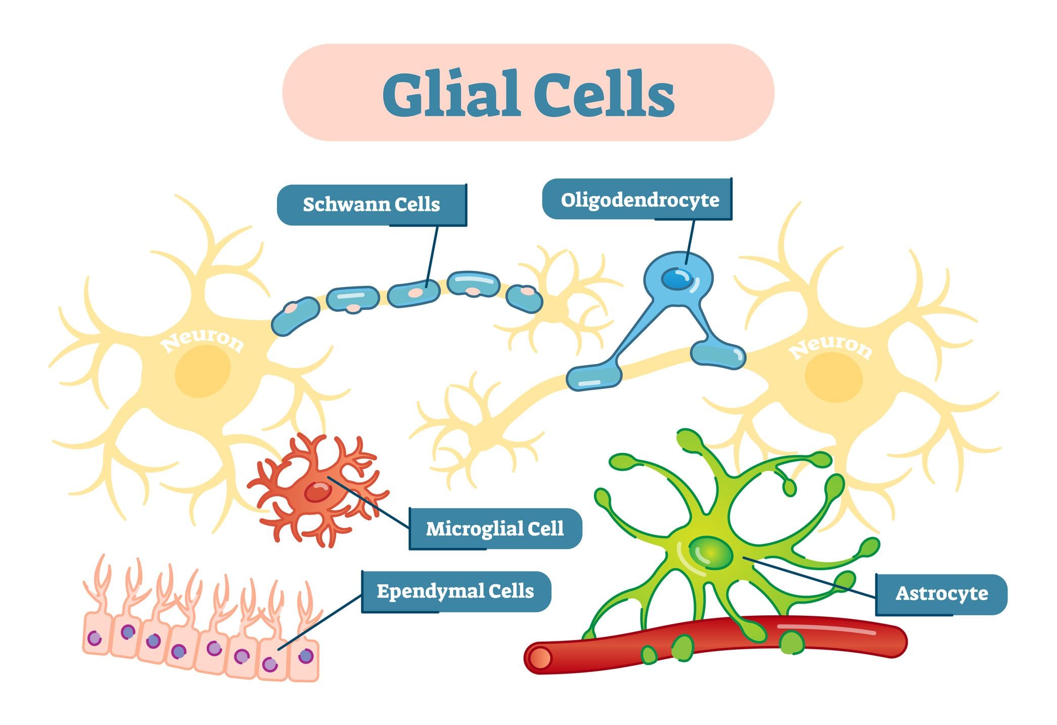 �
�