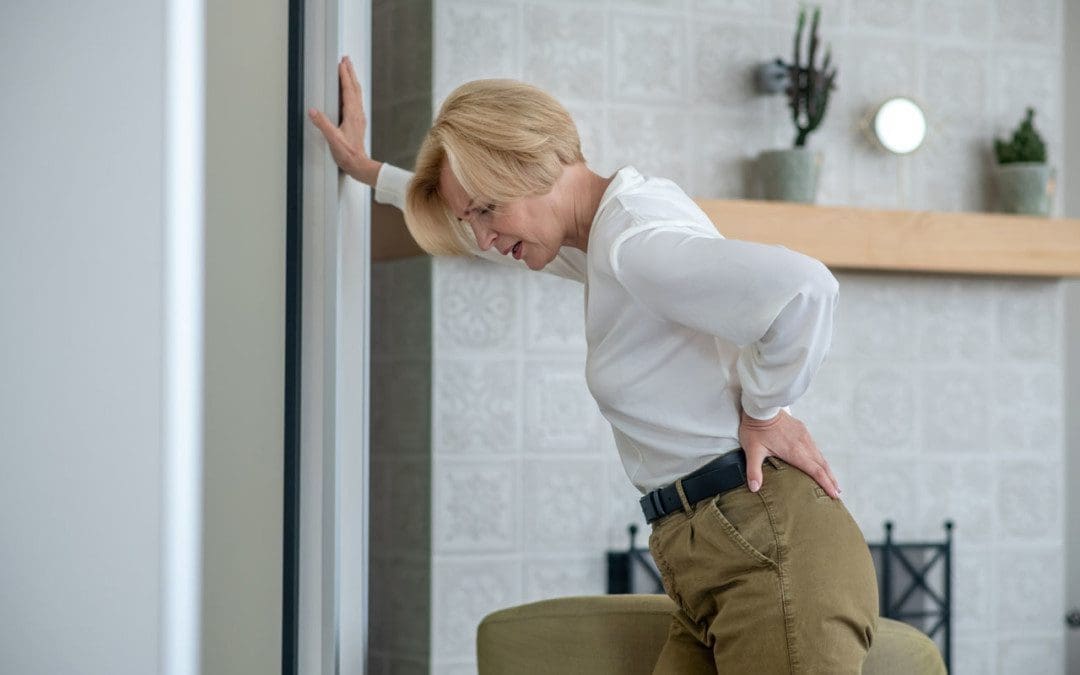
It is one thing to wake up with back pain, but another when the pain is combined with a fever, body aches, and chills. It could be the flu or another infection. However, after checking the body’s temperature and fever is present with no other symptoms than back pain unless it is the flu; the fever could be another issue that may or may not be related as there are a variety of causes for back pain like:
Muscle or ligament strain – If in poor physical condition, repeated and constant tension on the back can cause muscle spasms. Repeated heavy lifting or a sudden awkward movement can strain the back muscles and spinal ligaments.
Bulging or ruptured discs – Discs act as cushions between the bones/vertebrae in the spine. The soft material inside a disc can bulge or rupture and press on a nerve. However, a bulging or ruptured disc can present without back pain. Disc disease is often found by accident when spine X-rays are performed for another reason.
Arthritis – Osteoarthritis can affect the lower back. In some cases, arthritis in the spine can narrow the space around the spinal cord, a condition called spinal stenosis.
Osteoporosis – The spine’s vertebrae can develop painful fractures if the bones become porous and brittle.
Back pain without a fever is usually an indication of a misaligned spine.
Fever A Sign of Something Else
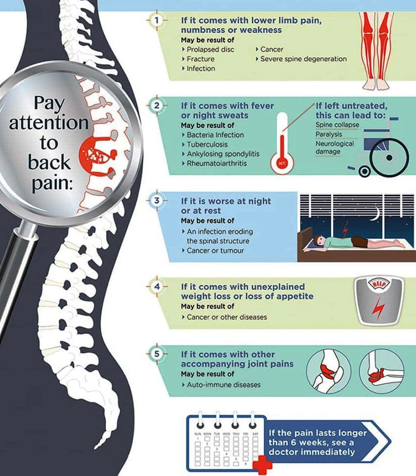
A fever is the body’s way of trying to raise its core temperature in an attempt to kill off a virus or a bacterial infection. Possible causes of back pain with fever include:
Kidney Infection
This type of infection often presents with low back pain and fever.
Spinal Epidural Abscess
This is an infection of the lower region of the spine, causing fever and lower back pain.
Vertebral Osteomyelitis
This is an infection of the lower spine that causes pain in the arms, lower back, and legs, along with a fever.
Meningitis
This causes swelling and inflammation of the brain and spine and needs to be addressed immediately.
Spinal Cord Abscess
This is an infection of the internal part of the spine. It is rare but can happen, causing low back pain and fever.
Symptoms
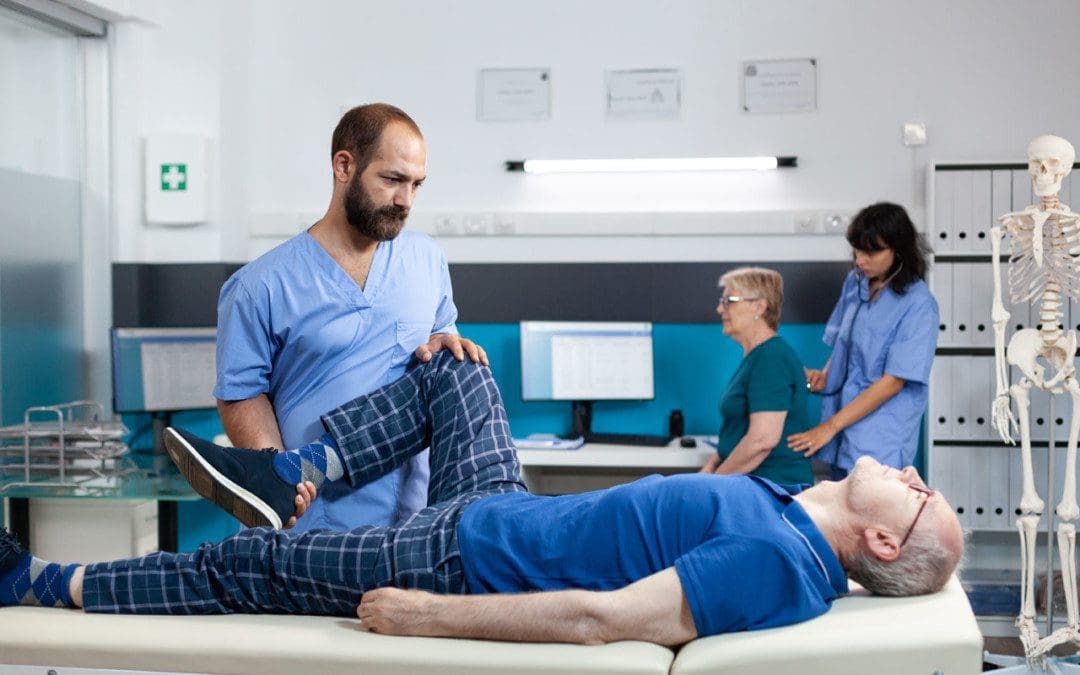
This is when seeing a chiropractor can help. A few signs that should not be ignored include:
Recently involved in an automobile accident.
Suffered a serious fall.
Feeling a tingling in the legs.
Having balance issues.
Having abdominal pain.
Pain is not going away, or it goes away for a while, then comes back.
Have weakness in the arms or legs.
Having bowel or urinary problems that were not present previously.
The pain is worse when sitting or standing up after sitting.
Have upper back pain after alcohol consumption.
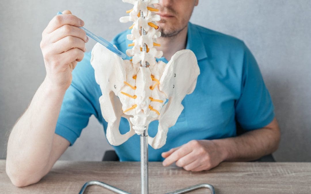
A chiropractor will take a complete medical history, X-rays, an MRI if necessary, and a thorough physical examination will be performed to determine the cause. After a diagnosis is reached, the chiropractor will perform adjustments to relieve the pain and open the nerve pathways to increase circulation to the area. A chiropractic massage will help reduce stress, relieve back pain, and reduce depression, which can also help reduce the fever unless it is from another issue.
Body Composition
Influenza
Influenza or the flu is a contagious respiratory illness caused by viruses that infect the nose, throat, and lungs. It can cause mild to severe illness and, in extreme cases, can lead to death. Like a common cold, the flu is spread primarily through tiny droplets that get expelled from an infected person when they sneeze, cough, or talk. Approximately 8% of the population gets the flu each season. Flu symptoms are sudden, causing the following:
Fever
Chills
Muscle or body aches
Headaches
Sore throat
Runny or stuffy nose
Cough
Fatigue
Vomiting and diarrhea which is more common in children.
Most individuals with healthy immune systems will recover around seven days. However, the elderly, pregnant women, individuals of any age with certain chronic medical conditions like asthma, diabetes, or heart disease, and children under the age of five have an increased risk of developing complications. Flu vaccination is currently recommended for anyone older than six months in the U.S. and effectively prevents infection in 50 – 80% of the population. The primary treatment method for the flu is to support the immune system with plenty of rest, proper nutrition, and hydration.
References
Ameer MA, Knorr TL, Mesfin FB. Spinal Epidural Abscess. [Updated 2021 Feb 11]. In: StatPearls [Internet]. Treasure Island (FL): StatPearls Publishing; 2021 Jan-. Available from: www.ncbi.nlm.nih.gov/books/NBK441890/
Kehrer, Michala et al. “Increased short- and long-term mortality among patients with infectious spondylodiscitis compared with a reference population.” The spine journal: official journal of the North American Spine Society vol. 15,6 (2015): 1233-40. doi:10.1016/j.spinee.2015.02.021
Rubin, Devon I. “Epidemiology and risk factors for spine pain.” Neurologic clinics vol. 25,2 (2007): 353-71. doi:10.1016/j.ncl.2007.01.004
Tsantes, Andreas G et al. “Spinal Infections: An Update.” Microorganisms vol. 8,4 476. 27 Mar. 2020, doi:10.3390/microorganisms8040476
The sciatic nerve is formed through a combination of motor and sensory fibers based on the spinal nerves of the lower back L4 to S3, known as the lumbosacral plexus. It is the largest and longest nerve in the human body and about as wide as an adult thumb. It begins at the base of the spine, runs along the back of each leg, and ends at the foot supplying the areas with fresh blood and nutrients. There are sciatic nerve branches that consist of primary branches and smaller branches.
Sciatic Nerve Branches
The nerve splits into two main branches near the back of the knee called the popliteal fossa.
This fossa is located slightly above the joint behind the knee.
The popliteal fossa is a diamond-shaped space that acts as the conduit for the blood vessels and nerves.
Primary branches
From the popliteal fossa:
The tibial nerve continues down the back of the calf to the heel and bottom of the foot.
The common peroneal nerve, aka common fibular nerve, travels sideways along the outer part of the knee to the outer border of the lower leg and foot.
Both nerves convert into small sensory nerves in the calf that supply the outer side of each foot.
The sciatic nerve breaks off into smaller branches, known as collaterals, that include:
These are muscle branches that supply the muscles in the thigh, including the hamstring group and the adductor magnus muscles along the inner thigh.
Other small branches supply the leg and foot muscles.
Articular branches supply the back of the hip joint, the back and side of the knee joint.
The sciatic nerve does not supply structures in the buttocks; however, pain commonly radiates/spreads into this area when the nerve is impaired, impinged, and inflamed.
Blood Supply
The delivery of nutrients to the sciatic nerve is done through blood vessels that also contribute to the nerve’s function. Any interruption of blood flow to the sciatic nerve can cause pain and dysfunction. The sciatic nerve and the sciatic nerve branches receive their blood supply from two sources that include:
The extrinsic system is made up of nearby arteries and veins.
The intrinsic system includes arteries and veins that run along the nerve and are embedded deep in a sheath known as the epineurium of connective tissue that envelops the nerve.
The intrinsic blood supply can be affected by conditions like diabetes, which can contribute to symptoms associated with diabetic neuropathy.
Both systems connect at various junction points.
Nerve Function
The combination of sensory and motor fibers that make up the sciatic nerve provides the essential functions in the lower limbs allowing the body to:
Stand
Walk
Run
Climb
Lift
A healthy sciatic nerve is well protected around the low back and buttock muscles where it starts, and it cannot be palpated or felt by touching or pressing on the area. When the nerve gets inflamed, injured, or pinched, the leg can feel stiff and inflexible when trying to move and can lead to pain, weakness, and tingling in the lower back, buttock, leg/s, and feet.
Anatomical Variations of the Nerve
Individuals can have variations in the anatomical structure of the sciatic nerve. These variations are considered normal, but they can increase the risk of developing sciatica brought on by impingement, entrapment, or irritation of the nerve root/s. Variations in sciatic nerve branches include:
The nerve divides above the piriformis muscle; one portion passes through the piriformis, with the other portion exiting the pelvis below the muscle. This is the most common variation.
The nerve divides above the piriformis muscle; one portion passes through the piriformis, with the other portion exiting the pelvis above the muscle.
The nerve divides above the piriformis, with one portion traveling in front while the other travels behind it.
Undivided sciatic nerve exits through the piriformis muscle.
Undivided sciatic nerve exits from behind the top part of the piriformis.
Around 10% of individuals have a nerve that divides above the popliteal fossa and does not merge but courses down in two separate branches.
The sciatic nerve and the sciatic nerve branches are significant components of the body. It supplies motor functions to move the legs and feet and provides sensory functions along the nerve path. Keeping the sciatic nerve healthy is key in helping to prevent back and spinal issues. Chiropractic can help realign the sciatic nerve and educate on maintaining the nerve’s health.
Body Composition
Fitness Motivation
New workout routine
Individuals that don’t feel like returning to previous workout routines are recommended to try out other fitness options. If the gym isn’t cutting it or there is burnout with the current routine, switch things up. This can include:
Virtual group classes.
1-on-1 personal training.
Outdoor activities.
All are valid options to explore if in a rut with the current routine.
The important thing is to find what works for you.
Allow the body to rest
Individuals may want to push it to the limit to get back into shape, but rest days are essential for healthy muscle development and improved performance.
Noticing the body is more sore and exhausted after a workout is an indication that the body needs rest. This also includes:
Maintaining proper hydration.
Stretching out the muscles regularly.
Taking days off from exercising are necessary to:
Prevent muscle fatigue.
Reduce the risk of injury.
Allow for adequate muscle recovery.
Long term commitment is key
It can be discouraging to commit to a workout schedule only to notice minor changes to strength and fitness.
However, small improvements do accumulate over time.
Small increases over time can have a huge impact on overall strength and fitness.
Keep the bigger picture in mind to remain positive.
References
Davis D, Vasudevan A. Sciatica. [Updated 2019 Feb 28]. In: StatPearls [Internet]. Treasure Island (FL): StatPearls Publishing; 2019 Jan-. Available from: www.ncbi.nlm.nih.gov/books/NBK507908/
Barral J, Croibier A. Manual Therapy for the Peripheral Nerves. Elsevier Health Sciences; 2007.
Ryan MM, Jones HR Jr. Mononeuropathies. In: Neuromuscular Disorders of Infancy, Childhood, and Adolescence. Elsevier; 2015:243-273. doi:10.1016/b978-0-12-417044-5.00014-7
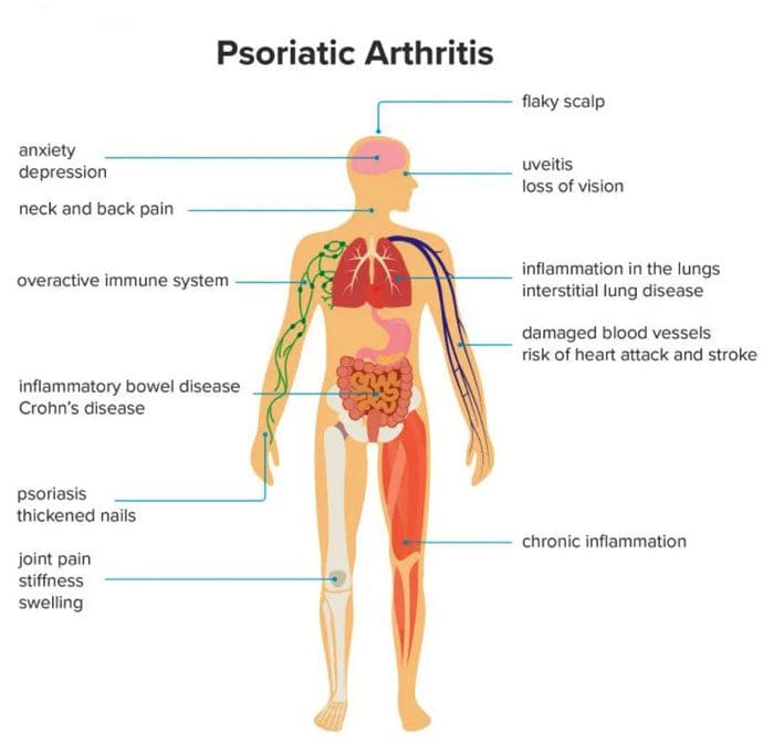
Psoriatic arthritis can develop in individuals who have psoriasis, affecting various joints, especially the knees. Psoriasis is a skin condition that causes skin cells to build up and form patches of itchy, dry skin known as plaques. Psoriatic arthritis is a long-term inflammatory disease that can cause inflammation, stiffness, and pain. Symptoms can progressively worsen over time without treatment. Early diagnosis is vital to minimize damage to the joints and slow the condition’s progress with treatment.
Psoriatic Arthritis
Psoriatic arthritis symptoms like stiffness and swelling can present differently from person to person. For example, some individuals with psoriatic knee arthritis will experience stiffness or pain in one knee, while others experience symptoms in both knees. Psoriatic arthritis in the knee can also cause swelling in the surrounding:
Ligaments
Tendons
Synovial membranes
Symptoms can also present in the:
Elbows
Feet
Hands
Symptoms
Symptoms usually begin between the ages of 30 and 50. Common symptoms include:
Stiffness after resting or sleeping.
Swelling.
Inflammation in the knee and surrounding area.
Warm or hot skin on the knee from the inflammation.
Pain in and around the joints, tendons, or ligaments.
Joint sticking, difficulty moving, or reduced range of motion.
Other symptoms include:
Back pain
Fatigue
Pain and redness in the eyes
Swollen fingers or toes
Difficulty walking from pain in the feet or Achilles tendon.
The severity of psoriasis does not determine psoriatic arthritis symptoms. Symptoms can go through a pattern of relapses and remissions. Individuals can have a sudden attack where symptoms get worse over a short time. After the flare-up, symptoms can improve as the condition goes into remission. Symptoms may not present for a long time until another flare-up. For example, an individual may have severe psoriasis but only mild psoriatic arthritis.
Causes
Psoriatic arthritis develops when the body’s immune system mistakenly attacks healthy cells and tissues. The faulty immune response causes the body to quickly generate new skin cells that stack on top of each other forming plaques. When the condition affects the joints, it leads to inflammation. While there is no apparent cause for psoriatic arthritis, researchers have found connections to genetics and the environment, as well as, individuals with close relatives that have psoriatic arthritis could be more likely to develop the condition. Other factors that could influence the development include:
Severe psoriasis
Traumatic injury/s
Obesity
Nail disease
Smoking
The condition can happen at any age, but according to the National Psoriasis Foundation, most individuals first notice symptoms about ten years after their psoriasis begins. However, only 30% of individuals with psoriasis develop psoriatic arthritis.
Diagnosis
Doctors use imaging tools to diagnose psoriatic arthritis in the knee. They will use:
MRI
X-rays
Ultrasound
To help them check for irregularities or signs of inflammation in the joint and surrounding tissues.
Additional tests are used to rule out other common forms of arthritis like rheumatoid and osteoarthritis.
Blood tests check for inflammation and specific antibodies.
In some cases, a small amount of fluid from the joint is taken to help eliminate the possibility of other underlying conditions like an infection.
Treatment
There is currently no cure for psoriatic arthritis, but treatments are being developed and show promise for long-term management. Current treatments focus on managing symptoms and improving the quality of life for the individual.
Biologics
Biologic medications like tumor necrosis factor or TNF inhibitors are recommended as the first-line therapy for most individuals with a new diagnosis of psoriatic arthritis. These meds help block TNF, which plays a crucial role in inflammation. They have shown to be effective at reducing the severity of symptoms and the frequency of flare-ups. Biologics can cause unwanted side effects, especially in individuals that experience frequent infections and need routine monitoring.
Small Molecule Medications
Individuals that cannot use biologic medications may be recommended a new class of medication called oral small molecules or OSMs. Examples include apremilast – Otezla and tofacitinib – Xeljanz.
Disease-modifying Antirheumatic Drugs
Disease-modifying antirheumatic drugs – DMARDs are a long-term option. They are used to slow the progression of psoriatic arthritis, and examples include methotrexate and cyclosporine. DMARDs work best when an individual begins taking them as early as they can take time to work. However, individuals are encouraged to continue taking them, even if symptoms do not improve right away.
Easing Inflammation
A doctor may prescribe nonsteroidal anti-inflammatory drugs – NSAIDs and corticosteroid injections when knee symptoms flare-up. These are short-term treatments that provide immediate relief, as long-term use can lead to side effects. Individuals can find relief with combined self-care that includes:
Taking over-the-counter NSAIDs like ibuprofen/Advil or naproxen/Aleve.
Applying ice and heat packs.
Gentle exercise to promote a full range of motion.
Gentle stretching or yoga can help relax tight muscles.
However, chiropractic is not the primary treatment for arthritis but is intended to be used in combination to relieve pain, loosen and stretch the muscles and balance the body.
InBody
Strength, Balance, and Improved Body Composition
Functional fitness is the ability to move comfortably every day. The benefits of physical activity also contribute to improved body composition. Working to reach a certain level of functional fitness can help the aging process that has been shown to reduce metabolic rate. Inactivity is why individuals lose Lean Body Mass as they age, leading to increased body fat. Lean Body Mass contributes to the body’s overall Basal Metabolic Rate or BMR, also known as metabolism. This is the number of calories the body needs to support essential functions. Everyone is encouraged to engage in strength training or resistance exercises, but specifically older adults. This can help regain muscle loss which can lead to an increase in lean body mass. The increase in Lean Body Mass increases BMR, which helps prevent fat gain.
References
Chang, K. L., et al. (2015). Chronic pain management: Nonpharmacological therapies for chronic pain [Abstract]. www.ncbi.nlm.nih.gov/pubmed/25970869
Chiropractic care for arthritis. (n.d.). arthritis.org/health-wellness/treatment/complementary-therapies/physical-therapies/chiropractic-care-for-arthritis
Bursitis types: This is a condition that affects the bursae, which are the small, fluid-filled sacs that provide cushion for the:
Muscles
Tendons
Bones near joints
The bursae make it easier for tissues to slide over each other. The body has around one hundred and sixty bursae. However, only a few become clinically affected. These include the:
Wrist
Elbow
Shoulder
Hips
Knees
The base of the big toe and heel
The condition typically presents near joints constantly being used repetitively, like a job, sports, house/yard chores, etc. What happens is one or more of the bursae sacs become inflamed, resulting in pain.
Causes
Inflamed or irritated bursae typically cause it from overuse or intense/vigorous activity.
It can also be caused by bacterial infection.
Arthritis and gout can also cause bursitis.
Another cause is age.
As tendons age, they can tear easily, lose their elasticity, and can’t take too much stress.
Intense physical activities can lead to bursitis. These include:
Gardening
Typing
Working with a computer mouse
Throwing
Golf
Tennis
Manual tasks
Carpentry
These types of activities can lead to incorrect posture, overuse, and injury/damage.
Symptoms
The main symptom is pain in and around the affected area that worsens with movement. Depending on the severity of the strain and the length of time it has been going on, the pain can be intense with active and passive movements. Other symptoms include:
Tenderness
Stiffness
For some individuals, it can present as acute, with the intensity increasing.
This happens when movement aggravates the condition.
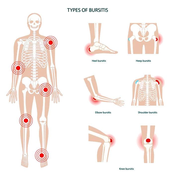
Bursitis Types
Four major types include:
Prepatellar
Trochanteric
Olecranon
Retrocalcaneal
Prepatellar Bursitis
Prepatellar is an inflammation of the sac situated between the skin and the patella/kneecap. The most common causes are trauma from a fall and direct pressure/friction from repetitive kneeling. This is one of the bursitis types that can get infected. Overproduction of liquid places pressure on the other areas of the knee, causing swelling. Most individuals report swelling and knee pain just over the front of the knee.
Trochanteric Bursitis
This bursitis type goes over the lateral area of the hip. There is a distinctive tenderness and aching pain. This type is more common for individuals with arthritis conditions and fibromyalgia. This condition is also seen after surgery, mainly osteotomies. The bursa can become inflamed in case of injury or overuse. It tends to affect middle-aged or older folks. Common causes include:
Muscle tears
Hip injuries
Tight hip or leg muscles
Disc disease of the low back
Leg-length inequality
Improper walking technique from a minor injury or strain
Overuse of the gluteal muscles
Flat feet
Improper footwear
Olecranon Bursitis
Olecranon is a common bursitis type. It is diagnosed by the appearance of swelling over the elbow. The swelling happens just behind the olecranon process of the ulna. The bursa can become infected. This bursitis does cause blood to rupture out, and fluid could be present. Individuals are advised to avoid leaning or resting on the elbows.
Retrocalcaneal Bursitis
This is characterized by pain in the Achilles tendon. Chronic inflammation of the bursa is brought on by friction, supination, and overpronation. The flexibility of the calf muscles can be significantly reduced. Severe pain and swelling of the posterior soft tissue in front of the Achilles tendon are common symptoms. This bursitis type is often accompanied by mid-portion insertional tendinosis.
Risk Of Getting Bursitis
Anybody at any age can develop bursitis, but older individuals, specifically those in their forties and beyond, are more susceptible. This comes from all the wear and tear of the muscles and bones.
Risk Factors
Overpronation of the foot
Leg length deviation
Osteoarthritis
Obesity
Tight hamstring muscles
Incorrect physical training
Not stretching properly
Body Composition
When Inflammation Becomes Permanent
When white blood cells cause inflammation, it’s signaling that the body’s immune system works properly. The process works like this:
Inflammation activates
White blood cells attack the foreign invader
The invader is neutralized
The inflammation deactivates
This is how the body’s defense system naturally works. But, white blood cells are not the only type of cell that emit cytokines. Adipocytes or fat cells are another type of cell that can emit cytokines and cause inflammation. Scientists have learned that fat is an active endocrine organ that secretes various proteins and chemicals, including inflammatory cytokines. The body stores excess calories as fat to be used later for energy. When the body keeps adding more adipose tissue, cytokines are released by the fat cells, triggering inflammation. Obesity is characterized as a state of low-grade, chronic inflammation. Increased fat cells place the body in a constant state of stress activating immune responses. This means the body is in a constant state of inflammation with the immune system switch permanently on.
References
Aaron, Daniel L et al. “Four common types of bursitis: diagnosis and management.” The Journal of the American Academy of Orthopaedic Surgeons vol. 19,6 (2011): 359-67. doi:10.5435/00124635-201106000-00006
Coelho, Marisa et al. “Biochemistry of adipose tissue: an endocrine organ.” Archives of medical science: AMS vol. 9,2 (2013): 191-200. doi:10.5114/aoms.2013.33181
Khodaee, Morteza. “Common Superficial Bursitis.” American family physician vol. 95,4 (2017): 224-231.
IFM's Find A Practitioner tool is the largest referral network in Functional Medicine, created to help patients locate Functional Medicine practitioners anywhere in the world. IFM Certified Practitioners are listed first in the search results, given their extensive education in Functional Medicine